


Abstract
The translation of radiolabeled tumor-targeting peptides into clinical routine is often hampered by an enhanced accumulation into the excreting organs. It has recently been reported that the (EH)3 purification tag is able to improve the biodistribution of Affibody molecules. Therefore, the aim of this study was to prove the positive influence of (EH)3 on the biodistribution of 2 peptidic radiopharmaceuticals, Glu-urea-Lys(Ahx)-HBED-CC and TATE-PEG2-HBED-CC (HBED-CC is N,N′-bis [2-hydroxy-5(carboxyethyl)benzyl] ethylenediamine-N,N′- diacetic acid, TATE is octreotate, and PEG2 is 8-amino-3,6-dioxaoctanoic acid spacer). Methods: Both compounds were compared with their respective (EH)3-conjugated variants in cell-based in vitro assays and organ distribution. Results: The introduction of (EH)3 to HBED-CC significantly changed the biodistribution profiles. In both cases, the uptake in several organs was reduced whereas tumor uptake was not affected. Most importantly, (EH)3 lowered the kidney and liver uptake of the prostate-specific membrane antigen inhibitor each by a factor of 2.8 and, in the case of octreotate, the liver accumulation by a factor of 51. Conclusion: The biodistribution data suggest that (EH)3 is able to improve the pharmacokinetic properties of peptidic radiopharmaceuticals, leading to reduced uptake in organs such as the liver, an important site of metastatic disease.
The success of radionuclide imaging and therapy of cancer lesions depends on the tumor-to-background contrast. Besides high tumor uptake, fast background clearance of radiolabeled molecules from critical organs such as the liver or kidney is of high importance and often correlates with molecular characteristics of the tracer. The kidney uptake is influenced by the interactions of the radiopharmaceutical with renal compartments such as the glomerulus and proximal tubules. Because filtration through the glomerular membranes occurs at sizes below 60 kDa, peptides and small molecules often interact with cells of the proximal tubules, resulting in effective reabsorption and renal trapping of the radiopharmaceutical. The reabsorption is mainly mediated by endocytosis and results in subsequent cleavage of the peptides. Thus, high radioactivity accumulation in the kidney is often caused by a long residence time of radiolabeled metabolites generated by intracellular degradation (1,2). The uptake of the radiolabeled compound into cells of the proximal tubules is subject to different mechanisms depending on the total charge, distribution of charge, and structural size (3,4). As a consequence, conjugation of the chelator to a tag of charged amino acids might potentially influence the kidney uptake and reabsorption.
Because the liver is a frequent metastatic site, accumulation of radiopharmaceuticals in this organ is problematic as well. Radiolabeled peptides can be taken up via several pathways, which are influenced by the lipophilicity of the peptide, the composition and exact position of hydrophobic and hydrophilic amino acids, and the charge of the chelator (5,6). Recently, Hofstrom et al. reported the positive effect of using a histidine/glutamine purification tag instead of a hexahistidine tag on biodistribution of Affibody molecules (5). The introduction of the tag resulted in a reduced liver uptake while maintaining high affinity for the respective target.
The aim of this work was the preliminary evaluation of the effect of (EH)3 on the biodistribution of the somatostatin receptor binding peptide octreotate-8-amino-3,6-dioxaoctanoic acid spacer (TATE-PEG2) and the peptidomimetic small-molecule inhibitor of the prostate-specific membrane antigen (PSMA) Glu-urea-Lys both labeled with 68Ga. Whereas somatostatin-binding peptides are the most widely used radiometal-labeled radiopharmaceuticals for the detection and therapy of neuroendocrine tumors (7), the 68Ga-labeled pharmacophore Glu-urea-Lys represents a promising imaging agent for the detection of recurrent prostate cancer (8–10).
MATERIALS AND METHODS
All commercially available chemicals (analytic grade) were used without further purification. 68Ga (half-life, 68 min; positron intensity, 89%; mean positron energy, 830 keV) was obtained from a 68Ge/68Ga generator based on a pyrogallol resin support (11,12). The compounds were analyzed using reversed-phase high-performance liquid chromatography (RP-HPLC) (Chromolith RP-18e, 100 × 4.6 mm; Merck). HPLC runs were performed using a gradient (0.1% aqueous trifluoroacetic acid [TFA] [A] to 0.1% TFA in CH3CN [100% B] in 6 min; 4 mL/min). The system (L6200 A; Merck-Hitachi) was equipped with a variable ultraviolet and a γ-detector (Bioscan). Ultraviolet absorbance was measured at 214 and 254 nm. Mass spectrometry was performed with a matrix-assisted laser desorption ionization mass spectrometry Daltonics Microflex (Bruker Daltonics).
Syntheses
The trityl- and tert-butyl–protected (EH)3 sequence p[(EH)3] was synthesized according to standard Fmoc protocols using 2-chlorotrityl-resin (Merck) and the respective trityl- and tert-butyl–protected amino acids. After cleavage from the resin using 1% TFA in dichloromethane, an RP-HPLC purification yielded the peptide building block (44%).
Octreotate-PEG2 was synthesized protecting all side-chain groups. The solid-phase synthesis was performed on a 2-chlorotrityl-resin (Merck) using the corresponding tert-butyloxycarbonyl (Boc) or t-Bu–protected amino acids. Subsequent cleavage from the resin with Hexafluoro-2-propanol (Merck) in dichloromethane (1:4) followed by RP-HPLC purification resulted in the peptidic building block (21% yield). Glu-urea-Lys(Ahx) was synthesized as previously described (8).
The monoactive tetrafluorophenyl (TFP) ester of N,N′-bis [2-hydroxy-5(carboxyethyl)benzyl] ethylenediamine-N,N′-diacetic acid (HBED-CC) and the bis-activated ester (HBED-CC)TFP2 were synthesized as previously described (12,13). The purified protected TATE-PEG2 or Glu-urea-Lys(Ahx) was reacted with 1.2 equivalents of HBED-CC-TFP-ester in the presence of 2 equivalents of N,N-diisopropylethylamine in N,N-dimethylformamide. After HPLC purification, the protecting groups were cleaved by TFA treatment, resulting in the monomeric reference structures TATE-PEG2-HBED-CC and Glu-urea-Lys(Ahx)-HBED-CC. Mass spectrometry confirmed the identity (Table 1).
Analytic and Binding Data of Investigated Conjugates
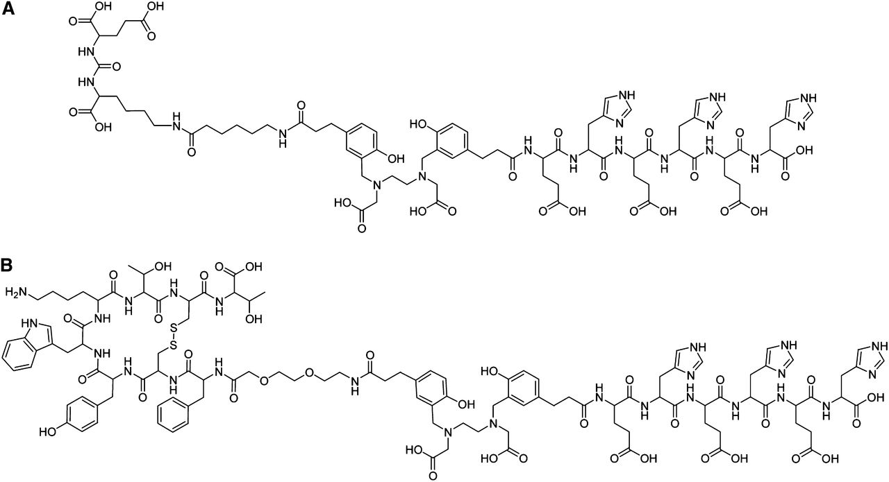
The (EH)3-conjugates were synthesized by incubating the bis-activated ester with 1 equivalent of purified protected TATE-PEG2 or Glu-urea-Lys(Ahx). After 2 h at room temperature, an excess of p[(EH)3] was added and incubated over night at room temperature. TATE-PEG2-HBED-CC-(EH)3 and Glu-urea-Lys(Ahx)-HBED-CC-(EH)3 (Fig. 1) were purified by HPLC. The remaining protecting groups were removed by TFA treatment. Mass spectrometry confirmed the identity (Table 1).

Chemical structures of (EH)3-conjugates investigated: Glu-urea-Lys(Ahx)-HBED-CC-(EH)3 (A) and TATE-PEG2-HBED-CC-(EH)3 (B).
68Ga Labeling
The HBED-CC and HBED-CC-(EH)3-conjugates (1 nmol in 0.1 M N-2-hydroxyethylpiperazine-N′-2-ethanesulfonic acid [HEPES] buffer, pH 7.5, 100 μL) were added to a mixture of a 10-μL HEPES solution (2.1 M) and 20-μµL [68Ga]Ga3+ eluate (∼10 MBq). The pH was adjusted to 4.2 using NaOH. The reaction mixture was incubated at room temperature for 2 min. The radiochemical yield was determined by HPLC.
Cell Culture
PSMA-positive LNCaP cells (CRL-1740; American Type Culture Collection) and somatostatin receptor–positive AR42J cells (European Collection of Cell Cultures) were cultured in RPMI medium supplemented with 10% fetal calf serum and l-glutamine (2 mmol/L) (all from PAA). Cells were grown at 37°C in humidified air with 5% CO2 and were harvested using trypsin–ethylenediaminetetraacetic acid (0.25%, trypsin; 0.02% ethylenediaminetetraacetic acid [Invitrogen]).
Cell Binding and Internalization
The competitive cell-binding assay and internalization experiments were performed as described previously (8). Briefly, the cells (105 per well) were incubated with a 0.2 nM solution of 68Ga-labeled radioligand (TATE-DOTA or Glu-urea-Lys(Ahx)-HBED-CC) in the presence of 12 different concentrations of analyte (0–5,000 nM, 100 μL/well). After incubation, the binding buffer was removed using a multiscreen vacuum manifold (Millipore). Cell-bound radioactivity was measured using a γ-counter (Packard Cobra II; GMI). The 50% inhibitory concentration values were calculated by fitting the data using a nonlinear regression algorithm (GraphPad Software).
For internalization experiments, 105 cells were seeded in poly-l-lysine–coated 24-well cell culture plates 24 h before incubation. After being washed, the cells were incubated with 25 nM of the radiolabeled compound for 45 min at 37°C and at 4°C. Cellular uptake was terminated by washing 4 times with 1 mL of ice-cold phosphate-buffered saline. To remove surface-bound radioactivity, cells were incubated twice with 0.5 mL of glycine-HCl in phosphate-buffered saline (50 mM, pH 2.8) for 5 min. The cells were washed with 1 mL of ice-cold phosphate-buffered saline and lysed using 0.3 N NaOH (0.5 mL). The surface-bound and the internalized fractions were measured in a γ-counter. The cell uptake was calculated as percentage of the initially added radioactivity bound to 106 cells (%ID/106 cells).
Biodistribution
Cells (5 × 10⁶) of either AR42J or LNCaP (in 50% Matrigel; Becton Dickinson) were subcutaneously inoculated into the right trunk of 7- to 8-wk-old female (AR42J) and male (LNCaP) BALB/c nu/nu mice (Charles River). The tumors were allowed to grow until they were approximately 1 cm3. The 68Ga-radiolabeled compounds were injected into a tail vein (1–2 MBq; 0.1–0.2 nmol). At 1 h after injection, the animals were sacrificed. Organs of interest were dissected, blotted dry, and weighed. The radioactivity was measured using a γ-counter and calculated as percentage injected dose per gram (%ID/g). All animal experiments complied with the current laws of the Federal Republic of Germany.
Statistical Aspects
All experiments were performed at least in triplicate. Quantitative data were expressed as mean ± SD. If applicable, means were compared using Student’s t test. P values less than 0.05 were considered statistically significant.
RESULTS
The 68Ga complexation resulted in high radiochemical yields of 99% after a 2-min reaction time at room temperature. The influence of the (EH)3 conjugation on the binding properties of Glu-urea-Lys(Ahx)-HBED-CC and TATE-PEG2-HBED-CC was investigated by analyzing the internalization and cell-binding properties. The results are summarized in Table 1. Although the binding properties of the TATE-PEG2-HBED-CC-(EH)3 are nearly identical with the parental TATE-PEG2-HBED-CC, the affinity of the (EH)3-conjugated PSMA inhibitor was reduced from 10.33 ± 1.2 to 31.80 ± 1.2 nM. On the other hand, the total cell uptake and internalization were slightly enhanced after conjugation.
The (EH)3-tag significantly changed the biodistribution profile and tumor-to-organ ratios of the 2 investigated radiopharmaceuticals (Fig. 2). The kidney uptake of the PSMA inhibitor Glu-urea-Lys(Ahx)-HBED-CC was reduced by a factor of 2.8 from 139.44 ± 21.40 to 49.03 ± 16.34 %ID/g (P = 0.004). Significant reduction was also observed in the liver (1.43 ± 0.19 to 0.51 ± 0.19 %ID/g; P = 0.004) and the blood (0.53 ± 0.04 to 0.31 ± 0.04 %ID/g; P = 0.003). The tumor uptake was not affected by the (EH)3-tag (4.40 ± 0.72 vs. 4.89 ± 1.34 %ID/g for the untagged; P = 0.61).
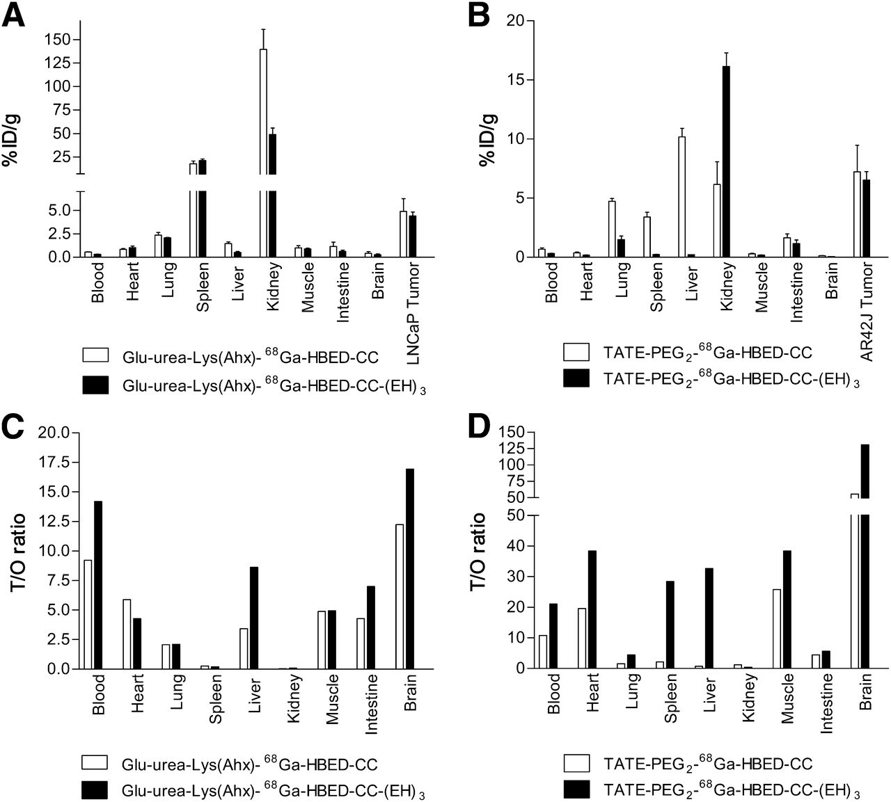
Organ distribution at 1 h after injection of 0.1–0.2 nmol of PSMA inhibitor Glu-urea-Lys(Ahx) labeled with either 68Ga-HBED-CC or 68Ga-HBED-CC-(EH)3 (A) and octreotate-PEG2 (TATE-PEG2) labeled with either 68Ga-HBED-CC or 68Ga-HBED-CC-(EH)3 (B). Data are expressed as mean %ID/g tissue ± SD (n = 3). Graphs in C and D show respective tumor-to-organ (T/O) ratios at 1 h after injection.
In the case of the octreotate construct, an increased kidney uptake (6.15 ± 1.92 to 16.14 ± 1.99 %ID/g; P = 0.003) and a reduced liver accumulation (10.18 ± 0.72 to 0.20 ± 0.01 %ID/g; P < 0.0001) were observed after conjugation of (EH)3. The uptake in the lung (P = 0.001), blood (P = 0.007), and spleen (P = 0.0001) was significantly reduced as well. The tumor uptake of both compounds was nearly identical (7.22 ± 2.26 vs. 6.53 ± 1.24 %ID/g for the (EH)3-conjugate; P = 0.67), resulting in an enhancement of most tumor-to-organ ratios (Fig. 2D).
DISCUSSION
The clinical translation of promising peptidic radiopharmaceuticals is often hampered by radioactivity accumulation in excreting organs. Recently, Hofstrom et al. (5) presented a strategy leading to a significant reduction of the liver uptake of Affibody molecules. The transfer of these findings to peptidic radiopharmaceuticals might further improve their pharmacokinetics and help to facilitate their clinical translation.
In this initial study, the effect of (EH)3 on the biodistribution profile of the somatostatin receptor–binding peptide TATE-PEG2 and the small-molecule inhibitor of PSMA Glu-urea-Lys was investigated. In vitro analyses of the (EH)3-conjugates in comparison to their respective non-(EH)3 counterparts showed similar binding and internalization properties. Remarkably, the affinity of the (EH)3-conjugated PSMA inhibitor was reduced by a factor of 3 whereas the specific cell uptake and internalization were slightly enhanced after conjugation. Former studies (8,13,14) showed that mainly the internalization properties and to a lesser extent the binding affinity determined the in vivo tumor uptake. Consequently, the slight reduction of binding affinity of Glu-urea-Lys(Ahx)-HBED-CC-(EH)3, compared with Glu-urea-Lys(Ahx)-HBED-CC, should not influence the validity of this study in terms of assessing the influence of (EH)3 on biodistribution.
The (EH)3-tag significantly changed the biodistribution profile of the 2 investigated radiopharmaceuticals. In the case of the PSMA inhibitor, the kidney and liver accumulation were significantly reduced. Because the kidney uptake is partly associated with a specific uptake via PSMA expression in the murine organ (15,16), it is essential to compare the tumor uptake of the (EH)3-conjugated PSMA inhibitor with the untagged reference. It was found that the tumor uptake was not significantly affected by the conjugation, indicating that (EH)3 influences a nonspecific process in the kidneys. These data indicate a considerable influence of (EH)3 on the biodistribution profile of the PSMA inhibitor without affecting the tumor-targeting properties. Considering the high clinical impact of PSMA inhibitors, the advantages of gallium-labeled variants for diagnostic purposes, and the limiting high kidney uptake, the present (EH)3-conjugated molecule represents a promising advancement. In particular, Glu-urea-Lys(Ahx)-HBED-CC-(EH)3 might be interesting for prostate cancer therapy with radiometals where low uptake in the liver and kidney is crucial. This result warrants further studies on, for example, different positions of the (EH)3 sequence in the molecule.
The pharmacokinetic properties of HBED-CC–conjugated octreotate-PEG2 were considerably influenced by (EH)3 as well, but in another way. Although both the liver and the kidney uptake of the PSMA inhibitor was reduced, an increased kidney uptake and a considerably reduced liver accumulation were observed. The fact that the liver uptake of TATE-PEG2-HBED-CC was enormously affected by the conjugation of (EH)3 is in good agreement with the findings of Hofstrom et al., who reported the positive effect of the tag on the liver uptake of Affibody molecules (5).
The increase in kidney uptake might be attributed to the extent of the reduction of the hepatic uptake resulting in the redistribution of the activity from the liver to the kidneys. In contrast to Glu-urea-Lys(Ahx)-HBED-CC, (EH)3 was not able to reduce the nonspecific kidney uptake of TATE-PEG2-HBED-CC. Because membranes of renal tubular cells expose negatively charged sites (17), the positively charged lysine in the octreotate sequence might facilitate the accumulation of radioactivity in the kidneys and reduce the effect of the (EH)3-tag. These preliminary findings suggest that predominantly the hepatic accumulation is affected by conjugating (EH)3. The reduction of renal uptake observed with the (EH)3-conjugated PSMA inhibitor Glu-urea-Lys(Ahx)-HBED-CC is potentially restricted to molecules lacking positively charged amino acids.
CONCLUSION
Taken together, the results obtained with 2 important radiopharmaceuticals clearly show the potential of (EH)3 for the improvement of pharmacokinetic properties of peptides and small peptidomimetic molecules. Further studies are required to prove this positive effect on the clearance of a broader selection of radiopharmaceuticals. The availability of a tool facilitating the clearance from critical organs would have high impact in the future development of radiopharmaceuticals and would consequently support the clinical translation process, in particular of those molecules that are limited because of their high liver or kidney uptake.
DISCLOSURE
The costs of publication of this article were defrayed in part by the payment of page charges. Therefore, and solely to indicate this fact, this article is hereby marked “advertisement” in accordance with 18 USC section 1734. We gratefully acknowledge a grant from the DFG (Deutsche Forschungsgemeinschaft; ED234/2-1). No other potential conflict of interest relevant to this article was reported.
Acknowledgments
Biodistribution was kindly performed by Ursula Schierbaum (German Cancer Research Center, Heidelberg, Germant).
Footnotes
Published online Jun. 26, 2013.
- © 2013 by the Society of Nuclear Medicine and Molecular Imaging, Inc.
REFERENCES
- Received for publication September 21, 2012.
- Accepted for publication February 28, 2013.


