


Abstract
Radiolabeled amino acids (AAs) are an important class of imaging agents for PET and SPECT that target the increased levels of AA transport by many tumor cells. System L AA transport has been a major focus of tracer development, and work in this field has led to several tracers that are effective for imaging brain tumors. Emerging data also support the use of certain radiolabeled AAs for neuroendocrine tumors and prostate cancer. Recently, new 18F-labeled AAs have been developed that target transporters other than system L, including system A, glutamine, xCT, and cationic AA transporters. This review provides an overview of this class of molecular imaging agents and highlights the current status of oncologic imaging with radiolabeled AAs in terms of tracer considerations and key clinical applications.
Radiolabeled amino acids (AAs) have been used for human imaging studies for decades, and there is active investigation in this field in part due to recent insights into the role of AA transporters and AAs in tumor metabolism. Beginning in the late 1990s, the genes coding for individual AA transporters have been cloned and characterized. There is now abundant evidence that certain transporter proteins are upregulated in a wide range of neoplastic cells, confer prognostic information, and are potential targets of therapy. The importance of certain AAs, such as leucine, glutamine, and arginine in neoplastic cells, has guided the development of new radiolabeled AAs to probe cancer metabolism.
GENERAL CONSIDERATIONS
Natural α-AAs are an important class of small molecules that play vital roles in many cellular processes including protein synthesis, energy metabolism, cell signaling, carbon sources for cell growth, and neurotransmission. In oncologic imaging applications, radiolabeled AAs target the increased levels of AA transport that occur in many tumor cells compared with normal tissues, thought to be mediated by increased cell surface levels of AA transporters. Although some radiolabeled AAs are incorporated into proteins or have other metabolic fates, tumor uptake and imaging properties reflect primarily the rate and mechanism of transport of the AA in most instances.
AA transporters are membrane-associated proteins coded by family members of the solute carrier (SLC) series of genes that mediate the transfer of AAs across cell membranes. AA transporter families have traditionally been characterized functionally as transport systems (e.g., system L, system A, system N, system ASC, and system xCT), many of which contain multiple family members. Over 20 distinct AA transport systems have been identified, with different substrate specificities, tissue expression patterns, dependence on cotransport of ions such as sodium, hormonal regulation, mechanisms of transport, and biologic significance in cancer. Two AA transporters, the system L transporter type 1 (LAT1, SLC7A5) and the system ASC transporter type 2 (ASCT2, SLC1A5), are upregulated in a wide range of human cancers, are associated with worse prognosis, and are potential targets for therapy (1,2). There is growing evidence that these and other AA transporters and their substrates interact with the mammalian target of rapamycin pathway, which regulates protein synthesis and cell proliferation (3,4). Radiolabeled AAs could potentially be used to monitor response to therapies targeting mammalian target of rapamycin signaling.
Structural and radionuclide considerations often influence the design of novel radiolabeled AAs and the selection of specific AA tracers for a study. Natural AAs or their close analogs may be preferred if specific aspects of their transport or biochemistry are important for a particular application. In many instances, nonnatural, nonmetabolized AAs such as O-(2-18F-fluoroethyl)-l-tyrosine (FET) and anti-1-amino-3-18F-fluorocyclobutane-1-carboxylic acid (FACBC) have advantages over natural AAs, including the ability to incorporate longer-lived radionuclides such as 18F and 123I, simplified radiosynthetic methods, and lack of radiolabeled metabolite formation, which simplifies kinetic analysis and avoids potentially confounding accumulation of activity in nontarget tissues. All AAs theoretically can be labeled with 11C, and the 20-min half-life of 11C is well suited to the time to peak tumor uptake, which typically occurs within 15–20 min after administration. The use of 18F takes advantage of the longer 110-min half-life of this radionuclide, which makes batch production and remote distribution feasible. However, 18F is not present in natural mammalian AAs and in some cases substantially alters the biologic properties of an AA. 123I (half-life, 13.2 h) has been used less frequently than 11C or 18F for labeling AAs, but several 123I-labeled AAs have been developed for SPECT imaging, including 3-123I-iodo-α-methyl tyrosine (IMT).
APPLICATIONS OF RADIOLABELED AAS FOR ONCOLOGIC IMAGING
A wide range of radiolabeled AAs has been developed and used for preclinical and clinical oncologic imaging studies (5). The structures of selected radiolabeled AAs for oncologic imaging are depicted in Figure 1. Much of this work has focused on aromatic AA derivatives, including 18F-FET, 6-18F-fluoro-3,4-dihydoxy-l-phenylalanine (FDOPA), 123I-IMT, and 3-18F-fluoro-α-methyltyrosine (FMT), which target system L AA transport. Other nonaromatic system L substrates include 11C-MET and the cycloleucine analog, 18F-FACBC. Tracers targeting other AA transport systems, including system A, glutamine, glutamate, and cationic AA transporters, have been developed but have not been as extensively evaluated in human studies.
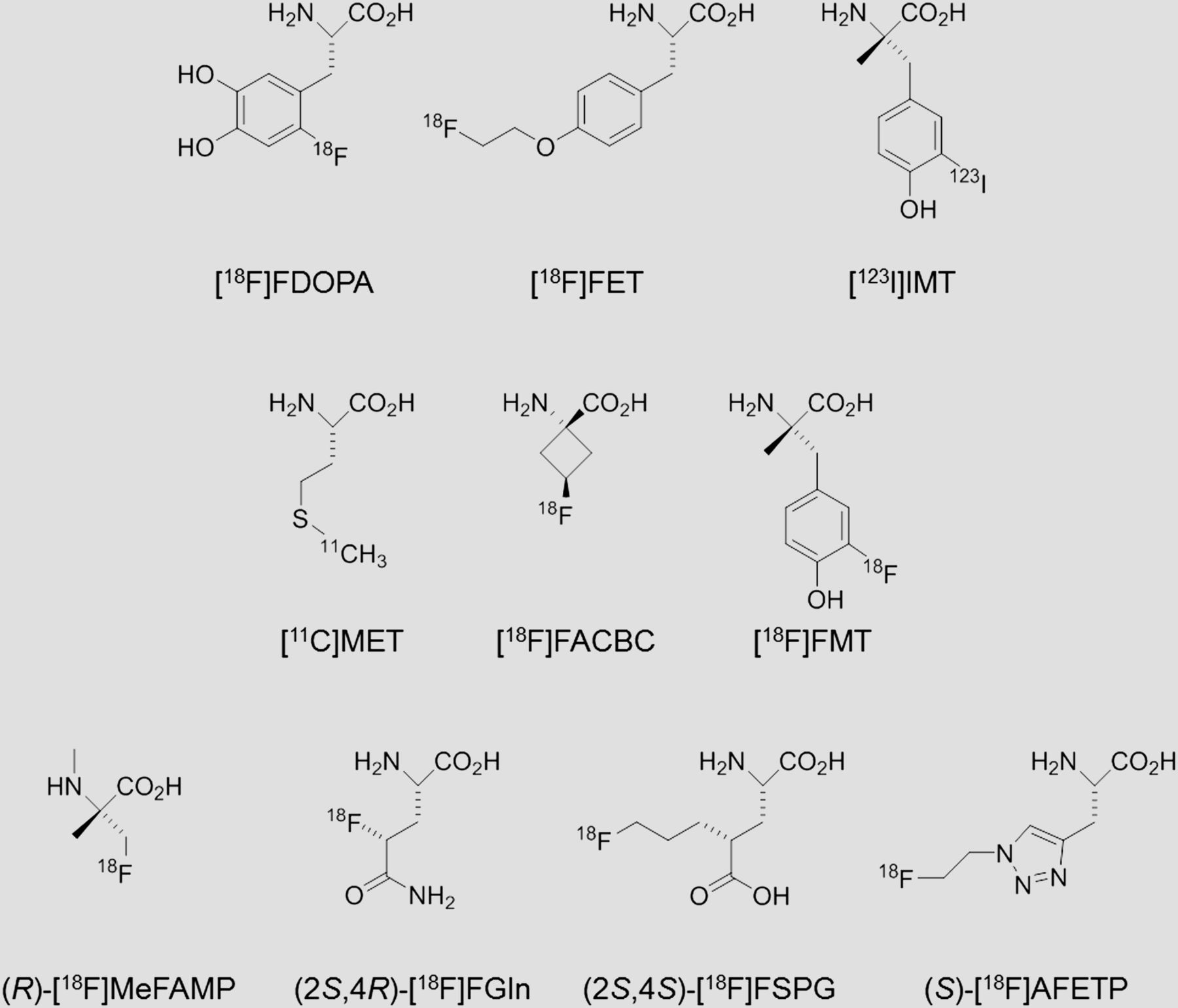
Structures of selected radiolabeled AAs. System L substrates: 18F-FDOPA, 18F-FET, 123I-IMT, 11C-MET, and 18F-FMT. Systems L and ASCT2 substrate: 18F-FACBC. System A substrate: (R)-18F-MeFAMP. Glutamine transporter systems: (2S,4R)-18F-FGln. System xCT substrate: (2S,4R)-18F-FSPG. Cationic and neutral AA transporter systems: (S)-18F-AFETP.
Brain Tumors
Neuroimaging plays a critical role in primary and metastatic brain tumors, including diagnosis, treatment planning, monitoring response to therapy, and assessing for recurrence. Contrast-enhanced MR imaging is the primary imaging modality for evaluating brain tumors but has limited accuracy for defining tumor boundaries and volumes, evaluating the nonenhancing portions of gliomas, and differentiating treatment effects such as radiation necrosis from viable tumor. 18F-FDG PET is limited by high physiologic uptake in normal brain, and both MR imaging and 18F-FDG PET have limited specificity for distinguishing recurrent tumor from radiation necrosis. Two major advantages of system L AA transport substrates for brain tumor imaging are their ability to cross the intact BBB and visualize the entire tumor volume and their relatively low uptake and retention in normal brain, compared with 18F-FDG. These properties of radiolabeled AAs targeting system L transport complement contrast-enhanced MR imaging by providing a more accurate visualization of the entire tumor volume.
Of the radiolabeled AAs targeting system L, 11C-MET and 18F-FET have the most data supporting their use for imaging brain gliomas (6,7). An example of 11C-MET uptake in recurrent glioblastoma is shown in Figure 2. Other tracers used for this application include 123I-IMT and 18F-FDOPA (8). There are relatively few studies directly comparing these tracers for tumor imaging, but the available data suggest they behave similarly in terms of tumor visualization. Both 18F-FET and 123I-IMT are metabolically stable, which may be advantageous for the analysis of tracer kinetics. There is a substantial body of data showing that 11C-MET and 18F-FET PET studies can improve the diagnostic yield of brain tumor biopsy, more accurately define gross tumor volumes and margins than MR imaging alone, and follow response to chemotherapy in nonenhancing gliomas (6,9). Changes in tumor volumes are typically observed earlier with AA PET than with MR imaging, particularly in nonenhancing gliomas, and appear to be more predictive of response to therapy than changes in absolute tracer uptake. There are also studies suggesting that the kinetics of 18F-FET uptake in gliomas can distinguish high-grade from low-grade tumors (10). 18F-FET and 11C-MET may also be useful for distinguishing recurrent brain tumors from radiation necrosis after radiation therapy (11,12), but additional data are needed to better define the accuracy of these tracers for this purpose in clinical practice.

Glioblastoma imaging with 11C-MET. Contrast-enhanced T1-weighted MR (A) and 11C-MET PET (B) images in patient with glioblastoma treated with surgical resection and radiation therapy, now with suspected recurrence. 11C-MET PET image demonstrates focally increased uptake within area of contrast enhancement on MR, and this lesion was pathologically proven to be recurrent glioblastoma on resection. In patients with radiation necrosis, lower 11C-MET uptake was observed (images not shown). (Reprinted from (12).)
Neuroendocrine Tumors
Neuroendocrine tumors encompass a range of neoplasms, including carcinoid tumors, pancreatic islet cell tumors, pheochromocytomas and paragangliomas, neuroblastoma, and small cell lung cancer. A common feature of these tumors is the potential for the production of biologically active substances that can lead to severe symptoms, morbidity, and even death. The accurate localization of the primary tumor and metastases is important for selecting the appropriate therapeutic approach in these patients. Uptake of 18F-FDG in neuroendocrine tumors is variable, and well-differentiated neuroendocrine tumors often have low levels of 18F-FDG uptake. Of the radiolabeled AAs, 18F-FDOPA has been used the most extensively in neuroendocrine tumors. The enzyme aromatic AA decarboxylase (AADC) is active in many neuroendocrine tumors, and metabolism of 18F-FDOPA by AADC likely contributes to its favorable imaging properties because other system L substrates are not as effective for neuroendocrine tumor imaging (13).
Of the neuroendocrine tumors, 18F-FDOPA appears to best suited for imaging carcinoid tumors, pancreatic insulinomas, and neuroblastoma and can detect lesions not identified by CT, MR imaging, or other tracers used clinically for neuroendocrine tumor imaging (13,14). Studies comparing the utility of FDOPA with that of somatostatin receptor scintigraphy have demonstrated that somatostatin receptor scintigraphy typically demonstrates higher sensitivity for neuroendocrine tumor detection on a per-lesion basis, although 18F-FDOPA can detect some neuroendocrine tumors that are not detected with somatostatin receptor scintigraphy (15,16).
Prostate Cancer
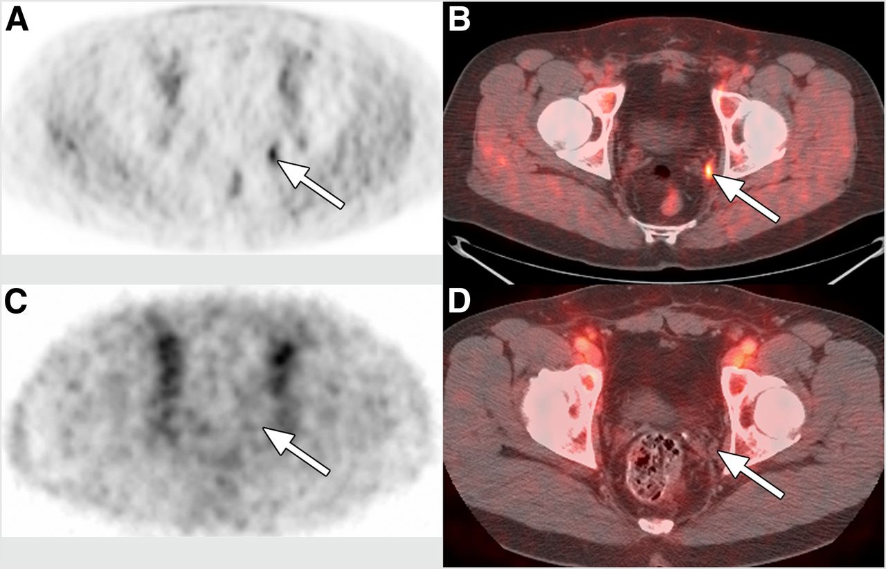
Prostate cancer is a common malignancy in men and the second leading cause of cancer-related mortality in the United States. The management of prostate cancer is challenging, because many prostate cancers are indolent whereas others are aggressive. Conventional imaging with CT and MR imaging has limited accuracy for the detection of the primary tumor and locoregional lymph node metastases. Unlike many other cancers, 18F-FDG uptake in prostate cancer is often low. The clinically available monoclonal antibody 111In-capromab, targeting the prostate membrane–specific antigen, is used for the detection of recurrent prostate cancer but has limited accuracy. In response to the great need for improved imaging techniques, several classes of PET tracers have been developed for prostate cancer imaging. Recent studies with anti-18F-FACBC have shown that this AA tracer can accurately detect prostate cancer within the gland itself and in pelvic lymph node metastases with specificity and sensitivity superior to 111In-capromab SPECT/CT (17). An example of 18F-FACBC PET/CT in a patient with recurrent prostate cancer is shown in Figure 3. One advantage of anti-3-18F-FACBC over other radiolabeled AAs is that the relatively low levels of urinary excretion simplify imaging of the pelvis. In vitro uptake studies on prostate cancer cell lines indicate that 18F-FACBC is a substrate for both LAT1 and ASCT2 (18).

Prostate cancer imaging with 18F-FACBC. 18F-FACBC PET (A) and 18F-FACBC PET/CT (B) images from a patient with suspected recurrence of prostate cancer demonstrate focal uptake in left pelvic lymph node metastasis that measures less than 1 cm in short-axis diameter (indicated with arrows). Biopsy demonstrated recurrent prostate cancer in this location. In contrast, 111In-capromab pendetide SPECT (C) and SPECT/CT (D) images do not show uptake in this metastatic lymph node (false-negative). (Reprinted with permission from (17).)
Other Cancers
In general, radiolabeled AAs have not performed as well as 18F-FDG PET for detection and staging of cancers other than those of the brain, neuroendocrine system, and prostate. The main limitation is decreased sensitivity for the detection of primary tumors and metastases, compared with 18F-FDG. One of the potential limitations of system L substrates is the ability of these transporters to mediate both influx and efflux of substrates, limiting the tumor-to-background ratios that can be achieved. There is growing interest in using radiolabeled AAs to better characterize the biologic behavior of cancers. These types of applications have the potential to provide information relevant to diagnosis, prognosis, and therapy. For example, the amount of uptake of the system L substrate 3-18F-FMT in lung adenocarcinoma is negatively correlated with survival (19). These results fit with the observation in many human cancers that higher levels of LAT1 messenger RNA and protein are associated with higher mortality.
EMERGING CLASSES OF RADIOLABELED AAS
The roles of radiolabeled AAs that are substrates for transporters other than system L are less well defined. Recently, AA tracer development has explored several different transport systems for oncologic imaging, including system A (20), system xCT (21,22), glutamine transporters (23,24), and cationic AA transporters (25,26). The availability of these new tracers will provide important tools to probe the roles of these AA transport systems in specific cancers and for particular applications.
Each of these transport systems has unique biologic features and the potential to provide clinically useful information through PET and SPECT imaging studies. System A transport is upregulated during cell growth and by a variety of cell stressors. Additionally, the fact that system A can concentrate its substrates intracellularly may provide higher tumor uptake than can be achieved with system L substrates. Glutamine is a key metabolic substrate that can provide energy, tricarboxylic acid cycle intermediates, and carbon for cell growth (3). As mentioned previously, the ASCT2 transporter plays important roles in tumor biology and is thought to be a key mediator of glutamine uptake by many tumor cells. System xCT is a cystine–glutamate exchange transporter that is upregulated in many cancers and is a biomarker for oxidative stress. Cationic AAs, including arginine, lysine, and histidine, are essential nutrients in proliferating cells. Arginine serves as a substrate for polyamine synthesis during cell proliferation and for nitric oxide synthesis. Additionally, therapies that deplete extracellular arginine are in clinical trials for cancers such as melanoma and hepatocellular carcinoma, and radiolabeled cationic AA transport substrates could serve as biomarkers for predicting and monitoring response to therapy.
CONCLUSIONS AND FUTURE DIRECTIONS
A range of radiolabeled AAs has been developed for tumor imaging. System L substrates such as 11C-MET and 18F-FET are well established for imaging brain tumors and provide better estimates of gross tumor volumes and margins, better visualization of nonenhancing gliomas, and potentially earlier and more accurate assessment of response to therapy than is possible with MR imaging alone. Other important imaging applications with radiolabeled AAs include neuroendocrine tumor imaging with 18F-FDOPA and prostate cancer imaging with 18F-FACBC. Tracers targeting other AA transport systems, including system A, glutamine, xCT, and cationic AA transporters, are under development, but their clinical utility remains to be established. Optimal application of this class of tracers for oncologic imaging will require matching the transport and metabolic properties of a given radiolabeled AA with the relevant tumor biology and clinical question.
DISCLOSURE
No potential conflict of interest relevant to this article was reported.
Footnotes
Published online May 24, 2013.
- © 2013 by the Society of Nuclear Medicine and Molecular Imaging, Inc.
REFERENCES
- Received for publication January 15, 2013.
- Accepted for publication May 2, 2013.


Jump to section
Related Articles
Cited By...
- 18F-Branched-Chain Amino Acids: Structure-Activity Relationships and PET Imaging Potential
- Prospective Clinical Trial of 18F-Fluciclovine PET/CT for Determining the Response to Neoadjuvant Therapy in Invasive Ductal and Invasive Lobular Breast Cancers
- 18F-FET PET Uptake Characteristics in Patients with Newly Diagnosed and Untreated Brain Metastasis
- Metabolic Imaging of Glutamine in Cancer
- Evaluation of Prostate Cancer with Radiolabeled Amino Acid Analogs
- 18F-Fluciclovine (FACBC) and Its Potential Use for Breast Cancer Imaging
- Development of a Widely Usable Amino Acid Tracer: 76Br-{alpha}-Methyl-Phenylalanine for Tumor PET Imaging
- Anti-1-Amino-3-18F-Fluorocyclobutane-1-Carboxylic Acid: Physiologic Uptake Patterns, Incidental Findings, and Variants That May Simulate Disease