


Abstract
When antisense oligomers are intracellular, they migrate to and are retained in the nucleus of tumor cells and therefore may be used to carry Auger electron–emitting radionuclides such as 111In for effective tumor radiotherapy. Methods: Our nanoparticle consists of streptavidin that links 3 biotinylated components: the antiHer2 antibody trastuzumab (to improve pharmacokinetics), the tat peptide (to improve cell membrane transport), and the 111In-labeled antiRIα messenger RNA antisense morpholino (MORF) oligomer. Results: As evidence of unimpaired function, tumor cell and nuclear accumulations were orders of magnitude higher after incubation with 99mTc-MORF/tat/trastuzumab than after incubation with free 99mTc-MORF and significantly higher with the antisense than with the sense MORF. In mice, tumor and normal-tissue accumulations of the 99mTc-MORF/tat/trastuzumab nanoparticle were comparable to those of free 99mTc-trastuzumab, confirming the improved pharmacokinetics due to the trastuzumab component. Although kidneys, liver, and other normal tissues also accumulated the nanoparticle, immunohistochemical evaluation of tissue sections in mice receiving the Cy3-MORF/tat/trastuzumab nanoparticle showed evidence of nuclear accumulation only in tumor tissue. In a dose escalation study, as measured by the surviving fraction, the nanoparticle significantly increased the kill of SK-BR-3 breast cancer Her2+/RIα+ cells, compared with all controls. Conclusion: Significant radiation-induced antisense-mediated cytotoxicity of tumor cells in vitro was achieved using an Auger electron–emitting antisense MORF oligomer administered as a member of a 3-component streptavidin-delivery nanoparticle.
When administered intravenously, free antisense DNAs and other oligomers accumulate at much higher levels in normal tissues than in tumor tissues (1). Thus, poor delivery, when added to inefficient intracellular transport and entrapment in tumor cells, remains a serious obstacle to the use of radiolabeled antisense oligomers (2). Recognizing that antisense oligomers will require chemical modification to address these issues, our laboratories are developing streptavidin-delivery nanoparticles. If oligomers are biotinylated, they may be combined with cell-transporting peptide carriers (3) and with a variety of antitumor antibodies via streptavidin without the need for covalent conjugation (4). We have already demonstrated that the presence of streptavidin apparently does not adversely influence the properties of the cell-transporting carrier peptides tat, polyarginine, or cholesterol (3); the antisense function of the radiolabeled morpholino (MORF) oligomer (3,4); or the function of the antitumor antibody (4). In the course of these and other studies, we have also shown that the antisense oligomer migrates to the nucleus of tumor cells when incubated alone (5) and as the nanoparticle (4).
Although 2-component nanoparticles consisting of the radiolabeled oligomer and carrier peptides such as tat are under development here for imaging purposes, 3-component nanoparticles in which an antitumor antibody has been included are under development for radiotherapy purposes. The antitumor antibody improves pharmacokinetics by providing tumor targeting; the tat peptide improves cell accumulation without entrapment; and the antisense oligomer, radiolabeled with an Auger electron–emitting radionuclide such as 111In, carries its radiolabel to the nucleus and retains it therein.
These laboratories have previously reported that the pharmacokinetics of a radiolabeled antisense DNA oligomer were not markedly changed by linking to streptavidin (1). However, the intent of this investigation was to intentionally alter the pharmacokinetics of a radiolabeled antisense MORF in a favorable direction by the addition of the antitumor antibody trastuzumab (Herceptin; Genentech Inc.). The question of whether the pharmacokinetics were improved therefore required investigation and was approached by comparing the pharmacokinetics of the 99mTc-MORF 3-component nanoparticle with that of the free 99mTc-labeled trastuzumab.
Because the nanoparticle will certainly accumulate to some degree in normal organs such as liver, spleen, and kidneys, the question of whether the antisense MORF within the nanoparticle will accumulate in the nucleus of these normal tissues needed to be addressed. For this aspect of the investigation, the MORFs were labeled with the Cy3 fluorophore, and immunohistochemistry was applied to tissue sections.
This investigation was conducted to determine whether the pharmacokinetics and nuclear migration properties of the nanoparticle are favorable and whether the antisense MORF labeled with the Auger electron–emitter 111In, when incubated with tumor cells in culture as a 3-component nanoparticle, will provide effective radiation-induced cytotoxicity by an Auger electron–mediated antisense mechanism.
MATERIALS AND METHODS
Chemicals and Cell Lines
The antisense MORF (base sequence 5′-GCG TGC CTC CTC ACT GGC) was directed against the RIα messenger RNA (mRNA) (GeneBank accession number NM_002734) (4). The antisense and the corresponding sense MORFs were obtained (from GeneTools Inc.) biotinylated on the 3′ equivalent end via a 6-amino hexanoic acid linker and with a primary amine on the 5′ equivalent end. Streptavidin was purchased from Sigma. The biotinylation reagent sulfo-NHS-LC-biotin was purchased from Pierce and used to biotinylate the trastuzumab antibody. The trastuzumab (Herceptin) was obtained from Genentech Inc. The tat peptide (GRKKRRQRRR) was obtained (from 21st Century Biochemicals) biotinylated on the N-terminal end via a 6-amino hexanoic acid linker as the native L isomer. The S-acetyl NHS-MAG3 was synthesized in house (6). The NHS-Cy3 was obtained from GE Healthcare. The SK-BR-3 (Her2+/RIα+) cell line was purchased from ATCC and was cultured and maintained in the McCoy 5a medium with 10% fetal bovine serum (FBS). The SUM190 (Her2+/RIα+) cells were obtained from Asterand Co. The 99mTc-pertechnetate was eluted from a Bristol-Myers Squibb Medical Imaging Inc. 99Mo–99mTc generator. The 111InCl3 was from Perkin Elmer Life Science Inc. All other chemicals were of reagent grade and used without purification.
Labelings and Nanoparticle Preparations
The amine-derivatized and biotinylated MORFs were conjugated with NHS-MAG3 as previously described (3) and with NHS-Cy3 in the identical manner (4). The preparation and testing of the nanoparticles was as previously described (3). Preparation of the diethylenetriaminepentaacetic acid (DTPA)–coupled and 111In-labeled MORFs was also described previously (7). The antibody was conjugated with NHS-MAG3 at a 3:1 MAG3:trastuzumab ratio and, after purification, was radiolabeled with 99mTc as previously described (6).
Subcellular Distribution
The SK-BR-3 cells were plated (4 × 10⁶ cells per well) in 6-well plates and incubated with 99mTc-labeled antisense or sense MORF as the 3-component nanoparticles and as the free MORFs, each at 50 nM, in McCoy 5a medium with 1% FBS for 3 h at 37°C. The percentage of added radioactivity that became incorporated in the cells was first measured. Thereafter, the nuclear and other fractions were separated by Nuclei EZ Prep kit (Sigma) according to the manufacturer's instruction, and each was counted separately for radioactivity in an automatic NaI (T1) well counter along with a standard. These measurements were made with 99mTc rather than 111In as radiolabel for convenience.
Cell Viability Assay
To help ensure that cytotoxicity would be specifically due to an Auger electron–mediated antisense mechanism, before performing the escalating dose trial we first selected a concentration of unlabeled nanoparticle that provides minimal nonspecific cytotoxicity as measured by reduced cell viability (WST-1; Roche Molecular Biochemicals). In triplicate, SK-BR-3 cells were seeded (104 cells per well) into 96-well plates and incubated with concentrations of the unlabeled study nanoparticle from 100 to 1,000 nM in McCoy 5a medium with 1% FBS for 1 d at 37°C. Control cells were cultured with phosphate-buffered saline (PBS) in place of the nanoparticle. After the incubation period, cells were washed with 100 μL of fresh medium before 10 μL of the formazan reagent were added to each well and the plates were incubated for another 3 h at 37°C. The absorbance of the colored formazan was determined using a SpectraMax M5/M5e Microplate reader (Molecular Devices Corp.) at 440 nm. An increasing concentration of the formazan dye signifies increasing cell proliferation and therefore cell viability and is a sensitive method of measuring cytotoxicity. Because a minimum concentration of about 50 nM was considered necessary to carry sufficient 111In in the escalating dose trial, and because the cell viability assay showed that a 50 nM concentration would provide minimal cytotoxicity, this concentration was selected for all subsequent studies. The cell cytotoxicity as measured by this assay was determined over 3 d at this concentration.
Membrane Integrity Assay
Because only a few percent of the radiolabeled nanoparticles during each cell incubation become incorporated, a concern is that the 111In remaining in the medium may provide nonspecific cytotoxicity by irradiating the cell membrane. Therefore, an assay of membrane integrity was used on cells incubated with the unlabeled nanoparticle at 50 nM but mixed with 111In as the DTPA chelate. In this chemical form, 111In is not incorporated into cells, because of its negative charge and hydrophilicity. The assay of membrane integrity was performed according to the manufacturer's instructions. Briefly, the cells were cultured in 96-well plates (104 cells per well) in the McCoy 5a medium with 10% FBS. On the following day, the cells were treated with nanoparticles and controls, all at 50 nM in the McCoy's 5a medium with 1% FBS. The cells were also incubated with PBS as a negative control and with 1% Triton X-100 as a positive control. After 1 d, the plates were centrifuged at 200g for 5 min, 100 μL of the medium from each well were transferred to fresh 96-well plates, 100 μL of lactate dehydrogenase (LDH) assay mixture (Roche) were added, and the plates were incubated at room temperature for up to 30 min before the LDH release was measured at 492 nm. Thus, the LDH released from untreated cells into the culture medium was compared with cells incubated with both the antisense and the sense MORFs added freely, added as the MORF 1-component, added as the MORF/tat 2-component, and added as the MORF/tat/trastuzumab 3-component nanoparticles and streptavidin alone, all unlabeled and all at 50 nM.
Because cells incubated with a sufficiently high dose of 111In-labeled nanoparticles in the medium will certainly suffer nonspecific cytotoxicities related to effects such as cell membrane disruption, the membrane integrity assay was then applied to cells incubated at 50 nM, in this case with the 111In-labeled antisense 3-component nanoparticle at 3 concentrations (1.11, 2.04, and 6.48 MBq/well). The assay was also applied to cells incubated with the antisense 3-component nanoparticle, unlabeled, and mixed with 111In-DTPA at the same concentrations. After treatment for 1 d, 100 μL of the culture supernatant were collected and transferred into new 96-well flat-bottom plates and stored at 4°C for 2–3 wk for complete 111In radioactivity decay. Thereafter, the supernatants were assayed for LDH release as described above.
Dose Escalation Trial
As described above, the initial studies required to establish a safe concentration (50 nM) of nanoparticle and a safe dose of 111In (1.11 MBq/well) free of nonspecific cytotoxicity were performed with the cell viability and membrane integrity assays. Thereafter, the cytotoxicity of the 111In nanoparticles could be evaluated with some assurance that the results would be due specifically to an Auger electron irradiation–mediated antisense mechanism. For the dose escalation trial, a cell clonogenic survival assay was used that measures the capability of treated cells to survive and replicate after treatment and that is often considered the gold standard of cytotoxicity measurements (8). Dose escalations were performed on cells grown in 6-well plates (4 × 10⁶ cells per well) with regular medium to 70% confluence and incubated at 50 nM with the 111In-labeled antisense and sense 3-component nanoparticles and the unlabeled nanoparticles mixed with free 111In-DTPA. Measurements were made for 111In concentrations from 0 to 1.11 MBq/well for 1 d at 37°C before the cells were washed with PBS twice, trypsinized, and suspended in 10% FBS/McCoy 5a medium. An aliquot with about 3,000 cells from each well was added to a new 60-mm tissue culture dish and incubated at 37°C with fresh medium added once at day 7. At day 14 of incubation, the cells were fixed with 95% ethanol and stained with 0.1% crystal violet. The results are presented as the surviving fraction defined as the ratio between the number of viable colonies with a particular incubate, compared with the number for cells incubated with PBS as control. In a second study, the unlabeled nanoparticle was used as the control. In this manner, the cytotoxicity of the identical nanoparticle could be compared both labeled and unlabeled to provide further assurance against nonspecific cytotoxicity due to the nanoparticles themselves in evaluating the cytotoxicity of the 111In-labeled antisense and sense nanoparticles.
Animal Studies
The pharmacokinetics of the 99mTc-labeled antisense MORF nanoparticle and 99mTc-labeled trastuzumab were compared in tumor-bearing nude mice. Eight female nude mice (NIH Swiss, 30–40 g; Taconic Farms) at 7 wk old were each injected subcutaneously in the left thigh with a 50-μL suspension containing 106 SUM190 cells mixed with 5 mg of Matrigel per milliliter and were used for imaging and biodistribution studies 4 wk later when the tumors reached about 1 cm in diameter. Four mice each received 14.8 MBq of 99mTc-antiRIαMORF/tat/trastuzumab nanoparticle containing 23 μg of trastuzumab in 200 μL of PBS via a tail vein. The 4 remaining mice each received the identical weight of trastuzumab radiolabeled with 14.8 MBq of 99mTc in 200 μL of PBS but as the free antibody. One animal was selected from both groups for imaging on a NanoSPECT/CT camera (Bioscan). The mice were sacrificed by cervical dislocation after anesthesia with inhalation of isoflurane at 32 h. The radioactivity that had accumulated in tissues or organs of interest was measured by removing the tissues or organs, followed by counting on a NaI (Tl) automatic γ-counter.
Because the nanoparticle was expected to accumulate to some degree in normal organs such as liver, spleen, and kidneys, it was necessary to determine whether the antisense MORF accumulated in these tissues as the nanoparticle also migrated to the nucleus. For this aspect of the investigation, the nanoparticles were prepared with the antisense and sense MORFs labeled with the Cy3 fluorophore, and immunohistochemistry was applied to tissue sections removed at 24 h. In groups of 3, female nude mice prepared as before received either the antisense or the sense Cy3-MORF as the MORF/tat/trastuzumab nanoparticles containing 1 μg of MORF in 100 μL of PBS via a tail vein. An additional 3 animals received only 100 μL of PBS. Mice were sacrificed 24 h thereafter, and tumor and organs were removed. Tissues were fixed in 10% buffered formalin for 24 h and then embedded in paraffin. Paraffin sections were cut to 5 μm and deparaffinized by 2 washes of xylene for 10 min each and then in 100% ethanol twice for 2 min each. The slides were heated in a microwave at 90°C for 10 min in Retrievagen B solution (pH 9.5; BD Pharmingen) and then cooled to room temperature. After rinsing in PBS, endogenous peroxidase activity was blocked by incubation with dual endogenous enzyme block buffer for 10 min. The slides were then washed with 2 to 3 changes of distilled water and, because of difficulties in observing the Cy3 fluorescence, incubated with sheep horseradish peroxidase–conjugated anticyanine antibody from Abcam (dilution of 1:50 of 1 mg/mL stock) overnight at room temperature. After being stained with horseradish peroxidase developing solution, all samples were counterstained with hematoxylin (blue stain) to stain the nucleus and were mounted in water-soluble medium.
RESULTS
Subcellular Distribution
After a 3-h incubation of cells with 99mTc-labeled antisense and sense MORFs added as the nanoparticles or alone at 50 nM, the percentage of radioactivity added to the well that accumulated in the intact cell and the percentage added to the well that accumulated in the nucleus were determined. As shown in Table 1, because of the trastuzumab, the percentage of 99mTc-MORF accumulating in cells was orders of magnitude higher when added as the nanoparticle, compared with alone, and a remarkable 90%−93% of this accumulation was in the nucleus after only 3 h. Consistent with multiple observations from these laboratories, the cellular accumulation was significantly higher (P < 0.05) for the antisense than for the sense nanoparticle. The MORF accumulation in the nucleus was 13-fold higher than in the cytoplasm when incubated as the antisense nanoparticle and 10-fold higher when incubated as the sense nanoparticle.
Percentage of 99mTc in SK-BR-3 Cells and in Nucleus of Cells When Incubated for 3 Hours as Radiolabeled AntiRIα Antisense or Sense MORF and as 3-Component Nanoparticles or Free
Cell Viability Assay
The WST-1 assay measures cell proliferation as an indication of cell viability and may therefore be used as a sensitive measure of cytotoxicity. When incubated with the unlabeled antisense MORF/tat/trastuzumab nanoparticle over 1 d at 100, 500, and 1,000 nM, the percentage viability of SK-BR-3 cells was 82.9 ± 0.40, 82.5 ± 6.05, and 59.4 ± 2.81, respectively (n = 4). Thus, cells incubated at the 50 nM concentration of this investigation will not confuse nonspecific with specific cytotoxicity by this assay.
As shown in absorbance-versus-time curves of Figure 1, there was no significant difference (P > 0.05) in cell viability at any time between cells incubated with unlabeled antisense compared with sense nanoparticle. Furthermore, whereas there was a significant difference between cells incubated with the nanoparticles and PBS at 1 d, this difference disappeared thereafter. The results of both studies reveal that the unlabeled nanoparticles show minimal nonspecific cytotoxicities by this assay at this concentration.
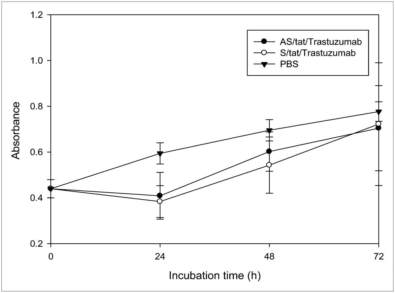
Absorbance as measure of cell viability for SK-BR-3 cells incubated with unlabeled antisense (AS) and sense (S) nanoparticles at 50 nM and PBS over 3 d. There was no significant difference between antisense and sense nanoparticles at any time and no significant difference (P > 0.05) between nanoparticles and PBS after 24 h. Error bars signify SD of mean (n = 4).
Membrane Integrity Assay
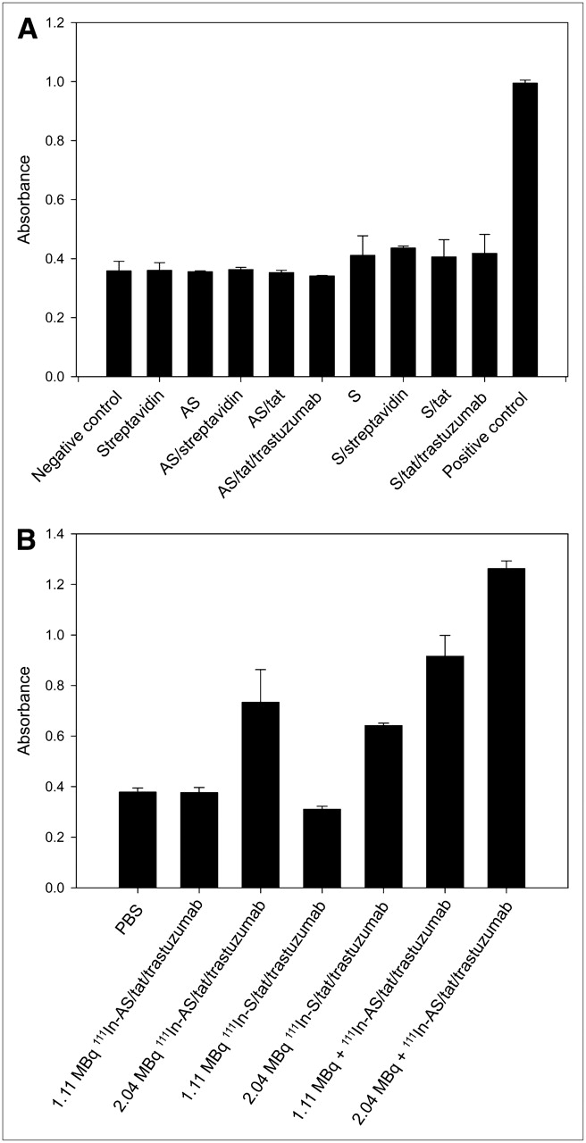
The membrane integrity (LDH) assay is reported to be less sensitive to cytotoxicity than the cell viability (WST-1) assay (9) but has the advantage of measuring the radiobiologic effects resulting from membrane irradiation with low-energy electrons emitted by extracellularly decaying 111In. The effect of the nanoparticles at 50 nM on the membrane integrity of the SK-BR-3 cells was determined by measuring the release of LDH into the cell medium by its absorption at 492 mm. Once again, it was first necessary to ensure that the unlabeled nanoparticle showed no nonspecific cytotoxicity by this assay at the 50 nM concentration. As shown in Figure 2A, when incubated unlabeled at 50 nM, neither the free MORFs nor the nanoparticles, whether antisense or sense, increased the LDH release, compared with PBS (negative control), and all samples showed significantly less release than cells incubated with 1% Triton X-100 (positive control) during the 1-d study. The slightly higher absorbances of the sense samples than of the antisense samples in the figure are not significant (P > 0.05).

(A) Histograms showing cytotoxicity related to membrane integrity in SK-BR-3 cells incubated for 1 d with unlabeled antisense (AS) or sense (S) MORF alone or as MORF 1-component, MORF/tat 2-component, MORF/tat/trastuzumab 3-component nanoparticles, or free MORF without streptavidin or streptavidin alone, all at 50 nM. Results are also presented for PBS as negative control and Triton X-100 as positive control. Error bars signify SD of mean (n = 3). (B) Histograms showing cytotoxicity related to membrane integrity in SK-BR-3 cells incubated for 1 d with 111In-labeled antisense and sense MORF as MORF/tat/trastuzumab 3-component nanoparticles at either 1.11 or 2.04 MBq/well. Results are also presented for cells incubated with unlabeled nanoparticles but mixed with free 111In-DTPA at both concentrations and for cells incubated with PBS as negative control. Error bars signify SD of mean (n = 3).
Because a sufficiently high dose of 111In in the cell medium will provide rapid nonspecific cytotoxicity by decreasing membrane integrity, cells were incubated for 1 d with the radiolabeled study and control nanoparticles but also with the unlabeled nanoparticles mixed with 111In-DTPA at the same concentration. Figure 2B shows that when compared with incubations with PBS, by LDH release, there was no significant difference in cytotoxicity among cells incubated with the 111In-labeled antisense or sense nanoparticles at 1.11 MBq/well. However, both labeled nanoparticles increased cytotoxicity (P < 0.01), compared with PBS, when incubated at 2.04 MBq/well and higher. Accordingly, 1.11 MBq/well was considered an upper limit on 111In dose for the subsequent dose escalation study. Surprisingly, the cytotoxicity of the unlabeled nanoparticles mixed with free 111In-DTPA was significantly higher (P < 0.01), compared with the labeled nanoparticles, at both 1.11 and 2.04 MBq/well as shown. These observations may be explained by synergy, possibly between the antibody and the radiation acting on the cell membrane (10).
Dose Escalation Trial
The results in Figure 3A are reported as the surviving fraction, the ratio of cells capable of forming colonies when incubated with the labeled nanoparticle, compared with cells incubated with PBS. As shown, the surviving fraction for both nanoparticles decreased with increasing 111In concentration and, when incubated at 1.11 MBq/well, was significantly lower (P < 0.01) in cells treated with the antisense than the sense nanoparticle. The results for the unlabeled nanoparticles (i.e., 0 MBq/well) in the figure show minimal nonspecific cytotoxicity for both the antisense and the sense nanoparticles.

(A) Surviving fraction of SK-BR-3 cells after incubation at 4 different doses of 111In as labeled antisense (AS) and sense (S) nanoparticles, compared with cells incubated with PBS. Error bars represent SD of mean (n = 3). Difference is significant at 1.11 MBq/well (P < 0.01). (B) Histograms showing surviving fraction of SK-BR-3 cells after incubation at 3 different concentrations of 111In as labeled antisense and sense nanoparticles and as unlabeled antisense nanoparticle mixed with free 111In-DTPA, compared with cells incubated with corresponding unlabeled nanoparticles. Error bars represent SD of mean (n = 3).
The results in Figure 3B are again presented as the surviving fraction but now compared with the identical nanoparticles added at the same 50 nM concentration but unlabeled. This ratio showed a significant decrease with increasing 111In concentration for the antisense nanoparticle, in contrast to the sense nanoparticle, which showed no change in cytotoxicity regardless of 111In concentration. The ratio is significantly lower, at 1.11 MBq/well, in cells incubated with the antisense nanoparticle than in cells incubated with the sense nanoparticle (P < 0.01).
Animal Studies
The biodistributions in SUM190 tumor-bearing mice of the 99mTc-labeled antiRIαMORF/tat/trastuzumab nanoparticle and the free 99mTc-labeled trastuzumab antibody administered intravenously at the same antibody dosage were compared by imaging and by necropsy. Figure 4 presents images obtained at 8 h after administration of the 99mTc-nanoparticle and the free 99mTc antibody. The biodistributions are remarkably similar, considering that the radiolabel is on the MORF oligomer in one case and on the antibody in the other. Although accumulations in liver, spleen, and kidneys are obviously higher in the animal receiving the nanoparticle, accumulations in tumor are obviously comparable.

Posterior projections of SPECT/CT acquisitions obtained on NanoSPECT/CT camera of 2 SUM190 tumor-bearing mice 8 h after administration. Biodistribution of 99mTc-antiRIαMORF/tat/trastuzumab nanoparticle (A) shows higher liver, spleen, and kidney levels but accumulation in tumor comparable to that of 99mTc-trastuzumab antibody (B).
Table 2 presents the biodistributions at 32 h obtained by necropsy for animals receiving each injectate. Although accumulations in most organs, in particular liver and kidneys, are higher by as much as a factor of 4 after administration of the nanoparticle, compared with administration of the labeled antibody, tumor accumulations are approximately equal.
Dosage Biodistribution at 32 Hours After Administration of 99mTc-Antisense MORF/tat/Trastuzumab Nanoparticle and 99mTc-Trastuzumab at Same Antibody Dose to SUM190 Tumor–Bearing Mice
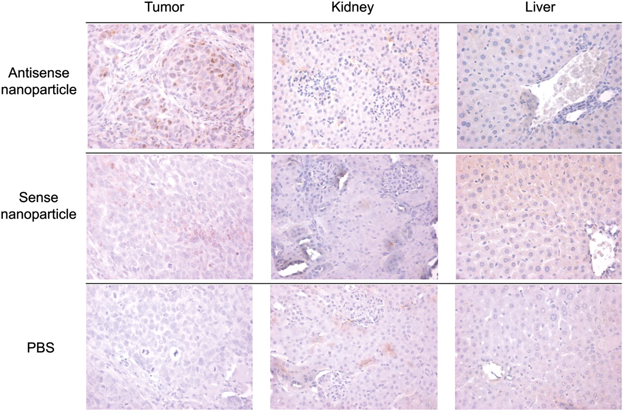
Figure 5 presents typical examples of tumor, kidney, and liver sections from mice receiving the antisense or sense Cy3-MORF/tat/trastuzumab nanoparticle or PBS. The animals were sacrificed 24 h later, and sections from tumor, liver, kidneys, bone marrow, heart, lung, muscle, spleen, stomach, and small and large intestines were prepared with sheep horseradish peroxidase–conjugated anticyanine antibody staining for the Cy3-MORFs and hematoxylin staining of the nucleus. Besides tumor, the other tissues of particular interest are the kidney and liver since, as the pharmacokinetic studies with radiolabeled nanoparticles show, levels of accumulation in these tissues are comparable to tumor. As evident from the figure, microscopic examination shows nuclear staining in approximately 20% of the tumor cells in animals injected with the antisense nanoparticle and 10% of the tumor cells in animals receiving the sense nanoparticle, whereas no nuclear staining is seen in the tumor cells of mice given the PBS injection as another control. Although nonspecific focal membranous staining is apparent, no nuclear staining is seen in the parenchymal cells of kidney and liver harvested from the mice receiving the antisense nanoparticle, sense nanoparticle, or PBS. No nuclear staining was observed in any sections from the other organs.

Immunohistochemical staining of formalin-fixed, paraffin-embedded tumor, kidney, and liver from SUM190 tumor–bearing mice administered Cy3-labeled antisense or sense MORF/tat/trastuzumab or PBS after 24 h (magnification is ×400).
DISCUSSION
Two significant observations led to the suggestion that the nanoparticles under development in these laboratories may provide effective radiotherapy of tumors. The first observation was evidence of unimpaired properties of the antibody, the cell-transporting peptide, and the antisense MORF when linked via streptavidin. The second observation was that antisense MORFs, when accumulated in tumor cells in culture, migrate rapidly and essentially quantitatively to the nucleus (1,3–5). Most of these properties were confirmed in the current investigation. Thus, Her2+ SUM190 cells in culture and as xenografts in mice were effectively targeted by the antiHer2 antibody trastuzumab component within the nanoparticle (Table 1), in the latter case accumulating in tumor at levels comparable to that of the free trastuzumab antibody itself (Fig. 4; Table 2). Furthermore, the antisense MORF within the nanoparticle directed its label to the nucleus of SK-BR-3 in culture (Table 1) and SUM190 in vivo (Fig. 5). Consistent with our previous findings in connection with free antisense oligomers, the antisense MORF within the nanoparticle showed significantly higher accumulations in tumor cells in culture than did the sense MORF (Table 1). Particularly encouraging are the observations by immunohistochemistry showing no evidence that the MORF within the nanoparticle accumulated in the nucleus of normal tissue after intravenous administration (Fig. 5).
Fortunately, the pharmacokinetics of the nanoparticle radiolabeled on the MORF component and linked to trastuzumab via streptavidin were found to be similar to those of the radiolabeled trastuzumab itself (Table 2; Fig. 4), providing further evidence that the function of the antibody had not been adversely influenced by the presence of streptavidin. Although this property of the MORF oligomer depends to some extent on its base sequence, all in vivo studies of radiolabeled free MORF in this laboratory have been characterized by a rapid clearance from circulation and important accumulation only in kidneys (11). The tumor accumulation of free MORF is negligible (12). Thus, the pharmacokinetics of the radiolabeled MORF when administered as the nanoparticle are far superior to those of the free radiolabeled MORF.
These results support the suggestion that the nanoparticles may be a useful vehicle to improve radiotherapy by carrying a radionuclide with short-range emissions to the nucleus of tumor cells. Of the short-range nuclear emissions that include α-particles and internal conversion electrons, only Auger electrons have ranges of less than the dimension of a nucleus (13). Therefore, the relative biologic efficacy of Auger emitters is significantly higher than that of α- or β-emitters when incorporated into the DNA of target cells (13). The radiation dose after the decay of an Auger electron emitter in the nucleus, compared with the cytoplasm of the cell, can be an order of magnitude higher (14).
Recently, alternative approaches using radiolabeled antibodies have been considered for this purpose. For example, an IgG antibody against an intranuclear protein was covalently conjugated to the tat peptide. Because tat peptides contain the membrane-translocation domain as well as a nuclear localizing peptide sequence, they have been observed to strongly promote the nuclear migration of cargo once internalized (15,16). The construct was radiolabeled and studied in vitro and in vivo in cells before and after upregulation of the protein. The results show conclusively that the tat peptide can encourage internalization and migration to the nucleus. Surprisingly, the results also indicate that the antibody, despite its size, crosses the nuclear membrane. Further studies will be needed to establish whether sufficient protein targets reside within the nucleus in this and other tumor cells and whether antibodies to these targets will be retained therein sufficiently to provide the needed radiation dose from the accompanying Auger emitter. In addition, with only an antibody against intranuclear determinants, presumably no element of tissue-specific targeting may be possible, with the result that the biodistribution of the label may be unfavorable.
As a result of the cell viability measurements, we selected for the dose escalation study a 50 nM concentration that would result in minimal nonspecific cytotoxicity by the WST-1 assay. A low concentration was also advisable to avoid the possibility of saturating the cell's Her2 determinants, its RIα mRNA target, and any receptors required for tat transport. A similarly low concentration of an antisense phosphorothioate DNA incubated freely did not saturate the mdr1 mRNA target mRNA levels in 3 other cell types (17). Therefore, lowering the concentration further was unnecessary but was also considered impractical since too low a concentration would limit the 111In dose carried by the nanoparticle below that required for the study. The results of the membrane integrity assay confirmed that unlabeled nanoparticles (Fig. 2A) and 111In-labeled nanoparticles, when added at 1.11 MBq/well or less (Fig. 2B), provide only modest cytotoxicity to SK-BR-3 cells when measured by these assays.
Because the clonogenic assay used in this investigation is a sensitive measure of the effects of cell damage, the assay is often regarded as the gold standard of cellular sensitivity assays (8). This assay was selected because the effects of Auger electrons emitted within the nucleus is expected to result in single- and double-strand breaks that will inhibit the ability of the cell to reproduce (18,19).
The results for the unlabeled nanoparticles (i.e., 0 MBq/well) in Figure 3A show minimal cytotoxicity for both the antisense and the sense nanoparticles and therefore support the results of Figures 1 and 2A, which also show minimal cytotoxicity for the unlabeled nanoparticles. The increase in cytotoxicity with 111In concentrations up to and including 1.11 MBq/well must certainly then be due to radiation, and since 111In concentrations of this magnitude have no effect on membrane integrity as shown in Figure 2, the cytotoxicity must be due to the decay of 111In within the cell. Because 111In-MORF, once internalized, migrates rapidly and almost quantitatively to the nucleus as shown in Table 1, the cytotoxicity probably occurs after intranuclear 111In decay. That the cytotoxicity is significantly higher for the antisense than for the sense nanoparticle at 1.11 MBq is probably related to the increased migration of the antisense oligomer, compared with the sense oligomer, to the nucleus (Table 1; Fig. 5) and is therefore evidence of antisense-mediated cytotoxicity.
The results in Figure 3B are presented as the surviving fraction for cells incubated with each 111In-labeled nanoparticle, compared with cells incubated with the identical nanoparticle added at the identical 50 nM concentration but unlabeled. This ratio decreases with increasing dose but only for cells incubated with the antisense nanoparticle such that the ratio is significantly lower at 1.11 MBq/well in cells incubated with the antisense nanoparticle than in cells incubated with the sense nanoparticle (P < 0.01). Because the results presented in this figure are now compared with the identical but unlabeled nanoparticle, significant differences must be due to the 111In. That the cytotoxicity of cells incubated with the sense nanoparticle showed no increase with dose is supporting evidence that when incubated as the sense nanoparticle, the 111In is not reaching the nucleus effectively. By contrast, the decreasing survival fraction in the same measurement for the antisense nanoparticle is strong evidence that in this case the 111In is reaching the nucleus.
The unexpected observations of this investigation are related to the nonspecific effects of the nanoparticles or the 111In. These observations are the surprisingly high cytotoxicity of cells incubated at both 1.11 and 2.04 MBq/well with the unlabeled nanoparticles mixed with 111In-DTPA as shown in Figure 2B; the lower surviving fraction in the clonogenic assay for cells incubated with the unlabeled antisense nanoparticle mixed with 111In-DTPA, compared with unlabeled antisense nanoparticle, as shown in Figure 3B; and the lower surviving fraction in the clonogenic assay for the nanoparticle incubated at 0 MBq/well, compared with incubations with PBS, as shown in Figure 3A. Studies investigating the role of Her2 in the radiosensitivity of cancer cells show that trastuzumab does not induce apoptosis when used alone but sensitizes the cells to radiation and enhances radiation-induced apoptosis of the cells in a Her2 level–dependent manner (10,20). This synergistic effect between trastuzumab and 111In may explain the first two nonspecific effects mentioned. For example, whereas the significantly higher cytotoxicity after incubation with free 111In may also be due to irradiation of the cell membrane by 111In in the cell medium itself, it may be more likely due to synergy with the trastuzumab on the cell membrane.
The remaining unexpected cytotoxicity does not involve radioactivity and therefore must be due solely to the unlabeled antisense and sense nanoparticles themselves. Because the fourth biotin binding site of streptavidin was not occupied, it is possible that the cytotoxicity observed for the unlabeled nanoparticle may be due to sequestration of endogenous biotin within the cell. To examine this possibility, SK-BR-3 cells were incubated at 400 nM with streptavidin, with biotin-saturated streptavidin, and with trastuzumab, streptavidin, or biotin alone or in various combinations. Surprisingly, by the WST-8 cell viability assay, significant proliferation inhibition was observed only for the biotin-saturated streptavidin. Because streptavidin itself showed no cytotoxicity by this assay, presumably sequestration of endogenous biotin may not explain the cytotoxicity observed by the clonogenic assay of Figure 3A. This result suggests that either the tat or, more likely, the antisense MORF, missing from these incubates, within the nanoparticle contributed to the observed cytotoxicity.
CONCLUSION
The 111In-MORF/tat/trastuzumab nanoparticle of this research was capable of tumor targeting, transmembrane transport, 111In nuclear migration, and specific radiation-induced cytotoxicity. These results suggest that tumor therapy may be effective using an Auger-emitting radionuclide on an antisense MORF oligomer administered as a member of a 3-component streptavidin-delivery nanoparticle.
Acknowledgments
This research was supported in part by the Office of Science (BER), U.S. Department of Energy (grant DE-FG02-03ER63602); the National Institutes of Health (grant R21CA129591); congressionally directed Medical Research Programs (grant W81XWH-06-1-0649) and Grants-in-Aid for Scientific Research (19659315, 19591434) from the Japanese Ministry of Education, Science, Sports and Culture.
Footnotes
-
COPYRIGHT © 2009 by the Society of Nuclear Medicine, Inc.
References
- Received for publication July 25, 2008.
- Accepted for publication December 8, 2008.


Jump to section
Related Articles
Cited By...
- Radiolabeled Oligonucleotides Targeting the RNA Subunit of Telomerase Inhibit Telomerase and Induce DNA Damage in Telomerase-Positive Cancer Cells
- ErbB-2 Blockade and Prenyltransferase Inhibition Alter Epidermal Growth Factor and Epidermal Growth Factor Receptor Trafficking and Enhance 111In-DTPA-hEGF Auger Electron Radiation Therapy