


Abstract
The repeatability of rest and hyperemic myocardial blood flow (MBF) measurements using 82Rb PET has not been evaluated. The aim of this study was to investigate the short-term repeatability of such measurements. Methods: Fifteen healthy volunteers underwent rest and pharmacologic stress 82Rb PET, repeated 60 min apart. Results: There was no significant difference in repeated rest MBF (0.77 ± 0.25 vs. 0.82 ± 0.25 mL/min/g, P = 0.31; mean difference, 6.18% ± 12.22%) or repeated hyperemic MBF (3.35 ± 1.37 vs. 3.39 ± 1.37 mL/min/g, P = 0.81; mean difference, 1.17% ± 13.64%). The repeatability coefficients were 0.19 mL/min/g for rest MBF and 0.92 mL/min/g for hyperemia. Conclusion: MBF using 82Rb is highly reproducible using a same-day short-term repeatability protocol. Serial MBF measurements with 82Rb PET should have the ability to quantify the acute effects of therapeutic interventions on MBF.
Assessment of coronary flow response has a potential role for serial evaluation of patients to determine the response to therapy and progression of disease (1,2).
82Rb PET has shown good diagnostic accuracy (3) and has prognostic value in patients with coronary artery disease (4). However, only limited data have been reported on myocardial blood flow (MBF) quantification, and there are no data evaluating repeatability using 82Rb (5,6).
Establishing the reproducibility of 82Rb MBF measurements is important for serial PET measurements of flow changes after various therapeutic interventions. The purpose of the current study was to investigate the same-day short-term repeatability of rest and hyperemic MBF as assessed by 82Rb PET.
MATERIALS AND METHODS
Study Protocol
Fifteen healthy volunteers (8 men and 7 women) with a mean age (±SD) of 29.4 ± 9.3 y participated.
MBF was measured at rest and with adenosine triphosphate stress (7,8) using 1,480 MBq of 82Rb (4,5). Ten-minute dynamic scans were obtained using a Siemens HR+ PET scanner (5). Adenosine triphosphate (160 μg/kg/min) was infused for 9 min, and image acquisition was started 3 min after the beginning of the infusion (7).
MBF was measured first at rest and 10 min later during stress; then, both scans were repeated with a test–retest interval of 60 min (9).
Quantification of MBF
MBF was measured using a previously described 1-tissue-compartment model (5).
The early-phase 82Rb images were used to define a region of interest in the left ventricular blood pool. The myocardial uptake images were calculated by adding the late-phase data from 4 to 6 min. A whole-myocardium region of interest was positioned with an algorithm we have developed (8).
The following equation was used to estimate the inflow rate (K1) of 82Rb into myocardium Ct(t) (5): where k2 is the outflow rate from myocardium into the blood Ca(t).
Conversion from K1 to MBF was estimated with the modified Renkin–Crone model (5,10).
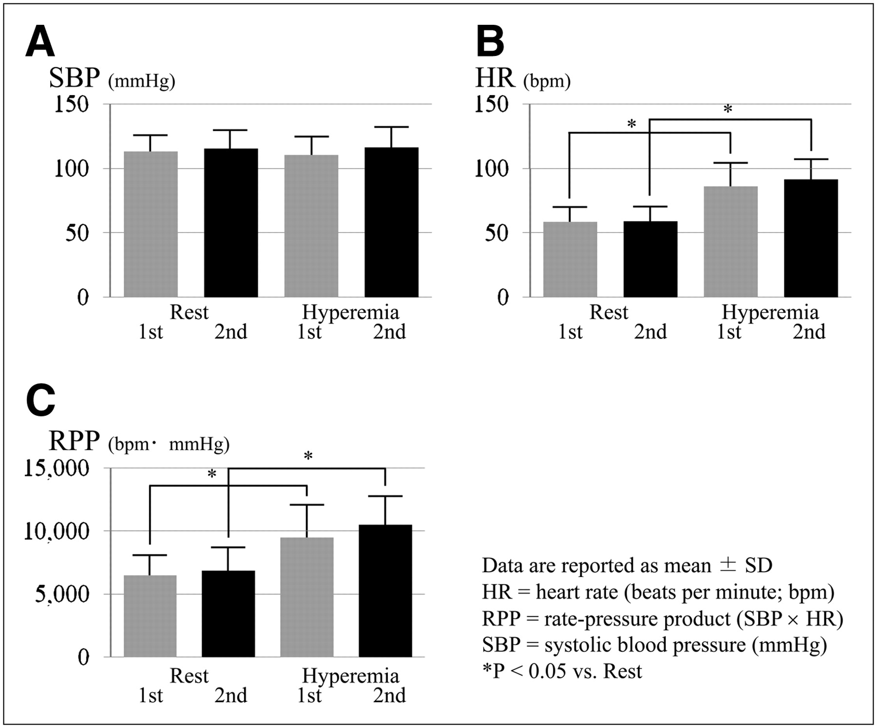
Coronary flow reserve (CFR) was calculated as the ratio of MBF during stress to MBF at rest (2). The rest MBF was corrected for the rate–pressure product (RPP) as rest MBF × (normal mean RPP/individual RPP) (7).
The normal mean RPP at rest in our institution was 8,150. RPP-corrected CFR was calculated as stress MBF/RPP-corrected rest MBF.
Statistical Analysis
The percentage difference between the first and second studies was calculated as (second MBF − first MBF)/mean MBF × 100%. A P value of less than 0.05 was considered statistically significant.
RESULTS
Physiologic Responses
In total, 13 of 15 (86.7%) participants had some adverse effects of adenosine triphosphate, but none of the participants had ischemic electrocardiography changes or chest pain.
The hemodynamic data did not significantly differ between the first and second studies (Fig. 1).

Hemodynamics.
MBF and CFR
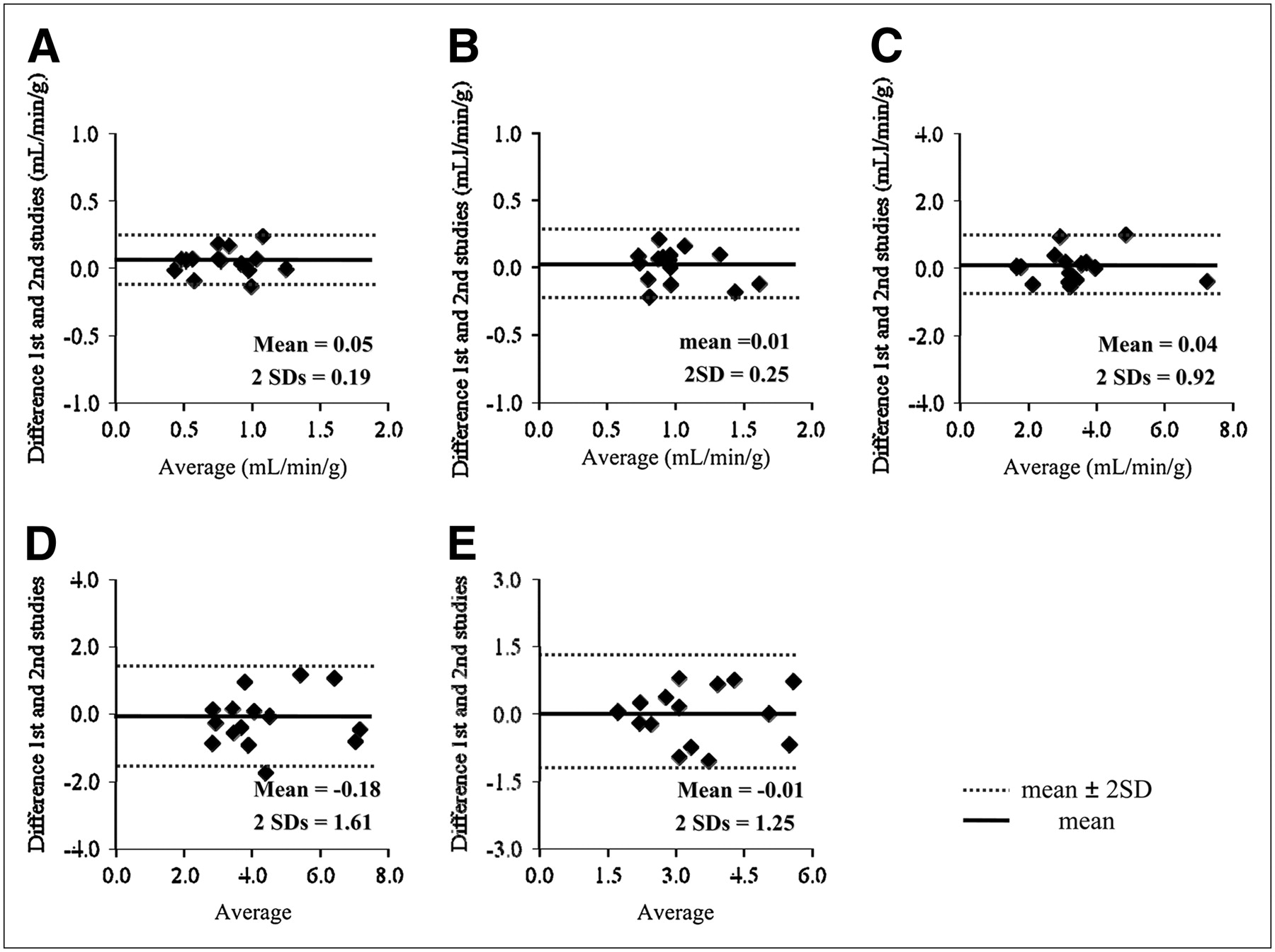
The repeatability coefficient of rest MBF was 0.19 mL/min/g, hyperemic MBF was 0.92 mL/min/g, and CFR was 1.61 mL/min/g (Table 1; Figs. 2 and 3). A good correlation was found between the first and second rest MBF (r = 0.93, P < 0.0001), hyperemic MBF (r = 0.94, P < 0.0001), and CFR (r = 0.86, P < 0.0001) (Fig. 4). RPP correction did not improve the repeatability of rest MBF (P = 0.08) or CFR measurements (P = 0.21).

MBF and CFR comparing first and second studies: repeated rest MBF (A), repeated hyperemic MBF (B), repeated CFR (C), and repeated RPP–corrected CFR (D).

Repeatability of MBF Bland–Altman plots: rest MBF (A), RPP-corrected rest MBF (B), hyperemic MBF (C), CFR (D), and RPP-corrected CFR (E).

Scatter plots comparing first and second MBF studies: rest MBF (A), RPP-corrected rest MBF (B), hyperemic MBF (C), CFR (D), and RPP-corrected CFR (E).
MBF
Both men and women had good repeatability of rest MBF and hyperemic MBF (Table 1).
DISCUSSION
To the best of our knowledge, this was the first study to evaluate short-term MBF repeatability using generator-produced 82Rb PET. Our study demonstrated that 82Rb PET rest and hyperemic MBF measurements and CFR had good repeatability. PET can quantify MBF and CFR using a suitable tracer kinetic model. In the current study, we applied a 1-compartment model for 82Rb MBF quantification similar to that of Lortie et al. (5), who reported good correlation with 13N-ammonia. Our rest and hyperemic MBF values were similar to those of the previous study (5,9,10), indicating that appropriate mathematic modeling was applied (7). It is known that there are sex differences in MBF (11), but in the present study both men and women had similar repeatability. Thus, this technique is applicable to both sexes.
82Rb PET has several advantages. Myocardial perfusion images can be obtained without a cyclotron and with a short interval because of the short, 76-s, half-life (4). 82Rb MBF quantification may be useful for the evaluation of therapeutic interventions. However, as a first step, it is important to estimate the reproducibility of the technique as reported in the present study (9,12).
The small mean difference in rest MBF in the current study indicated good repeatability and was slightly better than the previous findings of Kaufmann et al. using 15O-water (9) or of Nagamachi et al. using 13N-ammonia (12). Rest MBF is considered to be associated with the RPP. Nagamachi et al. reported that RPP correction significantly reduced MBF variability. However, there was no significant change after RPP correction in the present study because the initial data had sufficiently low variability.
Hyperemic MBF in the current study showed small differences between the 2 studies indicating good repeatability as well. Compared with the previous studies by Kaufmann et al. (9) or Nagamachi et al. (12), the present study showed a smaller difference and similar SD. The decreasing net extraction of 82Rb with increasing MBF (13) may be expected to increase variability during hyperemia. However, this relationship was not observed in the present study.
Our study had some limitations. It assessed the reproducibility of MBF and CFR in the whole left ventricular myocardium but not in regional myocardial segments. Further studies are needed to evaluate reproducibility in patients with coronary artery disease, in whom additional regional heterogeneity may be expected. We studied only short-term repeatability, not repeatability over longer intervals. However, an advantage of 82Rb is its short half-life, which enables various acute interventions to be evaluated in single-session studies similar to those using 15O-water. Thus, it is important to evaluate short-term repeatability in single-series studies (9). Nevertheless, additional prospective studies for longer-term reproducibility are warranted.
CONCLUSION
MBF and CFR using 82Rb were highly reproducible in this study of same-day short-term repeatability.
Acknowledgments
This study was supported in part by grant H19-C-068 from the Northern Advancement Center for Science and Technology (Sapporo, Japan).
Footnotes
-
COPYRIGHT © 2009 by the Society of Nuclear Medicine, Inc.
References
- Received for publication July 4, 2008.
- Accepted for publication October 8, 2009.


Jump to section
Related Articles
Cited By...
- Reply: Clarifying the Utility of Myocardial Blood Flow and Myocardial Flow Reserve After Cardiac Transplantation
- Clinical Quantification of Myocardial Blood Flow Using PET: Joint Position Paper of the SNMMI Cardiovascular Council and the ASNC
- SPECT Myocardial Perfusion Reserve in Patients with Multivessel Coronary Disease: Correlation with Angiographic Findings and Invasive Fractional Flow Reserve Measurements
- Single-Phase CT Aligned to Gated PET for Respiratory Motion Correction in Cardiac PET/CT
- Cardiac PET Imaging for the Detection and Monitoring of Coronary Artery Disease and Microvascular Health
- Cardiac Positron Emission Tomography