


Abstract
Recent studies on gene expression of β-cell mass (BCM) in the pancreas showed that vesicular monoamine transporter 2 (VMAT2) is highly expressed in the BCM (mainly in the islets of Langerhans). Imaging pancreatic BCM may provide an important tool for understanding the relationship between loss of insulin-secreting β-cells and onset of diabetes mellitus. In this article, 9-fluoropropyl-(+)-dihydrotetrabenazine (FP-(+)-DTBZ), which is a VMAT2 imaging agent, was evaluated as a PET agent for estimating BCM in vivo. Methods: Organ biodistribution after an intravenous injection of 18F-FP-(+)-DTBZ (active isomer) and 18F-FP-(−)-DTBZ (inactive isomer) was evaluated in normal rats. The specificity of uptake of 18F-FP-(+)-DTBZ was assessed by a pretreatment (3.8 mg of (+)-DTBZ per kilogram and 3.5 mg of FP-(+)-DTBZ per kilogram, intravenously, 5 min prior) or coadministration (2 mg of (+)-DTBZ per kilogram). PET studies were performed in normal rats. Results: The in vivo biodistribution of 18F-FP-(+)-DTBZ in rats showed the highest uptake in the pancreas (5% dose/g at 30 min after injection), whereas 18F-FP-(−)-DTBZ showed a very low pancreas uptake. Rats pretreated with FP-(+)-DTBZ displayed a 78% blockade of pancreas uptake. PET studies in normal rats demonstrated an avid pancreas uptake of 18F-FP-(+)-DTBZ. Conclusion: The preliminary data obtained with 18F-FP-(+)-DTBZ suggest that this fluorinated derivative of DTBZ shows good pancreas specificity and has the potential to be useful for quantitative measurement of VMAT2 binding sites reflecting BCM in the pancreas.
The pancreas comprises 2 tissue types: exocrine and endocrine. The endocrine tissue accounts for only a small percentage of the pancreatic mass, is found scattered in the islets of Langerhans, and comprises predominantly β-cells. β-cells produce insulin in response to metabolic demands, and loss of β-cells results in insulin deficiency and diabetes. In type 1 diabetes, common in juveniles, an autoimmune process dramatically destroys β-cells. In type 2 diabetes, β-cell mass (BCM) decreases as part of a slower, more insidious process that also involves peripheral insulin resistance and increased demand for insulin. Because of a significant BCM reserve, symptoms related to unstable glucose homeostasis do not result until BCM is reduced by more than 50%–60% (1). Unfortunately, most studies measuring BCM have relied on postmortem examination of the pancreas, and until recently (2,3), it has been impossible to prospectively measure BCM in vivo.
Although no cure for diabetes exists, several promising potential therapies for modifying the disease course are in clinical trials. These approaches focus mainly on preserving or replacing BCM and include pancreas or islet cell transplantation (4–8) or islet cell regeneration from stem cells. The ability to monitor BCM in vivo will facilitate development of disease-modifying therapies for diabetes mellitus (9–11). Indeed, in the past few years many attempts have been made to image BCM; most of this work has been based on specific binding sites or receptors, such as sulfonylurea receptors and other binding sites in the pancreas (12–15). Studies using labeled β-cell–specific antibody and the fragment as in vivo imaging agents have shown some promise, but they are not suitable for routine clinical use because of a relatively low cellular specificity (low pancreas accumulation vs. high liver and kidney accumulations) (16). Additional BCM ligands have been reported, but except for the vesicular monoamine transporter 2 (VMAT2) imaging agents, none has been successfully applied to image diabetes in humans (5–8,16–21).
Recently, immunohistochemical staining studies of gene expression in the endocrine pancreas showed that VMAT2 binding sites are expressed mainly on the β-cells in the islets of Langerhans (2,22). VMAT2 expression matches well with the insulin levels in human and monkey pancreas tissue (23). Therefore, VMAT2 could be an excellent target for mapping β-cell function. A VMAT2 ligand, (+)-11C-dihydrotetrabenazine (DTBZ), was developed previously for imaging VMAT2 in the striatum. It has been successfully applied for PET studies of Parkinson's and other neurodegenerative diseases (24,25) and was recently used for PET VMAT2 binding sites in the pancreas of monkeys and humans (1,2,26,27). This is, to our knowledge, the first use of PET with (+)-11C-DTBZ to measure the BCM in the pancreas, suggesting that PET of β-cells may be a useful tool to estimate the functional stage of these cells in living humans (1). It may also be possible to study the function of islet cells after transplantation (28,29). However, 11C has a relatively short half-life (t1/2 = 20 min), making clinical trials difficult and preventing longer imaging protocols that may be required to optimize imaging kinetics. In contrast, 18F-labeled analogs of DTBZ, with a longer half-life (t1/2 = 110 min), could be made more widely available through the same regional distribution network currently delivering 18F-FDG to nuclear medicine clinics.
While developing 18F-labeled analogs of DTBZ as imaging agents for Parkinson's disease, we have successfully tested a novel DTBZ derivative, an optically pure fluoropropyl derivative, 18F-labeled 9-fluoropropyl-(+)-DTBZ (FP-(+)-DTBZ) (Fig. 1), in rodents and nonhuman primates (30–32). It displayed excellent binding affinity (Ki = 0.11 nM) for VMAT2. The higher binding affinity (lower Ki value) of FP-(+)-DTBZ is superior to the value reported for (+)-DTBZ (Ki = 0.97 nM (33)). In addition to imaging the basal ganglia region of the brain, this fluorinated DTBZ derivative, similar to its congener (+)-11C-DTBZ (Fig. 1), could be a potential imaging agent to estimate the mass of β-cells related to the endocrine pancreas function in vivo. We report herein the initial evaluation of 18F-FP-(+)-DTBZ in normal rats and evaluate the selectivity of pancreas uptake by blocking studies.
MATERIALS AND METHODS
General
Optically pure 18F-FP-DTBZ was prepared by an 18F-fluoride displacement of the corresponding mesylate, and the product was purified by high-performance liquid chromatography as described previously (30,31). The radiochemical purity of 18F-FP-(+)-DTBZ was greater than 95%, and the specific activity was 55,500–74,000 GBq (1,500–2,000 Ci)/mmol at the end of synthesis. (+)-DTBZ and (+)9-O-desmethyl-α-dihydrotetrabenazine (used to prepare the mesylate precursor for 18F-labeling) were kindly provided by the National Institute of Mental Health under the Chemical Synthesis and Drug Supply Program. Male Sprague–Dawley rats weighing 220–350 g were used for normal distribution and imaging studies.
Biodistribution Studies
Animals were anesthetized with isoflurane and then injected (intravenously via the femoral vein) with 0.2 mL of a saline solution containing 18F-FP-(+)-DTBZ (active isomer) or 18F-FP-(−)-DTBZ (inactive isomer) (0.37–0.74 MBq [10–20 μCi]). The rats (n = 3–4 for each time point) were then sacrificed under isoflurane anesthesia at indicated time points after injection. Organs of interest were removed and weighed, and the radioactivity was counted with an automatic γ-counter. The percentage dose per organ was calculated by comparing tissue counts with suitably diluted aliquots of the injected material. The total activity of the blood was calculated under the assumption that it was 7% of the total body weight. The percentage dose per gram of samples was calculated by comparing the sample counts with the count of the diluted initial dose. Different regions corresponding to striatum, hippocampus, cerebellum, and cortex were dissected from the brain and counted to obtain the regional distribution of the tracer.
To further prove that the accumulation of 18F-FP-(+)-DTBZ in the pancreas was indeed due to the presence of VMAT2 binding sites, we pretreated the rats with (+)-DTBZ or FP-(+)-DTBZ (respectively, 3.8 and 3.5 mg/kg, intravenously), selective VMAT2 ligands, or FP-(−)-DTBZ (3.5 mg/kg, intravenously) at 5 min before tracer injection. Alternately, 18F-FP-(+)-DTBZ was injected together with (+)-DTBZ (2.0 mg/kg). At 30 min after the tracer (or tracer with added carrier) injection, the rats were sacrificed; organs or tissues were removed and counted as described above. The significance in reduction of tracer binding in selected organ or tissues was determined by a Student t test.
Imaging Studies
The University of Pennsylvania has a small-animal PET scanner (34), the MOSAIC PET (Philips), used in this study to perform the PET imaging on control rats (220–350 g). Each animal was anesthetized initially using 3% isoflurane in 1.0 L/min oxygen, in an acrylic induction chamber. When fully anesthetized, the animal was placed on the scanner bed, with a nose cone used to maintain anesthesia at 1.5% isoflurane in 1.0 mL of oxygen per minute. Body temperature was maintained by placing a heating pad under the animal. A dose of 30–37 MBq (0.8–1 mCi) 18F-FP-(+)-DTBZ (volume < 0.4 mL) was injected into the tail vein. A total of 24 dynamic scans (5 min/frame) were acquired over a course of 2 h. Images were reconstructed using a fully 3-dimensional iterative image reconstruction algorithm with system attenuation correction incorporated in the algorithm. Corrections were applied for scatter, randomness, and attenuation. Regions of interest (ROIs) were drawn, guided by a detailed rat atlas and micro-CT images. Visual analysis was performed using coronal, transverse, and sagittal reconstruction. ROIs were drawn over the pancreas as well as other organs or tissues. Mean counts per voxel in each region were calculated at various times.
RESULTS
The in vivo biodistribution study of 18F-FP-(+)-DTBZ in rats clearly showed that the pancreas displayed the highest uptake among all organs or tissues in the body (5.5% dose/g at 30 min after injection) (Table 1). The liver, an organ adjacent to the pancreas, showed a significant but lower uptake (2.8% dose/g) than the uptake observed in the pancreas. The washout of radioactivity from the pancreas was relatively slow, with 2.7% dose/g remaining at 2 h after injection. The high pancreas uptake observed with 18F-FP-(+)-DTBZ is consistent with the reported value of 6.2% dose/g in rats for (+)-11C-DTBZ (26). Other BCM ligands, such as sulfonylurea receptor ligand, glibenclamide, and fluorodithizone, a molecule exploiting the unique glucose-handling machinery of β-cells, displayed almost exclusive distribution in the liver with barely detectable amounts in the pancreas (35).
In Vivo Biodistribution of 18F-FP-(+)-DTBZ in Control Sprague–Dawley Rats
To confirm that pancreas uptake is specifically due to the VMAT2 signal, we performed blocking studies (via either pretreatment or coadministration with (+)-DTBZ or pretreatment with FP-(+)-DTBZ or FP-(−)-DTBZ) (31). In these experiments, the pancreas uptake was blocked by the competing dose of (+)-DTBZ (30% for pretreatment and 36% for coadministration). Pretreatment with FP-(+)-DTBZ (Ki = 0.11 nM) resulted in a greater blocking of the uptake in the pancreas (78%) (Table 2). As expected, the inactive isomer (Ki > 3,000 nM) showed very low inhibition of pancreas uptake (9%) (Table 3). In the same experiments, the striatal region (a brain region containing a high concentration of VMAT2 binding sites) showed a complete blocking of 18F-FP-(+)-DTBZ uptake by the active isomer (Table 2), whereas the inactive isomer (Table 3), as expected, showed a very low inhibition of striatal uptake (Table 2). The lesser blocking of pancreas uptake by (+)-DTBZ (30%–36% blockage) was probably because in vivo kinetics of (+)-DTBZ may be different from those of FP-(+)-DTBZ. Higher pancreas blockage of 18F-FP-(+)-DTBZ uptake by FP-(+)-DTBZ (78%) suggests matching kinetics between the cold and hot ligand binding to the same VMAT2 binding sites in the pancreas.
Blocking Studies of Pancreatic Uptake of 18F-FP-(+)-DTBZ at 30 Minutes After Tracer Injection in Rats
Biodistribution in Rats After Intravenous Injection of 18F-FP-(−)-DTBZ
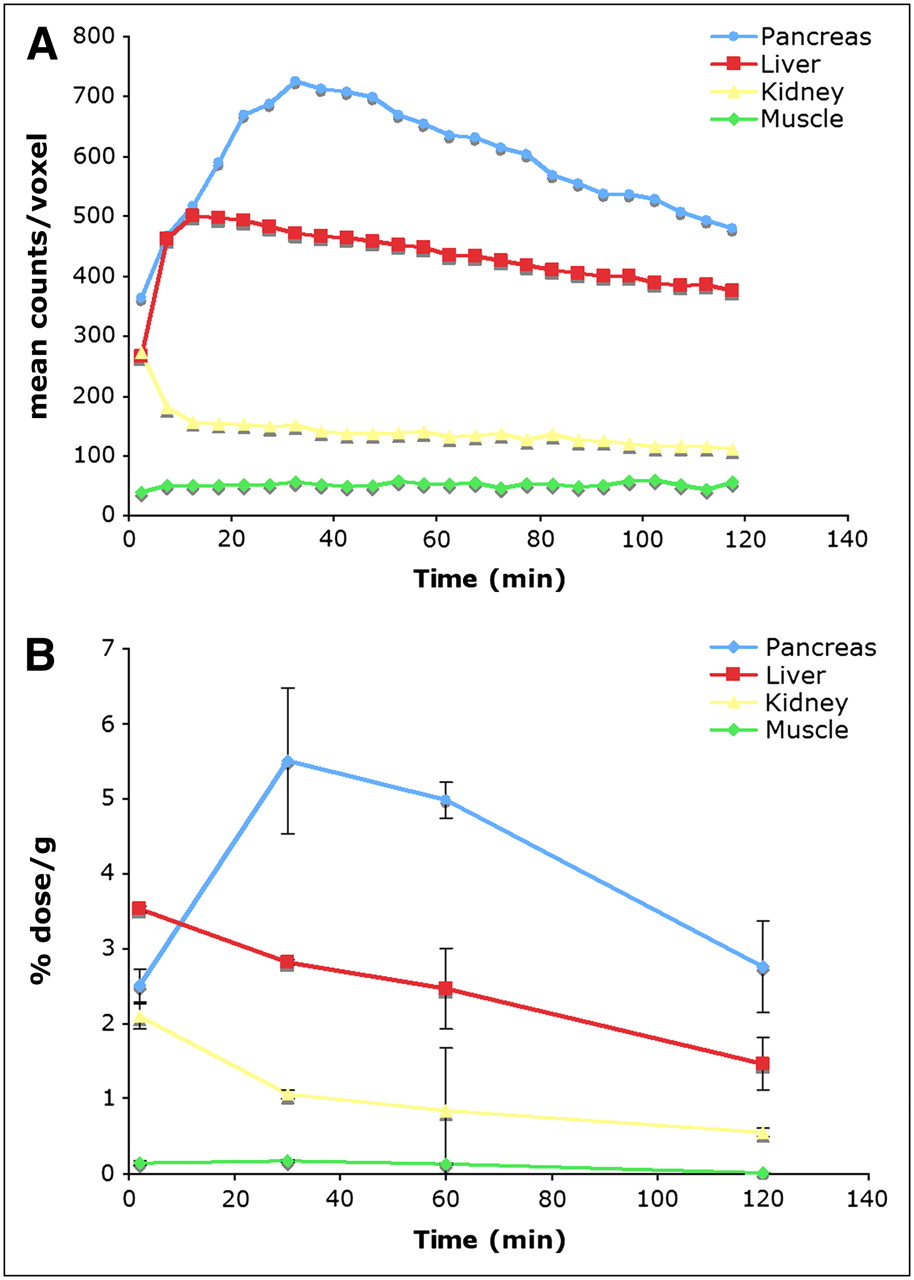
Small-animal PET was performed on Sprague–Dawley rats after an injection of 18F-FP-(+)-DTBZ. The pancreas was clearly visualized in control rats (transverse, coronal, and sagittal views), with the pancreatic activity greater than the uptake activity of any other organ (Fig. 2). Quantitative measurements of radioligand uptake were obtained by placing ROIs in the liver, kidney, and pancreas (Fig. 3A). It appeared that 18F-FP-(+)-DTBZ was removed from the blood by both liver and kidney. In the liver, 18F-FP-(+)-DTBZ is excreted into the bile duct, showing a steady increase of accumulation of activity over the 2-h scanning period. Pancreatic 18F-FP-(+)-DTBZ uptake increased from immediately after the intravenous injection to about 30–35 min after injection, followed by a slight washout. Because of lower accumulations of radioactivity in the muscle or kidney, these regions provide convenient reference regions. Our data obtained with 18F-FP-(+)-DTBZ compare with the results reported previously with (+)-11C-DTBZ and PET (26). The time–activity curves of 18F-FP-(+)-DTBZ for pancreas, liver, kidney, and muscles obtained from PET correlated well with curves from the biodistribution data obtained by dissection and counting of the tissues (Fig. 3B). Thus, the imaging data were consistent with in vivo biodistribution results.

Potential PET agents for VMAT2.
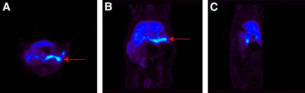
Representative in vivo PET images (data acquired at 30–35 min after tracer injection) of transverse, coronal, and sagittal views of abdominal planes of male Sprague–Dawley rat. 18F-FP-(+)-DTBZ (33.3 MBq [0.9 mCi]) was injected into rat, after which animal was scanned for 2 h. Images were reconstructed and ROIs were defined. Red arrows indicate pancreas uptake.

Quantitation of in vivo activity within pancreas, liver, kidney, and muscle ROIs during scan period is shown in time–activity curves. Consistently, observed time–activity curve via imaging (A) is very similar to results obtained with tissue dissection and counting method (B).
We have also performed preliminary binding studies with 18F-FP-(+)-DTBZ using purified human islet homogenates. Specific VMAT2 binding signal was detected in the islet cell homogenates. The binding appeared to be saturable with a high binding affinity (data not shown). However, because of a limited supply of human β-cells, we have not been able to investigate this further.
DISCUSSION
We report the results of an initial study of 18F-FP-(+)-DTBZ, a radiotracer targeting VMAT2 binding sites, as a potential imaging agent for BCM in the pancreas. Originally, the 18F derivative was designed to image VMAT2 binding sites in the brain and is now in clinical trials to assess reductions of VMAT2 binding sites in patients with Parkinson's disease and dementia with Lewy bodies (25). Our interest in studying VMAT2 binding sites in the pancreas is based on recent reports suggesting that neurons in the pancreas share expression of a large number of genes and display functional similarity with neurons in the brain (2,22). That report indicated that a high density of VMAT2 is present in the β-cells of the pancreas with little or no VMAT2 in surrounding organs. Therefore, VMAT2 binding sites in the pancreas may serve as markers for functional β-cells. To test this hypothesis, we have studied the localization of 18F-FP-(+)-DTBZ in the pancreas of normal rats. The specificity of 18F-FP-(+)-DTBZ for VMAT2 binding in the pancreas was further confirmed by the in vivo competition with the specific VMAT2 ligand (+)-DTBZ as well as FP-(+)-DTBZ. Significantly, pretreatment of FP-(−)-DTBZ did not show any inhibition of pancreas uptake, suggesting 18F-FP-(+)-DTBZ for VMAT2 binding in the pancreas is a highly selective and specific process.
The greater level of blocking by FP-(+)-DTBZ versus (+)-DTBZ suggests that the kinetics of the compounds are different. A significantly high nonspecific background distribution was also observed with (+)-11C-DTBZ (26). Whether this level of nonspecific distribution will impact the usefulness of the radioligand for PET studies of β-cell losses in disease remains to be seen. Increasing the signal with a higher specific VMAT2 binding in the pancreas will likely improve the potential for imaging BCM containing these VMAT2 binding sites.
While our work was in progress, an abstract describing 18F-labeled 9-FCH2-DTBZ for imaging BCM was presented at the 17th International Symposium on Radiopharmaceutical Sciences, April 30–May 4, 2007, in Aachen, Germany (36). The paper concluded that 18F-labeled 9-FCH2-DTBZ shows regional brain uptake and binding affinity similar to those shown by (+)-11C-DTBZ, and it may be feasible to use 18F-labeled 9-FCH2-DTBZ as a PET tracer for measuring BCM. The report further confirmed our contention that 18F-labeled DTBZ derivatives are excellent candidates for imaging BCM. Direct comparison studies will be needed to figure out which ligand shows better pancreas VMAT2 binding in vivo. Other imaging agents for imaging BCM have been reported, but none has demonstrated as promising properties as those demonstrated by VMAT2 imaging agents (16,21). The potential clinical application of various BCM imaging agents has been extensively reviewed (1).
CONCLUSION
18F-FP-(+)-DTBZ has the potential to be a useful marker for BCM. High-quality PET images of the pancreas in normal rats can be obtained using this radiotracer. In conjunction with the findings on (+)-11C-DTBZ, it seems clear that VMAT2 radioligand binding and PET imaging may be a useful way to evaluate BCM in the pancreas.
Acknowledgments
We thank Dr. Henry Wagner for his helpful discussion and in selecting Figure 3 as the small-animal image of the year at the 2007 annual meeting of the Society of Nuclear Medicine. We are grateful to the Chemical Synthesis and Drug Supply Program of the National Institute of Mental Health for providing the samples of resolved (+)9-O-desmethyl-DTBZ-(−)-TBZ and other TBZ analogs used in this project. We also thank Dr. Paul Harris for his helpful discussion and Dr. Carita C. Huang for her editorial assistance.
Footnotes
-
COPYRIGHT © 2008 by the Society of Nuclear Medicine, Inc.
References
- Received for publication February 11, 2008.
- Accepted for publication April 3, 2008.


Jump to section
Related Articles
Cited By...
- Radiomanganese PET Detects Changes in Functional {beta}-Cell Mass in Mouse Models of Diabetes
- Glucagon-Like Peptide-1 and Its Class B G Protein-Coupled Receptors: A Long March to Therapeutic Successes
- In Vivo Imaging of Endogenous Pancreatic {beta}-Cell Mass in Healthy and Type 1 Diabetic Subjects Using 18F-Fluoropropyl-Dihydrotetrabenazine and PET
- Multimodal image coregistration and inducible selective cell ablation to evaluate imaging ligands
- Unique sphingomyelin patches are targets of a beta-cell-specific antibody
- Reply: Beta-cell Imaging: Opportunities and Limitations
- Whole-Body Biodistribution and Radiation Dosimetry of 18F-FP-(+)-DTBZ (18F-AV-133): A Novel Vesicular Monoamine Transporter 2 Imaging Agent
- Assessment of Islet Specificity of Dihydrotetrabenazine Radiotracer Binding in Rat Pancreas and Human Pancreas
- 11C-Dihydrotetrabenazine {beta}-Cell Imaging
- 11C-Dihydrotetrabenazine PET of the Pancreas in Subjects with Long-Standing Type 1 Diabetes and in Healthy Controls