


Abstract
There have been few radiotracers for imaging adrenergic receptors in brain by PET, but none has advanced for use in human studies. We developed a radiosynthesis for the α2-adrenergic antagonist 11C-yohimbine and characterized its binding in living pigs. As a prelude to human studies with 11C-yohimbine, we determined the whole-body distribution of 11C-yohimbine and calculated its dosimetry. Methods: Yorkshire × Landrace pigs weighing 35–40 kg were used in the study. Baseline and postchallenge PET recordings of 11C-yohimbine in pig brain were conducted for 90 min, concurrent with arterial blood sampling, and with yohimbine and RX821002 as pharmacologic interventions. 15O-Water scans were performed to detect changes in cerebral perfusion. The PET images were manually coregistered to an MR atlas of the pig brain. Maps of the 11C-yohimbine distribution volume ([Vd] mL g−1) in brain were calculated relative to the arterial input function. Results: Whole-body scans with 11C-yohimbine revealed high accumulation of radioactivity in kidney, intestine, liver, and bone. The estimated human dose was 5.6 mSv/GBq, a level commonly accepted in human PET studies. Brain imaging showed baseline values of Vd ranging from 1.9 in medulla, 3.0 in cerebellum, and to 4.0 in frontal cortex. Coinjection with nonradioactive yohimbine (0.07 mg/kg) reduced Vd globally to approximately 1.5–2 mL g−1. A higher yohimbine dose (1.6 mg/kg) was without further effect on self-displacement. Very similar results were obtained by displacement with the more selective α2-adrenergic antagonist RX821002 at doses of 0.15 and 0.7 mg/kg. Cerebral blood flow was globally increased 43% after administration of RX821002. Notable features of 11C-yohimbine are a lack of plasma metabolism over 90 min and a rapid approach to equilibrium binding in brain. Conclusion: The new radiotracer 11C-yohimbine seems well suited for PET investigations of α2-adrenergic receptors in brain and peripheral structures, with the caveat that displaceable binding was present in cerebellum and throughout the brain.
Since the first report of external detection of dopamine D2-like receptors in living brain (1), specific radioligands have been developed for PET studies for receptors and reuptake sites of the biogenic amines dopamine, serotonin, and noradrenaline (2,3). Altered noradrenergic transmission is implicated in several neurodegenerative diseases and in the mechanism of action of some antidepressants. However, radiotracers for markers of noradrenaline innervations and receptors have become available only in recent years. Plasma membrane noradrenaline transporters on presynaptic terminals can now be imaged with 11C-labeled 2-[(2-methoxyphenoxy)phenylethyl]morpholine (11C-MeNER) (4). Whereas specific radiotracers are not available for imaging of brain β-adrenergic receptors, α2-adrenergic receptors in brain have been labeled with the antagonist 11C-mirtazapine. However, that compound suffers from incomplete selectivity for adrenergic receptors (5). Therefore, a more specific marker for PET studies of α2-adrenergic receptors is required.
Yohimbine is the major alkaloid from the bark of Pausinystalia yohimbe, a West African plant, and is also present in extracts of Rauvolfia sp. Yohimbine has a long history as an antihypertensive agent but generally increases blood pressure at rest, apparently mediated by a central antagonism of α2-adrenergic receptors (6). In addition to α2-adrenergic antagonist properties, yohimbine interacts with α1 and 5-hydroxytryptamine 1A (5-HT1A) receptors (7–9). Despite the complex pharmacology of yohimbine, we hypothesized that 11C-yohimbine at tracer concentrations might exhibit selectivity for α2-adrenergic sites in living brain. To test this hypothesis, we developed a radiosynthesis for 11C-yohimbine and characterized its cerebral uptake and binding in PET studies of living pigs. The equilibrium distribution volume ([Vd] mL g−1) of 11C-yohimbine was mapped in a baseline condition and after blockade with low and high doses of nonradioactive yohimbine. In other displacement studies, the pharmacologic specificity of 11C-yohimbine for α2 sites was tested by displacement with the selective α2 antagonist RX821002 (9). We also determined the whole-body distribution of 11C-yohimbine in a single pig and calculated organ dosimetry.
MATERIALS AND METHODS
Chemicals
Acetonitrile, dimethyl sulfoxide (DMSO), and iodine were from Bie & Berntsen. Yohimbine hydrochloride, yohimbinic acid, tetrabutylammonium fluoride (TBAF, 1 mol/L in tetrahydrofuran), and sodium phosphate monohydrate were from Sigma–Aldrich. RX821002 hydrochloride was from Tocris. Solutions for intravenous administrations of yohimbine (5% ethanol in sterile saline) and RX821002 (sterile saline) were prepared 30 min before use. The individual doses are expressed on the basis of the free base.
Radiochemistry
11C-Carbon dioxide was prepared by 14N(p,α)C proton bombardment with the GE Healthcare PETtrace 200 cyclotron and was converted to 11C-methyliodide using the GE MeI Box (reduction of CO2 to CH4, followed by gas-phase reaction with iodine). 11C-Methyl iodide (6–9 GBq after 30-min bombardment at 40 μA) was trapped in DMSO (300 μL) containing NaOH (1.5 μL, 3 mol/L), TBAF (5 μL, 1 mol/L), and yohimbinic acid (1.5 mg) in a 1-mL vial. This mixture was heated at 90°C for 5 min. Purification of 11C-yohimbine was performed by semipreparative high-performance liquid chromatography (HPLC) (PerkinElmer model 200) using a 5-mL injection loop. The mobile phase, consisting of 50% aqueous 70 mmol/L NaH2PO4 and 50% acetonitrile, was delivered at a rate of 8 mL/min to a Sphereclone ODS(2) C-18 (Phenomenex, 250 × 10 mm) semipreparative column. The reaction was quenched by adding 500 μL of HPLC eluent to the vial, and the mixture was transferred to the injection loop. Product elution was monitored with online γ-detection of an in-house design and ultraviolet (UV)–visible detection (model 759A, λ = 254 nm; Applied Biosystems). The fraction containing 11C-yohimbine (retention time, 4–5 min) was collected and the mobile phase was removed by evaporating to near-dryness at 90°C under reduced pressure. The product was reformulated with isotonic saline and was transferred through a sterile filter (0.22 μm) to the final product vial. A small sample (100 μL) was taken for quality control measurement.
Analytic Chemistry
The amount of yohimbine in the final product was determined by HPLC using a LUNA CN 5-μm 100 column (Phenomenex, 250 × 4.6 mm) with an eluent consisting of 60% aqueous 70 mmol/L Na2HPO4 and 40% acetonitrile with serial radioactivity and UV detection. Reference standards of yohimbinic acid (5 μg/mL) and yohimbine (5 μg/mL) were used for quantification, and a solution of yohimbine (100 μg/mL) was coinjected with the product solution to verify product identity on the basis of the elution position of the UV peak. The chemical stability of 11C-yohimbine productions was checked 1 h after the end of synthesis (EOS).
Biologic Procedures
This project was approved by The Danish Experimental Animal Inspectorate. Female pigs (Yorkshire × Danish Landrace crossbred, 35–40 kg) (n = 7) were prepared for scanning, as described in detail elsewhere (10). Anesthesia was induced with ketamine/midazolam and maintained with isoflurane/N2O/O2. Body temperature and blood chemistry (gases, glucose, and pH) of the pigs were maintained within the normal range, and isotonic saline was infused intravenously at a rate of 3 mL/min. Heart rate, blood oxygen saturation, and arterial blood pressure were continuously monitored. Arterial blood samples were drawn every 1–2 h and levels of glucose, partial pressure of oxygen (Po2), partial pressure of carbon dioxide (Pco2), and pH were determined. Blood glucose levels were maintained within the normal porcine range of 3.5–6 mmol/L by intravenous infusion of isotonic glucose as required. Hemodynamic stability was maintained by adequate levels of isoflurane anesthesia (i.e., 2%–2.5%). In addition, oxygen tension in the bloodstream was kept above 12 kPa, and CO2 levels were maintained between 5.5 and 6.5 kPa by manual adjustments of respiration rate, respiratory volume, and the composition of the gaseous mixture delivered to the pig. Levels of pH in the bloodstream were maintained between 7.35 and 7.45 primarily by adjusting the tidal volume of gases delivered via the tracheal tube. After the last scan, pigs were killed with an overdose of pentobarbital.
Whole-Body PET Recording and Dosimetry Recordings
A whole-body PET recording of 11C-yohimbine was performed in 1 pig using a Siemens ECAT EXACT HR47 tomograph. A whole-body transmission scan was obtained first. A series of 3 consecutive whole-body PET recordings were obtained after a single intravenous injection of 11C-yohimbine (223 MBq; specific activity of 120 GBq/μmol at time of injection). Seven horizontal bed positions were recorded at intervals of 2, 3, and 4 min; thus, the entire body was scanned during intervals of 0–14, 15–35, and 36–63 min after tracer injection.
Time–radioactivity curves were obtained from the repeated whole-body scans as follows: organs with relatively high accumulation of tracer relative to their surroundings were identified by visual inspection (bone, kidney, intestine, liver, and urinary bladder) and were included as individual source organs. Their tracer content was determined by extraction of the radioactivity concentration (Bq/mL) from a large volume of interest (VOI) defined in several consecutive transaxial planes through each organ. The measured concentrations were converted to total radioactivity content by multiplying by the relevant organ mass for the standard man (11) and dividing by the organ density. Because the urinary bladder was drained continuously during the PET procedure, the bladder radioactivity content was estimated from the difference between the injected dose and the decay-corrected total whole-body radioactivity. To estimate exposure after the last emission frame, the residual radioactivity was assumed to decrease thereafter only by physical decay. For each source organ, the percentage of injected dose (%ID) was plotted as a function of time, and the residence times were calculated as the areas under these curves from time zero to infinity.
Organ absorbed doses were based on the OLINDA/EXM scheme of a standard adult man, using the residence time of the source organs, calculated above. Results were multiplied by a correction factor (37/73.3), expressing the ratio of the pig body weight to that of the standard adult man. Thus, the dosimetry results in the pig were normalized to the expected human radiation doses. Finally, the doses for 25 target organs and the total effective dose were estimated using the OLINDA 1.0 software (12).
Dynamic Brain Imaging
Pigs (n = 6) were positioned with their head in the center of the field of view of the tomograph, with the head held in place using a custom-made holder. After a brief transmission scan, pigs underwent a series of 3 dynamic emission recordings initiated on intravenous bolus injection of 11C-yohimbine (240–593 MBq) at intervals of 120 min (corresponding to 6 11C-half-lives). The dynamic 11C-yohimbine emission recordings consisted of 26 frames increasing in duration from 15 s to 10 min, for a total of 90 min. Three pigs received 11C-yohimbine first in a baseline condition, and again after challenges, first with a low dose (0.07 mg/kg) and then with a high dose (1.6 mg/kg) of yohimbine. Excess cold yohimbine was delivered as a slow intravenous bolus infusion beginning 5 min before initiation of the dynamic (3-dimensional [3D]) PET recordings. Three other pigs received 11C-yohimbine in a baseline condition, and again after challenge, first with a low dose (0.15 mg/kg) and then with a high dose (0.7 mg/kg) of RX821002, administered as a 5-min bolus infusion beginning 30 min before the initiation of dynamic (3D) PET recordings.
One yohimbine-challenged pig and all 3 RX821002-challenged pigs also underwent a series of 3-min, dynamic emission recordings (2-dimensional) after the administration of 15O-water (500 MBq, intravenously), with continuous arterial sampling during the emission recording. Each 15O-water scan was initiated 10 min before the 11C-yohimbine scans, as described previously (13). In the present 4 animals with both baseline cerebral blood flow (CBF) and 11C-yohimbine recordings, the blood–brain clearance of 11C-yohimbine (K1, mL g−1 min−1) was estimated by linear transformation of the first 3 min of the data recorded in cerebellum. The first-pass extraction fraction of 11C-yohimbine was calculated relative to the CBF in the same animals as K1/CBF.
Blood Chemistry and Metabolite Determination
During the dynamic 11C-yohimbine recordings in baseline and under challenge conditions, a series of 40 blood samples were obtained from a femoral artery, at intervals increasing from 5 s to 10 min. Total plasma radioactivity was measure in a well counter cross-calibrated to the tomography. The fraction of untransformed 11C-yohimbine was measured by radio-HPLC in extracts of plasma from samples taken at 2, 5, 10, 20, 40, 60, and 90 min after injection. Different HPLC conditions were tested: LUNA CN 5-μm 100 column (Phenomenex, 250 × 4.6 mm) with an eluent consisting of 60% aqueous 70 mmol/L Na2HPO4 and 40% acetonitrile; Sphereclone Silica (Phenomenex, 250 × 4.6 mm) using 95% methanol and 5% acetic acid, pH 4, as eluent; and third, Sphereclone ODS(2) C-18 (Phenomenex, 250 × 4.6 mm) and a mixture of acetonitrile and aqueous 70 mmol/L Na2HPO4 (60:40) as eluent. Detection consisted of serial UV detection (λ = 254 nm) and radiodetection.
Image Analysis
Voxel-wise parametric maps of the Vd (mL g−1) of 11C-yohimbine were calculated in the native space relative to the arterial input by the method of Turkheimer et al. (14). In this method, Akaike-weighted averages of the estimates for 1- and 2-tissue compartment models obtained by fitting linearized equations (15) were used for the voxelwise estimation of the magnitude of Vd without division. In addition, we calculated Vd using the graphical method of Logan with arterial input (16), omitting the data from the first 10 min of each PET recording. Maps of CBF were calculated using an in-house (NYFLOW) implementation of the 3-weighted integration method (17). The resultant blood flow images were blurred with a 1-mm filter (full width at half maximum) for optimizing visual appearance.
Individual summed emission images were inspected for evidence of movement during the 6-h scanning sessions. Because there was inconspicuous movement in any case, the sum of all emission frames from each PET session was calculated and was manually registered to the MR-based common stereotactic space for pig brain (18), using 9 degrees of freedom and rigid-body linear transformation. The inverse of the calculated transformation matrix was used to resample the statistically defined anatomic templates for 10 brain regions into the native space, for calculation of the mean magnitude of 11C-yohimbine Vd for each region in the parametric maps.
Statistics
We used multivariate ANOVA (SPSS PC+) to determine whether drug challenge with either yohimbine or RX821002 significantly altered Vd values of 11C-yohimbine in porcine brain regions. A paired t test was used to determine whether the RX821002 challenge affected CBF values. Bonferroni correction was applied to determine whether Vd values of 11C-yohimbine differed significantly between brain regions and whether CBF values differed significantly.
RESULTS
Radiolabeling of Yohimbine
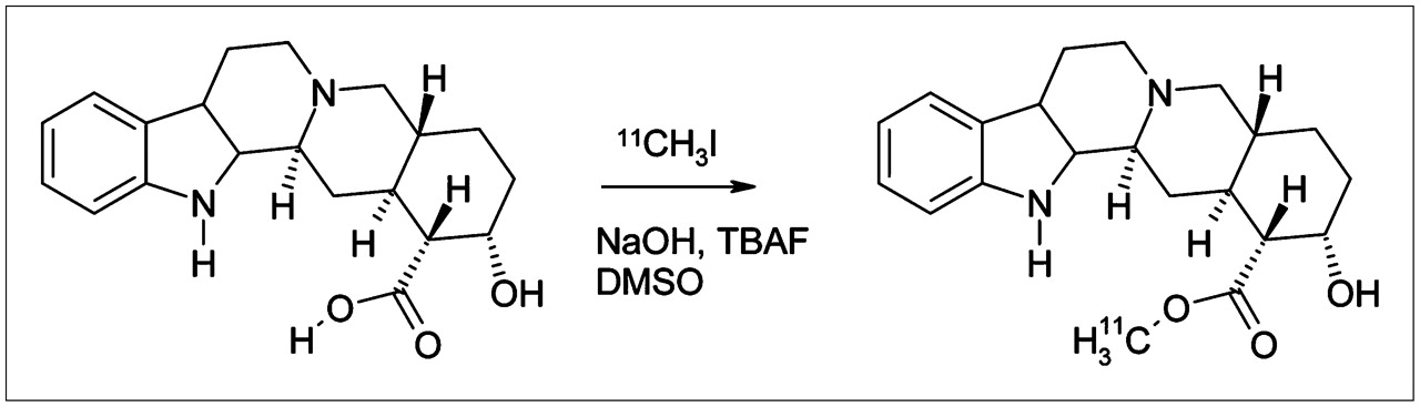
The radiosynthesis of 11C-yohimbine is depicted in Figure 1. The radiochemical purity of 11C-yohimbine was >95%, and the radiochemical yield was 60%–85%, calculated on the basis of the collected 11C-methyl iodide. In initial test productions using an excess of sodium hydroxide, the radiochemical yield was <25%, presumably due to product decomposition. The mean specific radioactivity of 11C-yohimbine was 40 GBq/μmol (range, 10–300 GBq/μmol), and the final content of yohimbinic acid was <1 μg/mL. Individual batch productions of 11C-yohimbine showed no alterations in appearance, pH, and chemical or radiochemical purity at 1 h after the EOS. The product 11C-yohimbine was chemically stable for at least an hour after the EOS. The metabolite analysis of the plasma samples showed only a single radioactive species—namely, untransformed 11C-yohimbine. Even at late time points, radioactive plasma metabolites were not detected.

Radiosynthesis of 11C-yohimbine from yohimbinic acid.
Whole-Body Dosimetry
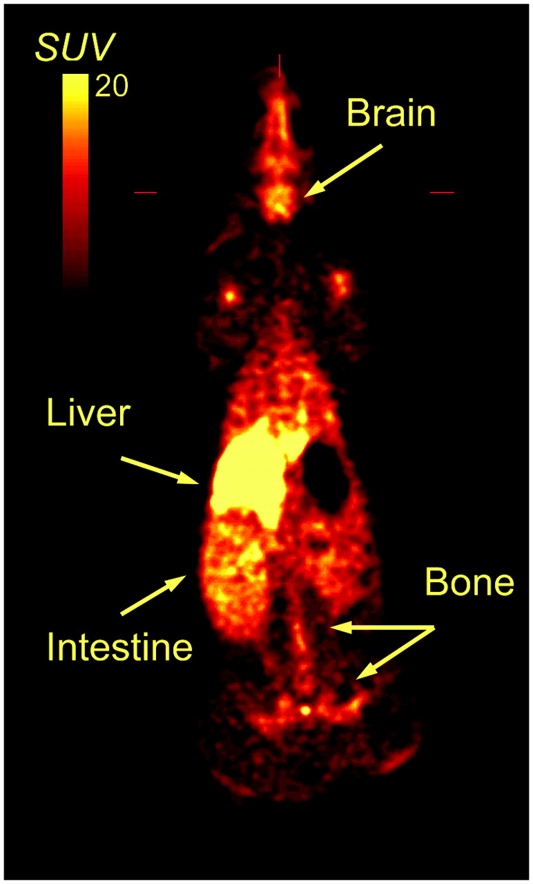
The summed whole-body PET recording of 11C-yohimbine in pig is shown in Figure 2. Table 1 presents the calculated dosimetry for humans, based on the data obtained in pig. The main part of the radioactivity was present in liver, with smaller amounts in brain and skeleton.

Whole-body distribution of 11C-yohimbine in pig. Image is average of 3 consecutive whole-body scans. Standardized uptake value (SUV) is defined as percent radioactivity relative to injected dose divided by body weight.
Radiation Dose Estimates to Human Target Organs Based on Biodistribution Data from Pig Given Injection of 11C-Yohimbine
Cardiovascular Effects of Drug Administrations
In the baseline condition for both groups of pigs with pharmacologic challenge, the mean heart rate was 110 min−1, and the mean systolic and diastolic arterial blood pressures were 118 and 76 mm Hg, respectively. The low dose of yohimbine (0.07 mg/kg) was without effect on cardiovascular function, whereas the high dose of yohimbine (1.6 mg/kg) produced a transient 30% decrease in mean arterial blood pressure, associated with a 25% increase in heart rate, which, nonetheless, remained within the normal range for anesthetized pigs for the duration of the experiment. The RX821002 challenge at the low dose (0.15 mg/kg) produced an immediate 60% increase in heart rate that gradually returned to baseline values within 2 h, with no effect on mean arterial blood pressure; the high dose of RX821002 (0.7 mg/kg) had no further effect on heart rate.
Brain Imaging
Representative time–radioactivity curves measured in frontal cortex and cerebellum during 90 min after injection of 11C-yohimbine in a baseline condition and after an intravenous bolus infusion of yohimbine (0.07 mg/kg) appear in Figure 3A. The radioactivity concentrations peaked at 15 min in cerebellum and at 25 min in frontal cortex, followed by a washout during the following 60 min. With RX821002 pretreatment, peak radioactivity concentrations occurred in cerebellum and frontal cortex at 8 min (Fig. 3B). The corresponding arterial input Logan plots at baseline, and after low-dose yohimbine challenge, are illustrated in Figure 3C, whereas the low dose of RX21002 is illustrated in Figure 3D.

Time–radioactivity curves and arterial input Logan plots of 11C-yohimbine studied by PET in living porcine brain. Time–radioactivity measurements in frontal cortex (•) and cerebellum (▪) in baseline condition, and in frontal cortex (○) and cerebellum (□) after challenge with low-dose yohimbine (A), and corresponding measurements before and after challenge with low-dose RX821002 (B) are presented along with fitting of a 1-compartment model to observed data (smooth lines). Corresponding arterial input Logan plots at baseline and after challenge with the low-dose yohimbine (C) and before and after challenge with low dose of RX821002 (D) are shown. Data are from representative experiment. CVOI = radioactivity concentration in VOI; Int[Cp] = integral of plasma radioactivity concentration.
In all brain regions and under all conditions, the mean voxelwise Vd values obtained by the Logan method were virtually identical to the results obtained by the method of Turkheimer et al. (14), the latter results are presented in Tables 2 and 3. The distribution of 11C-yohimbine in the baseline condition showed regional differences; highest binding was found in cortical structures and thalamus, intermediate binding was in mesencephalon, and lowest binding was in cerebellum, pons, and medulla. Administration of yohimbine at 0.07 mg/kg decreased the magnitude of Vd in all brain regions to approximately 2 mL g−1 (Fig. 4). The repeated-measures data analysis indicated that challenge with yohimbine significantly reduced Vd values of 11C-yohimbine in porcine brain (within-factor [scan], P < 001), with a generally greater decline in regions with relatively high baseline values than those with low values at baseline (scan × region interaction, P < 0.001). In addition, regional Vd values of 11C-yohimbine in pigs challenged with yohimbine tended to be lower in medulla and pons than in other brain regions (main effect of region, P < 0.01). The higher dose of yohimbine had no further effect on Vd; slight increases relative to the low dose of yohimbine were not significant. The administration of RX821002 at the low dose evoked a uniform displacement of 11C-yohimbine throughout the brain (Fig. 5), and the high dose was without further effect (Table 2). The repeated-measures data analysis indicated that challenge with RX821002 significantly reduced Vd values of 11C-yohimbine in porcine brain (within-factor [scan], P < 001). Moreover, the decline in 11C-yohimbine binding after challenge with RX821002 was generally greater in regions with relatively high baseline Vd values, such as thalamus and cortical regions, than in regions with relatively low baseline values (scan × region interaction, P < 0.001).
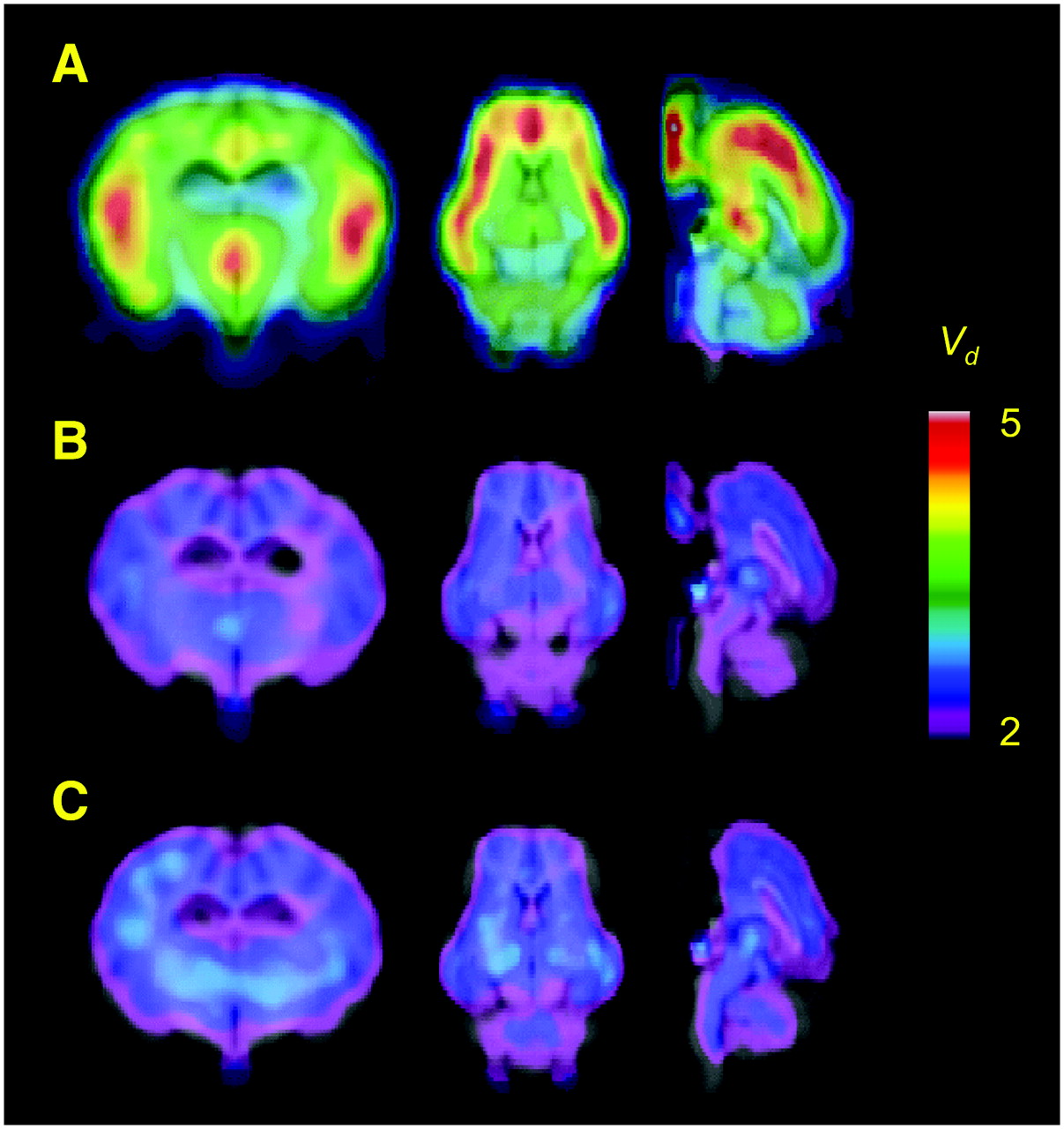
Parametric maps of 11C-yohimbine in living porcine brain. Pharmacologic condition: (A) baseline; (B) 0.07 mg/kg yohimbine; (C) 1.6 mg/kg yohimbine. Each map is mean of 3 separate determinations, resampled into MR-based common stereotactic space for pig brain, and maps are shown superimposed on the MR image.
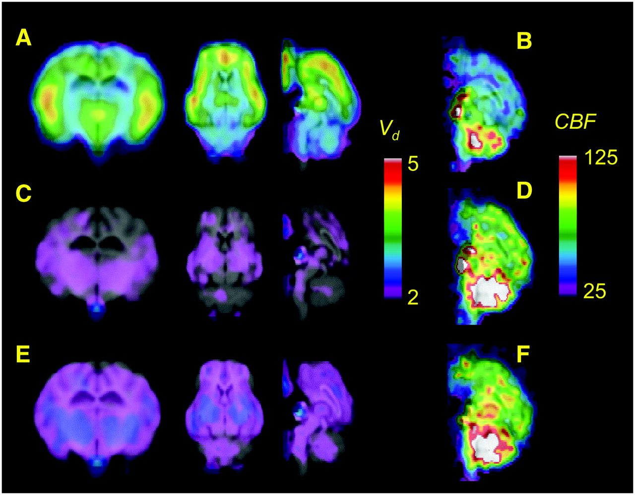
Parametric maps of 11C-yohimbine and 15O-water in living porcine brain. Pharmacologic condition: (A and B) baseline; (C and D) 0.15 mg/kg RX821002; (E and F) 0.7 mg/kg RX821002. Each map is mean of 3 separate determinations, resampled into MR-based common stereotactic space for pig brain, and maps are shown superimposed on MR image.
Magnitude of Vd (mL/g) of 11C-Yohimbine in Regions of Living Porcine Brain
Magnitude of Vd (mL/g) of 11C-Yohimbine Before and After Challenge with RX821002
Administration of 0.07 mg/kg yohimbine had no effect on CBF value, whereas the high dose of 1.6 mg/kg yohimbine resulted globally in a 30% reduction in CBF (data not shown). Pretreatment with the low dose of RX821002 evoked an increase in CBF in all brain regions examined (Fig. 5; Table 4; mean increase of 43% ± 8%). With regard to CBF, the data analysis indicated that RX821002 significantly increased values in porcine brain (within-subject factor [scan], P < 001), with the greatest change occurring in frontal cortex and putamen (scan × region interaction, P < 0.001). However, no reliable overall difference was observed between brain regions in the effects of RX821002 on CBF values (main effect of region, P > 0.2). No further increase was evoked by the high dose of RX821002. Baseline K1 for 11C-yohimbine was 19 ± 3 mL g−1 min−1 in cerebellum, corresponding to an extraction fraction of 27%.
Regional CBF Under Baseline Conditions and in Response to Increasing Doses of RX821002
DISCUSSION
Noradrenaline neurotransmission is implicated in brain functions such as attention, memory, and emotion (19,20). Abnormalities of noradrenaline function have been linked to several disorders such as anxiety and depression (21), and noradrenergic drugs have a long history as antidepressants and antihypertensive agents. Some symptoms of acute withdrawal from opiates and alcohol are associated with overactivity of noradrenergic neurons in the locus coeruleus, a cell nucleus that may be altered by chronic cigarette smoking. Finally, loss of noradrenergic forebrain innervations has been noted in degenerative diseases such as Alzheimer's disease and multisystem atrophy (22,23), and inhibition of noradrenaline transporter has some efficacy in the treatment of Parkinson's disease (24).
The role of sympathetic innervations on the control of cerebral circulation has been extensively studied since the 1970s (25). In the present study, the α2-adrenoceptor antagonist RX821002 globally increased CBF by approximately 40%. We have reported a similar increase in CBF in pigs treated with mirtazapine (5). In contrast, the high dose of yohimbine in the present study tended to decrease the magnitude of CBF in the anesthetized pig, as we have observed earlier (5). Thus, reduced CBF evoked by a high dose of yohimbine may be attributable to effects mediated by cerebrovascular receptors other than α2-adrenergic receptors in pig brain. Contraction of cerebral arteries is mediated by α2-adrenergic receptors in dogs but by α1-adrenergic receptors in monkeys and humans (26). Thus, yohimbine at high doses may mediate a decrease in CBF in pig by a mechanism distinct from the increases evoked by mirtazapine and RX821002 by antagonism of α2-adrenergic sites. On the basis of the pharmacology of yohimbine, we speculate that this action is linked to α1-adrenergic binding sites.
The α2-adrenergic receptors have been described so far by autoradiography only in cerebellum of the pig (27). However, we have developed 11C-mirtazapine for imaging studies of α2-adrenergic receptors in brain (5). 11C-Mirtazapine, like 11C-yohimbine in the present study, had notably high binding in the medial frontal and cingulate cortices of the pig, intermediate binding in the thalamus, and only traces of displaceable binding in the cerebellum, generally consistent with in vitro autoradiographic results obtained for α2-adrenergic receptors for other species (28,29). However, displacement of 11C-mirtazapine by yohimbine in living pig brain was incomplete; approximately 40% of the specific binding relative to cerebellum remained in diverse brain regions even after a high dose of nonradioactive yohimbine (3 mg/kg). Likewise, one third of 11C-mirtazapine binding throughout pig brain was displaced by a low dose (0.1 or 1 mg/kg) of RX821002, suggesting that yohimbine and RX821002 have comparable affinity or selectivity toward α2-adrenoceptors. In contrast, 11C-yohimbine binding was entirely and homogeneously self-displaced at a yohimbine dose of <0.1 mg/kg, indicating a very high-affinity interaction.
In spite of its long history of clinical use, there have been few studies reporting the metabolism of yohimbine in humans. We note both similarities and differences between yohimbine metabolism in humans and pigs. Le Corre et al. reported that the hepatic enzymes CYP3A4 and CYP2D6 degrade yohimbine to 10-hydroxyyohimbine (10-OH YOH) and 11-hydroxyyohimbine (11-OH YOH), with CYP2D6 playing the major role in the formation of 11-OH YOH (30), whereas no CYP2D6 activity could be detected in microsomes from pig liver (31). In addition, some individuals in the study of Le Corre et al. failed to form 11-OH YOH after injection of yohimbine. Similarly, no radioactive metabolites of yohimbine were found in plasma in the present pig study. In humans, 10-OH YOH is present in urine (32), whereas no studies of urinary metabolites have been performed yet in pigs. We view the lack of radioactive plasma metabolites of 11C-yohimbine in pigs as convenient for quantification of α2-adrenoceptors by PET, but human studies may require more sophisticated kinetic models.
In anticipation of future human studies with 11C-yohimbine, we have performed a whole-body dosimetry study. The whole-body distribution of 11C-yohimbine is similar to that of 11C-mirtazapine in pig, and we estimate the human dose to be 5.6 mSv/GBq, which also compares with our earlier estimates for rac-11C-mirtazapine (6.8 mSv/GBq) and the (S-) and (R)-enantiomers (4.8–5.9 mSv/GBq) (6). Thus, we predict whole-body dosimetry in the range commonly accepted for neuroimaging in human subjects (33). However, radiation dose data extrapolated from pigs to humans may have underestimated the effective dose from 11C-mirtazapine by 30%–35% (S.B. Hansen, unpublished data, September 2001). Consequently, we must measure whole-body dosimetry of 11C-yohimbine in humans to fulfill requirements of radiation health authorities before using 11C-yohimbine for PET neuroimaging in humans.
CONCLUSION
We have developed an efficient radiosynthesis for 11C-yohimbine of high specific activity. The whole-body dosimetry study in pig suggests that this tracer could be safely used in human studies. Labeled 11C-yohimbine metabolites were not detected in pig plasma, and the binding rapidly reached equilibrium in pig brain. The pattern of cerebral binding was consistent with the expected pig brain distribution of α2-adrenergic receptors. Binding of 11C-yohimbine was entirely self-displaced by a very low dose of yohimbine or by a low dose of RX821002. We suggest that 11C-yohimbine at tracer doses preferentially binds with high affinity to α2-adrenoceptors in brain and shows promise as a PET radioligand for in vivo studies.
Acknowledgments
This work was supported by grants from Denmark's National Science Foundation and The Danish Medical Research Council.
Footnotes
-
COPYRIGHT © 2006 by the Society of Nuclear Medicine, Inc.
References
- Received for publication July 13, 2006.
- Accepted for publication September 5, 2006.


