


Abstract
This study assessed the utility of scintimammography using a standard γ-camera and a dedicated breast camera as an adjuvant to mammography and clinical examination. Methods: The study population comprised 37 patients with dense breasts and a family or personal history of breast cancer. The subjects had no suggestive clinical or mammographic findings. After intravenous administration of 99mTc-sestamibi, the patients were imaged using a conventional γ-camera and a dedicated breast camera that allowed breast compression during image acquisition. Images were interpreted independently by 2 reviewers. All patients with positive scintimammography findings underwent biopsy. Results: Dedicated breast camera results were positive in 13.5% (5/37) of patients. Biopsy of these 5 patients yielded 3 carcinomas: an infiltrating lobular carcinoma, a ductal carcinoma in situ, and an infiltrating tubular carcinoma. These 3 carcinomas were undetectable by clinical breast examination or mammography, even on retrospective review. Only one of these, the tubular carcinoma, was readily detectable by the standard γ-camera. Conclusion: Scintimammography using a dedicated breast camera may augment mammography and clinical breast examination for the subset of women who have dense breast tissue and are at high risk of breast cancer.
The sensitivity of mammography decreases to less than 70% in women with radiographically dense breast tissue (1). These patients tend to be younger, and their breast cancer is often aggressive. In this group, the sensitivity of standard screening methods is compromised (1–7). Unfortunately, breasts that are radiographically dense may also be difficult to examine clinically (8). Scintimammography, a functional rather than an anatomic imaging technique, may be a useful adjunct to breast self-examination, clinical breast examination, and mammography in the subset of women with radiographically dense breasts.
The sensitivity of scintimammography, unlike standard screening modalities, is not compromised by inherent breast density (9,10). 99mTc-Sestamibi localizes in most tumors soon after injection because of their relatively increased blood supply, as well as higher metabolic activity, compared with surrounding tissues. Its accumulation is roughly proportional to mitochondrial concentration (11).
The sensitivity of scintimammography with a standard γ-camera is dependent on lesion size. Mekhmandarov et al. reported that the sensitivity for detection of malignancy in small lesions with a standard γ-camera was poor—less than 55% for nonpalpable tumors with a mean size of 1.34 cm (4). The dedicated breast camera (LumaGEM; Gamma Medica, Inc.) allows the breast to be compressed during image acquisition. Compression facilitates lesion detection with the dedicated breast camera, as it does in mammography. Because the breast tissue is spread over the camera face, the signal-to-noise ratio for lesions is increased and lesions are more easily detected.
MATERIALS AND METHODS
Subject Selection
The study population comprised 37 women (mean age, 54 y; range, 34–80 y) with normal findings on clinical breast examination, BI-RADS category I or II mammography findings, and a BI-RADS parenchymal pattern of “heterogeneously dense” or “extremely dense” breast tissue. Most of the women had a strong family history of breast cancer; 9 had a personal history of breast cancer. This prospective study was approved by a local institutional review board and was performed at a 436-bed medical center in northwestern Pennsylvania. Subjects were enrolled from August 2001 through mid November 2001. Informed consent was obtained from all patients before enrollment. Subjects were excluded if they were pregnant or lactating or were physically unable to lie prone with their arms above their head for 20 min or to sit upright and still for 1 h.
Dedicated Breast Camera
The dedicated beast camera was designed to optimize breast scintigraphy. The detector head was highly compact (15.0 × 12.5 × 8.5 cm) and weighed 10 kg. The dead space between the outside edge of the head and the active area of the field of view was less than 1 cm. The camera was attached to an adapter that fits on most upright mammography machines, replacing the radiographic Bucky (Fig. 1).
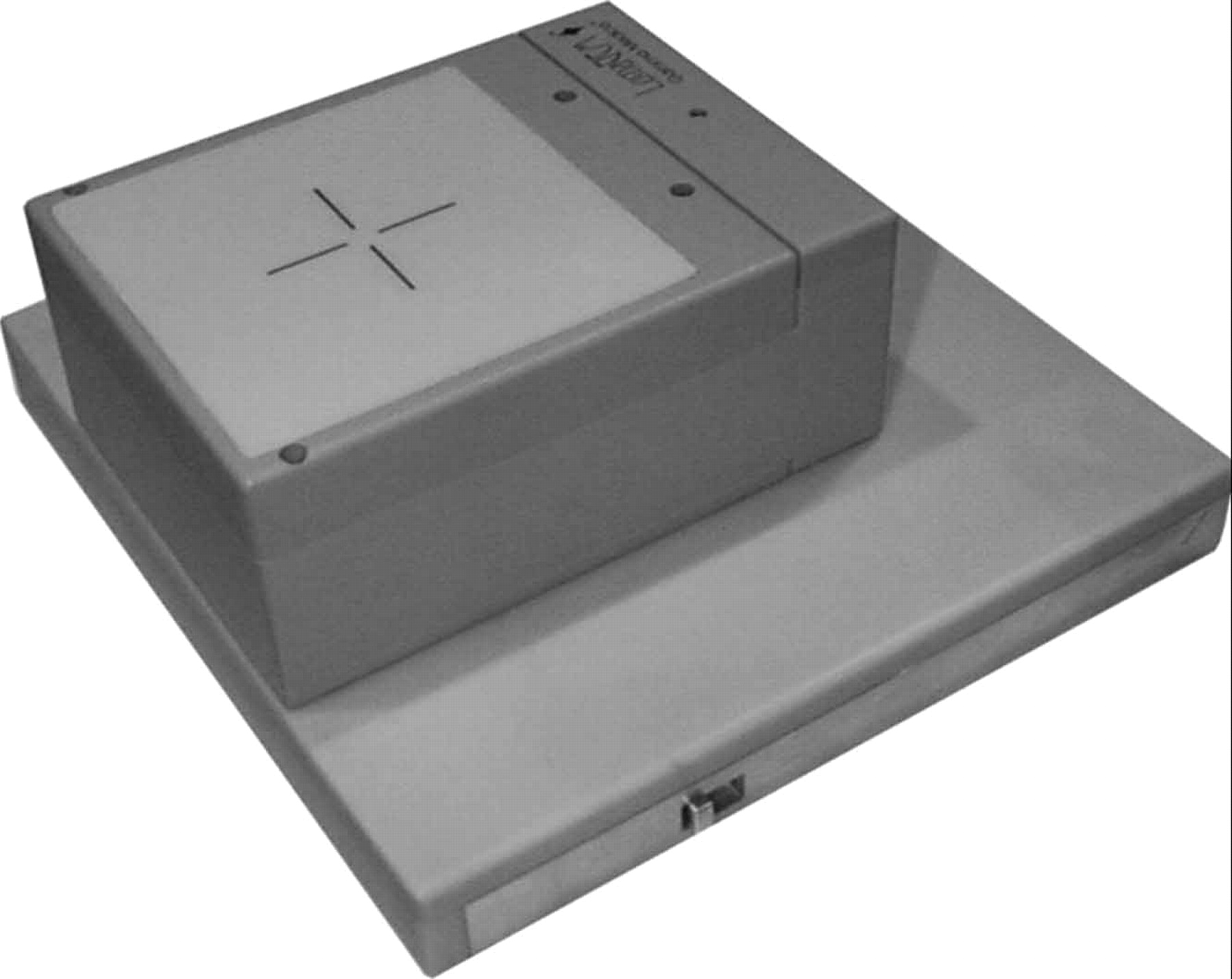
Dedicated breast camera attached to an adapter that allows the unit to be mounted on most mammography units.
The dedicated breast camera used a pixilated array of more than 3,000 discrete NaI (Tl) scintillation crystals, each of which was 2 × 2 × 6 mm. A reflective material surrounded each crystal so that scintillations were contained within the right rectangular, parallel-piped geometry of the crystals. The center-to-center pitch of these crystals was 2.2 mm. This array was coupled to an array of position-sensitive photomultiplier tubes that had some internal positioning capability. The readout circuit of the position-sensitive photomultiplier tubes indicated the resolution of the individual NaI (Tl) crystals. A look-up table was generated that corresponded to the position of the individual crystals, and events were positioned according to this table. The intrinsic spatial resolution of the camera was determined by the 2.2-mm pitch of the crystal array. This afforded an approximately 2-fold improvement in spatial resolution, compared with standard γ-cameras.
The standard γ-camera was modeled with a 3.5-mm intrinsic resolution and a low-energy high-resolution collimator with a 1.5-mm borehole diameter and 25.4-mm borehole length. Similarly, the dedicated breast camera was modeled with a 2-mm intrinsic resolution and the same low-energy high-resolution collimator (Fig. 2). The expected extrinsic resolution of the dedicated breast camera was better at all source depths than that of the standard γ-camera. The extrinsic resolution was the quadrature sum of the intrinsic resolution of the detector and the collimator-dependent resolution. As object-to-detector distance decreased, the resolution of each system asymptotically approached the intrinsic resolution of the detector, whereas as detector-to-object distance increased, the resolution of each system asymptotically approached that of the collimator. The resolution and corresponding source depth at which the 2 contributions were equal for various intrinsic resolution values were plotted (Fig. 2). Improved spatial resolution enhanced the contrast resolution of small lesions by increasing the target-to-background ratio.

The extrinsic resolution (Resolution) vs. source depth for a standard γ-camera (STD Gamma Cam) with 3.5-mm intrinsic resolution and a dedicated breast camera (LumaGEM) with 2-mm intrinsic resolution. The solid line represents extrinsic resolution vs. source depth when detector resolution is equal to collimator resolution (Coll Res = Det). The points representing the standard γ-camera (○) and the dedicated breast camera (▵) are shown.
Data Collection
Data were collected on patient demographics, past medical history, treatment (if past medical history indicated breast cancer), physical examination results, and current physical breast examination results. Data were also collected on the results of mammography, ultrasound (if performed), scintimammography, and biopsy.
Imaging Procedure
Into the dorsum of the patient’s foot, 740 MBq of 99mTc-sestamibi (Bristol-Myers Squibb Co.) was injected. The patient was asked to lie prone, with the imaged breast dependent through a table cutout (11). Imaging with the standard γ-camera commenced 10 min after injection. A lateral view of each breast was acquired for 10 min. A 5-min anterior acquisition followed, with the patient’s arms above her head to evaluate the axillae for lymphadenopathy.
Approximately 1 h after the initial 99mTc-sestamibi injection, an additional 740 MBq of 99mTc-sestamibi was injected into a vein on the dorsum of the patient’s foot. Imaging with the dedicated breast camera commenced 10 min after injection. Each breast was imaged in 2 projections: caudal (same positioning as for a craniocaudal mammogram, with the camera on the caudal surface of the breast) and lateral oblique (same positioning as for a mediolateral oblique mammogram, with the camera on the inferolateral aspect of the breast) (Fig. 3). The images were then reviewed by a physician, who determined if additional views were necessary.

Image acquisition using the dedicated breast camera in caudal (left) and lateral oblique (right) projections, with the detector on the undersurface and the inferolateral surface, respectively, of the breast.
The mammography gantry allowed flexibility in positioning. With the dedicated breast camera, the breast was compressed during image acquisition. Compression spread the breast tissue over the camera face, reducing background count density while maintaining the count density of the lesion. The resultant improvement in target-to-background ratio increased the ability of the dedicated breast camera to detect subtle lesions undetected by the standard γ-camera. If a lesion was suspected in an area that was not close to the camera, the gantry was repositioned and additional views were acquired. Breast compression and flexible positioning resulted in improved contrast resolution of suspected lesions.
For patient convenience, both standard γ-camera imaging and dedicated breast camera imaging were performed the same day. This introduced some bias, as the standard γ-camera imaging was performed first. Sestamibi has been shown to wash out from breast tumors at variable rates (12,13). As such, the target-to-background ratio of the dedicated breast camera images was theoretically less than if a single dose had been injected. However, count statistics improve after administration of a second dose.
Two investigators independently interpreted the images from the standard γ-camera and the dedicated breast camera. Both investigators knew the clinical and mammographic findings. The relative degree of sestamibi uptake was not considered. Findings were interpreted as positive or negative on the basis of whether an area of focally increased sestamibi uptake with definable borders was identifiable, even if the target-to-background ratio was minimal. Patients with positive findings underwent biopsy. The results of mammography, scintimammography with a standard γ-camera, and scintimammography with the dedicated breast camera were recorded. The imaging results of both cameras were compared with biopsy results.
By study design, detectable lesions were not palpable or mammographically visible and were detectable only scintimammographically; therefore, the dedicated breast camera was used for lesion localization before biopsy.
Scintimammographic Needle Localization
Scintimammographic localization was necessary for lesions that were detectable by scintimammography but not by physical palpation or mammography. Methods of lesion localization using the standard γ-camera have been described (14,15). A method of lesion localization using an open biopsy paddle with the dedicated breast camera was developed. The breast was compressed against the dedicated breast camera using a mammography biopsy paddle with a 4 × 8 cm opening. The patient received a 1,110-MBq injection of 99mTc-sestamibi. After 10 min, the breast was positioned such that the suspicious area was within the opening. Then, the biopsy paddle was used to compress the breast against the camera. Image acquisition was initiated, with the camera monitor in the persistence mode, until the lesion became visible on the monitor. Using a felt-tip pen, the physician marked the site of the lesion on the monitor. With the monitor still in the persistence mode, a 57Co point source was positioned on the patient’s breast. The physician viewed the monitor and moved the point source on the patient until the activity on the screen from the point source coincided with the dot previously placed on the screen. The location of the point source was then marked on the patient’s skin. The patient’s skin was then prepared with povidone–iodine (10% U.S. Pharmacopeia). A localization needle was inserted into the breast at the localized site, with the needle shaft perpendicular to the camera face.
The camera was rotated approximately 90°, and the localization procedure was repeated (Fig. 4). Because the needles were placed while the breast was compressed in different orientations, the site of needle intersection identified the lesion site. An open biopsy was performed on the area where the 2 needles intersected. After biopsy, the specimen was imaged to confirm that it contained the area of focally increased activity (Fig. 5).
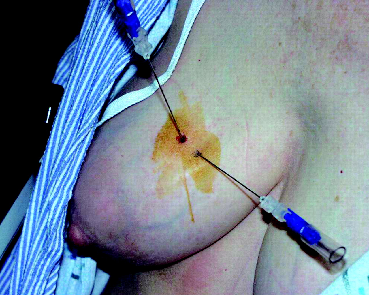
Localization needles placed using the dedicated breast camera.

Dedicated breast camera image of the biopsy specimen confirms that the localized lesion (infiltrating lobular carcinoma) was contained within the specimen.
RESULTS
Standard γ-camera results were positive in 8.1% (3/37) of patients. Dedicated breast camera results were positive in 13.5% (5/37) of patients, including the 3 patients whose lesions were detected by the standard γ-camera. In these 5 patients, biopsy yielded 3 carcinomas: infiltrating lobular carcinoma (patient 1), ductal carcinoma in situ (patient 2), and infiltrating tubular carcinoma (patient 3). Of these 3 carcinomas, only that of patient 3 was readily detected by the standard γ-camera. In patient 1, the mammographic results were negative (Fig. 6). The standard γ-camera detected a focal area of increased uptake in an anterior view of the chest. The finding would be interpreted as a positive area of indeterminate location; however, no abnormality was seen in the lateral view of the breast (Fig. 7). The dedicated breast camera provided a better signal-to-noise ratio, allowing lesion detection in the breast (Fig. 8).

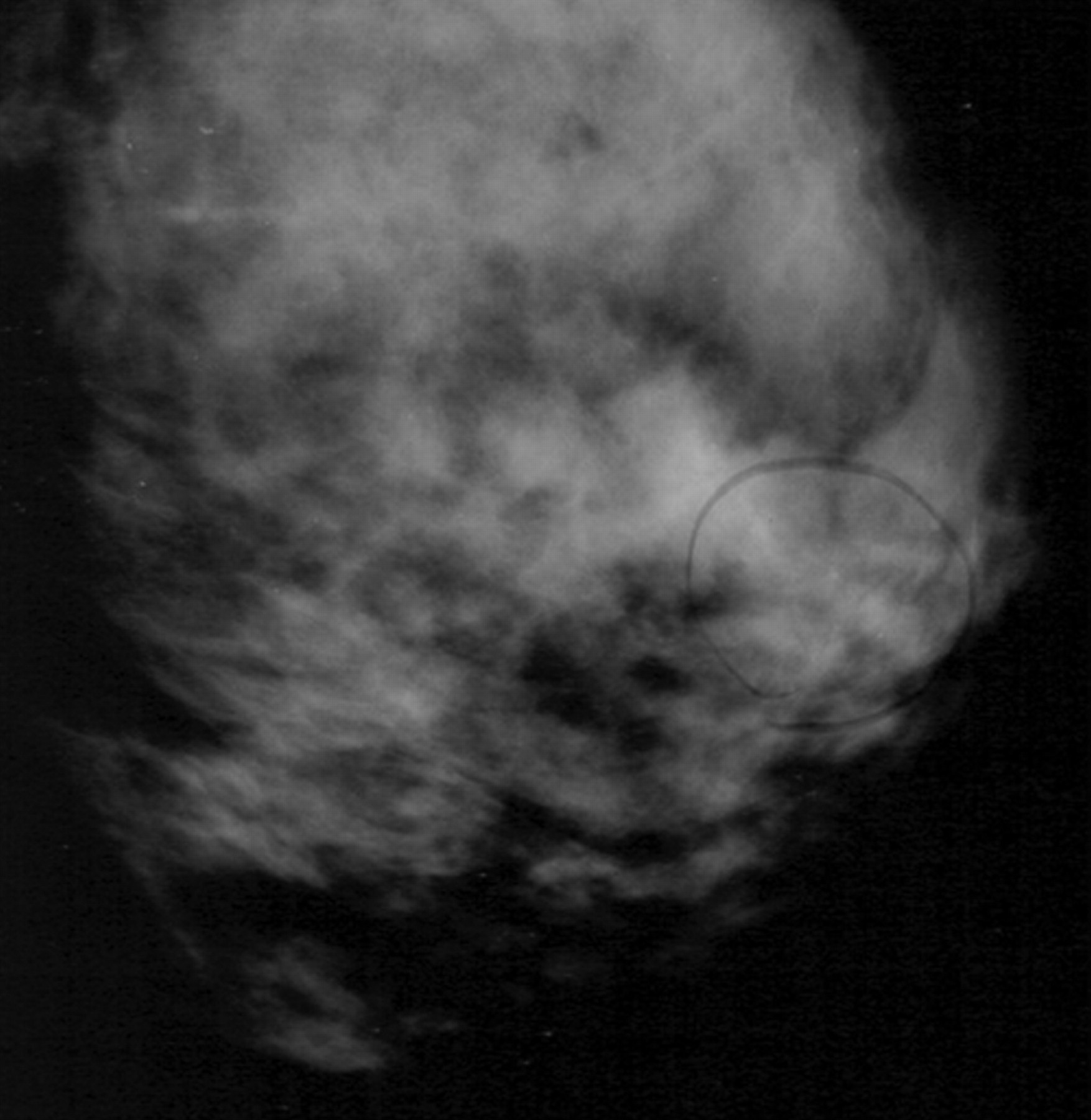
Patient 1, with infiltrating lobular carcinoma. Mediolateral oblique mammogram failed to demonstrate a nonpalpable lesion that was obscured by dense fibroglandular tissue. The circled, loosely grouped calcifications were anterior to the tumor and had been stable for years. The lesion was interpreted as BI-RADS category 2.

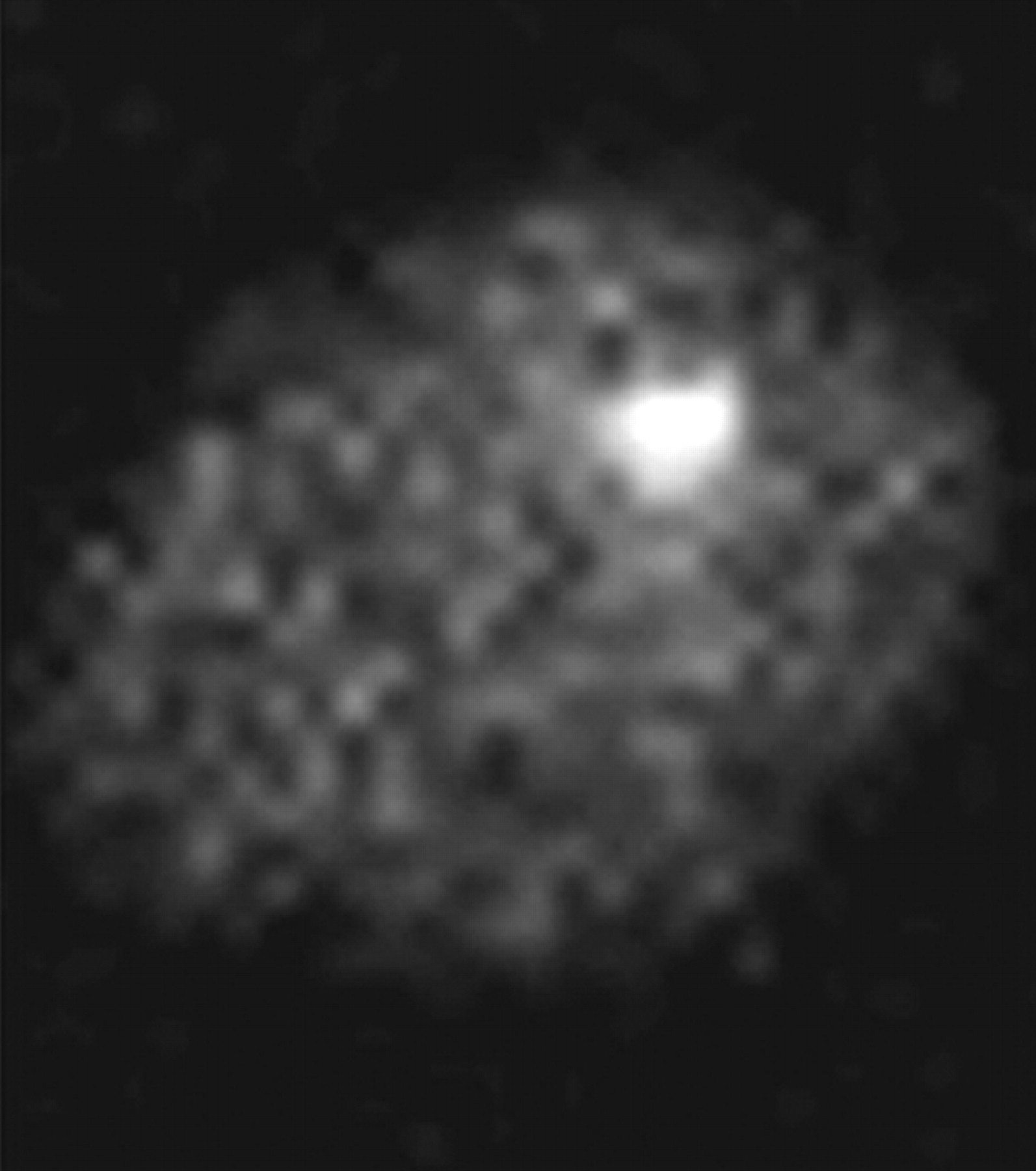
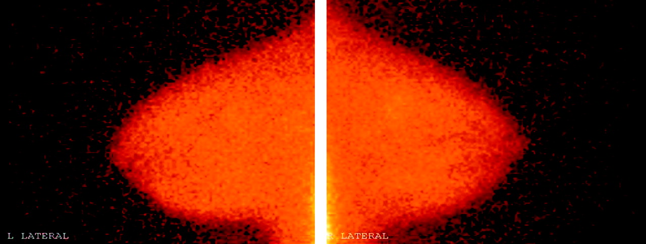
Lateral scintimammograms of patient 1 obtained with a conventional γ-camera. The images were acquired with the patient supine and with the breast dependent. These images were interpreted as showing normal findings. An anterior view of the chest showed a focal area of increased uptake, which would be interpreted as a positive finding of indeterminate location.
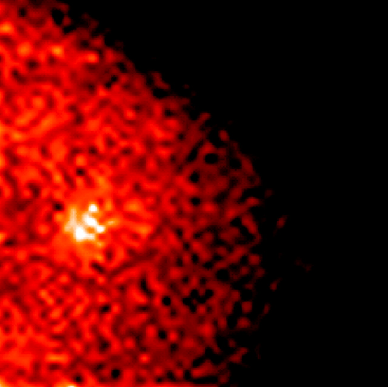
Scintimammograms of patient 1 obtained with a dedicated breast camera. Better signal-to-noise ratio allowed lesion detection. These images were interpreted as showing positive findings. Biopsy revealed infiltrating lobular carcinoma.
These 3 carcinomas were undetectable by clinical breast examination or by mammography, even on retrospective review. The remaining 2 biopsy results revealed a case of fat necrosis and a case of fibrocystic change (Table 1; Fig. 9).

Biopsy results for dedicated breast camera vs. standard γ-camera.
Comparison of Results
DISCUSSION
The value of scintimammography as an adjuvant to standard screening modalities (mammography and clinical examination) is in the early detection of breast carcinoma. Scintigraphy may be appropriate for the subset of women whose breasts are difficult to examine by conventional means, including those with increased mammographic density, fibrocystic changes, implants, or scarring from previous surgery or radiation.
Scintimammography using the dedicated breast camera detected carcinomas in 8.1% (3/37) of women with no palpable mass and no suggestive mammographic findings. The standard γ-camera detected only 1 of these 3. Of the 5 biopsies that were performed as a result of positive scintimammography results, 2 revealed benign findings. Of the 3 true positives, only 1 was readily detected by scintimammography with a standard γ-camera. We concur with Brem et al. (2) that more lesions were detected with a dedicated breast camera than with a standard γ-camera.
Selection bias will influence outcome. Subjects who met selection criteria were at an increased risk of breast carcinoma. They were screened by mammography and physical examination, both of which are known to be less sensitive for dense breasts. Because the study results were derived from a small, select patient population, larger studies should be undertaken to validate the potential use of scintimammography as an adjuvant screening modality for women whose breasts are difficult to examine by conventional screening modalities.
CONCLUSION
Scintimammography with a dedicated breast camera may augment mammography and clinical breast examination as an adjuvant diagnostic modality in the subset of women with dense breast tissue and a high risk of cancer. Dense breast tissue reduces the sensitivity of mammography and clinical examination. Of women with BI-RADS category I or II mammography results, dense breasts, and a family history or personal history of breast carcinoma, 13.5% (5/37) had positive findings from dedicated breast camera screening. Subsequent histologic examination revealed carcinoma that was otherwise undetectable by conventional methods in 3 of the 5 women with positive findings. Only 1 of the 3 carcinomas detected with the dedicated breast camera was easily detectable with the conventional γ-camera.
Acknowledgments
Financial support for this study was provided by Gamma Medica, Inc., Northridge, CA, and Bristol-Myers Squibb, New York, NY.
Footnotes
Received Jun. 23, 2003; revision accepted Jan. 5, 2004.
For correspondence or reprints contact: Leonard R. Coover, MD, Hamot Medical Center, 201 State St., Erie, PA 16550.
E-mail: Leonard.Coover{at}hamot.org
REFERENCES
In this issue


Jump to section
Related Articles
Cited By...
- Breast-specific gamma imaging as an adjunct modality for the diagnosis of invasive breast cancer with correlation to tumour size and grade
- SNM Practice Guideline for Breast Scintigraphy with Breast-Specific {gamma}-Cameras 1.0
- The SNM Practice Guideline on Breast Scintigraphy
- Dedicated Breast Camera: Is It the Best Option for Scintimammography?
- Scintimammography with a Pinhole Collimator