

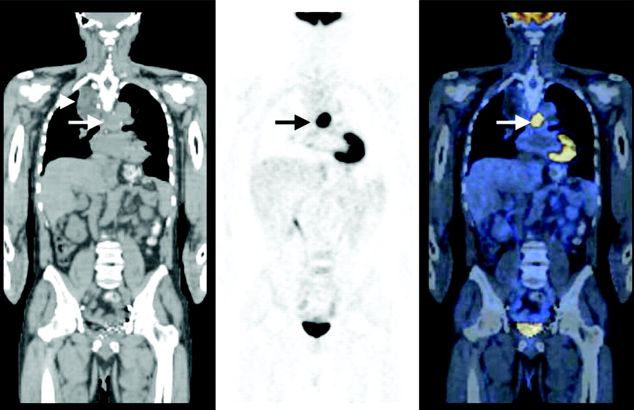
FIGURE 3.

A 62-y-old man with history of locally advanced adenocarcinoma of distal esophagus after neoadjuvant therapy, with partial response, determined by outside 18F-FDG PET study. Patient had undergone esophagectomy and gastric pull-up surgery and now was referred for 18F-FDG PET scan to evaluate disease status 6 mo after completion of neoadjuvant therapy and surgery. Coronal CT (left), PET (middle), and PET/CT (right) images demonstrate intense 18F-FDG uptake in midline in midchest, corresponding to tracheobronchial lymph nodes, consistent with metastatic disease (arrows). Patient’s disease progressed, and patient died within 3 mo after study. This study emphasizes that 18F-FDG PET after neoadjuvant therapy appears to predict prognosis with high accuracy; median survival time of nonresponders is much shorter than that of responders. Note postsurgical anatomic changes in right upper chest secondary to gastric pull-up surgery on CT image (left; arrowhead). PET/CT studies were obtained on a GE Discovery LS unit—a PET/CT fusion system combining GE LightSpeed multislice CT and Advance NXi PET (GE Medical Systems).
In this issue


Related Articles
Cited By...
- Diagnostic Value of Neck Node Status Using 18F-FDG PET for Salivary Duct Carcinoma of the Major Salivary Glands
- Preclinical and Clinical Evidence that Deoxy-2-[18F]fluoro-D-glucose Positron Emission Tomography with Computed Tomography Is a Reliable Tool for the Detection of Early Molecular Responses to Erlotinib in Head and Neck Cancer
- Use of PET for Monitoring Cancer Therapy and for Predicting Outcome