


Abstract
The preparation, characterization, and first biologic evaluation in rats of a novel class of monocationic 99mTc heart imaging agents are reported. The complexes are represented by the general formula [99mTc(N)(PNP)(L)]+, where L is the monoanionic form of a dithiocarbamate ligand of the type [R1(R2)-N-C(=S)S]−, PNP is a diphosphine ligand of the type [(R3)2P-(CH2)2]2-N(R4), and R1–R4 are organic functional groups. Methods: The new complexes were prepared by use of both liquid and freeze-dried formulations through a 2-step procedure. The first step involved the formation of the [Tc≡N]2+ group through the reaction of 99mTcO4− with succinic dihydrazide as a donor of nitride nitrogen atoms (N3−) in the presence of Sn2+ ions. This step was followed by the simultaneous addition to the reaction solution of the ligand PNP and of the sodium salt of the dithiocarbamate ligand (NaL) to afford the final products, [99mTc(N)(PNP)(L)]+. The chemical identities of the resulting 99mTc complexes were determined by comparing their chromatographic properties with those of the corresponding 99gTc analogs prepared by use of the long-lived isotope 99gTc and characterized by spectroscopic and crystallographic techniques. Ex vivo biodistribution studies were conducted in rats. In vivo tomographic images of the rat heart were obtained by use of a small-animal SPECT scanner. Results: The [99mTc(N)(PNP)(L)]+ complexes are monocationic and possess a distorted square-pyramidal geometry in which the Tc≡N multiple bond occupies an apical position and the diphosphine and dithiocarbamate ligands span the residual 4 coordination positions on the basal plane through the 2 phosphorus atoms and the 2 sulfur atoms, respectively. Imaging and biodistribution studies demonstrated that these radiopharmaceuticals localize selectively in the myocardium of rats and are retained in this region for a prolonged time. The kinetics of heart uptake and clearance were found to be influenced by variations in the lateral R1–R4 groups. Blood and lung washouts were extremely fast. Elimination occurred mostly through the kidneys and the liver. Surprisingly, at 1 h after injection, liver activity was almost negligible. Analysis of heart-to-liver and heart-to-lung uptake ratios showed that these values increased exponentially over time and became much higher than those determined for 99mTc-sestamibi and 99mTc-tetrofosmin. These findings were confirmed by analysis of high-quality SPECT images collected in rats for the new complexes and compared with images obtained with 99mTc-sestamibi and 99mTc-tetrofosmin. Conclusion: The high myocardial uptake and the very high heart-to-lung and heart-to-liver uptake ratios indicate that the [99mTc(N)(PNP)(L)]+ complexes exhibit very favorable distribution properties and could be used to obtain SPECT cardiac images with improved quality.
- 99mTc heart perfusion imaging agents
- asymmetric 99mTc-nitride heterocomplexes
- mixed diphosphino-dithiocarbamate 99mTc-nitride complexes
In the last 2 decades, cationic 99mTc tracers have been developed as 201Tl+ analogs for the imaging of myocardial perfusion. The most successful cationic, lipophilic 99mTc perfusion agents are the well-known compounds 99mTc-sestamibi and 99mTc-tetrofosmin (1–3). Some years ago, the bis(N-ethoxy,N-ethyl)dithiocarbamato nitride technetium-99m (99mTc-N-NOET) complex was introduced as the first example of a neutral heart imaging agent exhibiting prolonged retention in the myocardium (4–8). Surprisingly, the biologic behavior of this compound appeared to be more similar to that of the monocation 201Tl+ than to that of the above-mentioned cationic complexes (4,7).
Despite the great success of current heart imaging agents, their properties are still far from those expected for an ideal perfusion tracer. In particular, the relatively low first-pass extraction of 99mTc-sestamibi and 99mTc-tetrofosmin makes these tracers less suitable for measuring blood flow at high flux rates. Moreover, the persistent liver uptake observed for all cardiac imaging agents completely prevents the assessment of defects located at the inferoapical myocardial wall. As a consequence, there still exists considerable interest in the search for a myocardial perfusion agent having imaging characteristics much closer to those predicted for an ideal perfusion agent (9).
The Tc(V)-nitride complex 99mTc-N-NOET represents the first reported example of a heart imaging agent characterized by the presence of a terminal Tc≡N multiple bond. It has a square-pyramidal geometry in which 2 identical bidentate dithiocarbamate ligands are symmetrically bound to a Tc≡N group in the basal plane of the square-pyramid (5,10). We describe here the preparation and characterization of a new class of Tc(V)-nitride cardiac imaging agents. These complexes are monocationic and are represented by the general formula [99mTc(N)(PNP)(L)]+, where L is the monoanionic form of a dithiocarbamate ligand of the type [R1(R2)-N-C(=S)S]−, PNP is a diphosphine ligand of the type [R32(P)-(CH2)2]2-N(R4), and R1–R4 are organic functional lateral groups (Fig. 1). The unique structural feature of this class of 99mTc-nitride complexes is attributable to the fact that they are composed of 2 different bidentate ligands bound to the same Tc≡N group (Fig. 1). This composition gives rise to a new type of 5-coordinated heterocomplexes having an unusual asymmetric structure (11). These compounds were prepared after the development of a somewhat complex chemistry based on the chemical properties of the new metal nitride fragment [99mTc(N)(PNP)]2+. This fragment is formed by a Tc≡N group coordinated to a PNP ligand and is easily produced in an aqueous physiologic solution as the dichloride derivative [99mTc(N)(PNP)]Cl2. It was found that this precursor complex exhibits a strong electrophilic character and selectively reacts with electron-rich, soft, π-donor ligands, having S, O, and N as coordinating atoms, without removal of the Tc≡N group and of the ancillary PNP ligand (12,13). Thus, the formation of [99mTc(N)(PNP)(L)]+ heterocomplexes can be viewed as resulting from the selective coupling of the metal fragment [99mTc(N)(PNP)]2+ with the bidentate dithiocarbamate π-donor ligand L.

Chemical structure of [99mTc(N)(PNP)(L)]+ complexes. PNP = diphosphine ligand; L = monoanionic dithiocarbamate ligand; R1–R4 = lateral organic functional groups.
A biologic evaluation of the new asymmetric heterocomplexes was performed in rats. The results showed that these radiopharmaceuticals localized selectively in the rat myocardium and were retained in this region for a prolonged time. Liver washout was extremely fast and quantitative. Tomographic images of the rat heart were obtained by use of a small-animal SPECT scanner and compared with those collected with 99mTc-sestamibi and 99mTc-tetrofosmin. These data, along with the unprecedented high heart-to-lung and heart-to-liver uptake ratios, suggest that this class of perfusion agents could be conveniently used to obtain high-quality cardiac images. A brief account of the preliminary data on the preparation and biodistribution in rats of only 2 members of the large class of complexes described here has been communicated elsewhere (14).
MATERIALS AND METHODS
Reagents and Physical Measurements
The diphosphine ligands bis(dimethoxypropylphosphinoethyl)methoxyethylamine [PNP3; (CH3OC3H6)2P(CH2)2-N(C2H4OCH3)-(CH2)2P(C3H6OCH3)2], bis(dimethoxypropylphosphinoethyl)ethoxyethylamine [PNP5; (CH3OC3H6)2P(CH2)2-N(C2H4OCH2CH3)-(CH2)2P(C3H6OCH3)2], and bis(diethoxypropylphosphinoethyl)ethoxyethylamine [PNP6; (CH3CH2OC3H6)2P(CH2)2-N(C2H4OCH2CH3)-(CH2)2P(C3H6OCH2CH3)2] (Fig. 2) were prepared by following published procedures (13,15) or were obtained from Argus Chemical. The sodium salts of dithiocarbamate ligands, [R1(R2)-N-C(=S)S]Na (Fig. 2), were prepared by reacting the corresponding secondary amines with equivalent amounts of carbon disulfide in NaOH solutions as reported previously (5) or were obtained from Alchemy. Sodium diethyldithiocarbamate and sodium dimethyldithiocarbamate were commercially available (Aldrich Chimica). Succinic dihydrazide [SDH; H2NNH(OC)(CH2)2(CO)NHNH2], ethylenediaminetetraacetic acid (EDTA), SnCl2·2H2O, tetrabutylammonium bromide, triphenyphosphine (PPh3), and γ-cyclodextrin were purchased from Aldrich. All common solvents were reagent grade and were used without further purification. The radiopharmaceuticals 99mTc-sestamibi (Cardiolite; Dupont Radiopharmaceuticals) and 99mTc-tetrofosmin (Myoview; Amersham Healthcare) were obtained from commercial kit formulations.

Dithiocarbamate (L) and diphosphine (PNP) ligands and acronyms used in this study.
Electrospray ionization (ESI) mass (16) spectra of technetium-99 compounds (∼10−6 mol dm−3 solutions) were recorded on an LCQ instrument (Finnigan). The complexes were injected by use of a syringe pump at a flow rate of 5 μL/min.
Preparation of [99mTc(N)(PNP)(L)]+ Complexes
All preparations were made by excluding oxygen from vials and solutions by nitrogen filling and purging, respectively. The PNP and L ligands are illustrated in Figure 2.
Liquid Formulation.
Na99mTcO4 (0.250 mL, 50.0 MBq–4.0 GBq) was mixed in a vial containing 5.0 mg of SDH, 5.0 mg of EDTA, 0.10 mg of SnCl2·2H2O (suspended in 0.10 mL of saline), and 1.0 mL of ethanol. The mixture was kept at room temperature for 30 min. To the resulting solution, 1.0 mg of PNP (dissolved in 0.30 mL of ethanol) and 2.0 mg of the sodium salt of the appropriate dithiocarbamate ligand (dissolved in 0.10 mL of saline) were simultaneously added. The vial was heated at 100°C for 15 min. The yield was >95%.
Lyophilized Formulation.
A lyophilized kit formulation was developed for the preparation of the [99mTc(N)(PNP3)(DBODC)]+ (where DBODC is bis-N-ethoxyethyl-dithiocarbamato) and [99mTc(N)(PNP5)(DBODC)]+ complexes as described below.
One milliliter of generator-eluted Na99mTcO4 (50.0 MBq–4.5 GBq) was added to a vial containing 5.0 mg of SDH, 5.0 mg of EDTA, 0.1 mg of SnCl2·2H2O, and 1.0 mL of phosphate buffer (0.1 mol dm−3) in a freeze-dried form. The resulting solution was kept at room temperature for 30 min. The contents of a second lyophilized vial—3.5 mg of PNP3 (or PNP5), 3.5 mg of DBODC, and 3.5 mg of γ-cyclodextrin—were reconstituted with 1.75 mL of saline. Then, 1.0 mL of the resulting solution was withdrawn from the second vial and added to the first vial, which was heated at 100°C for 15 min. The yield was >95%.
Chromatography
Radiochemical purity (RCP) was determined by thin-layer chromatography (TLC) and high-performance liquid chromatography (HPLC). TLC was performed on silica gel plates (Merck) with ethanol:chloroform:toluene:NH4Ac (0.5 mol dm−3) (5:3:3:0.5) as the mobile phase and on reversed-phase C18 plates (Merck) with saline:methanol:tetrahydrofuran:glacial acetic acid (2:8:1:1) as the mobile phase. Thin-layer chromatograms were analyzed with a Cyclone instrument equipped with a phosphorImaging screen and OptiQuant image analysis software (Packard Instruments). HPLC was performed on a System Gold Instrument (Beckman Instruments) equipped with programmable solvent module 126, scanning detector module 166, and radioisotope detector module 170. Chromatographic runs were performed on a reversed-phase C18 column (Ultrasphere; 4.6 × 250 mm; Beckman) with a reversed-phase C18 precolumn (Ultrasphere; 4.6 × 45 mm; Beckman). A methanol:phosphate buffer mixture (0.02 mol dm−3) was used as the mobile phase at a flow rate of 1.0 mL min−1. Rf values and HPLC retention times (tr) for some selected complexes are reported in Table 1.
Rf Values, Retention Times (tr), and log k′0 Values for [99mTc(N)(PNP)(L)]+ Complexes
Purification
To avoid the interference of free reagents with evaluation of the stability and the biodistribution of 99mTc-nitride heterocomplexes, a purification procedure was carried out as follows. A reversed-phase SepPak C18 cartridge (Waters Corp.) was activated by passage of 5.0 mL of ethanol and 5.0 mL of deionized water. After preparation, the reaction solution containing the final [99mTc(N)(PNP)(L)]+ heterocomplex was diluted by the addition of 8.0 mL of deionized water. The resulting solution was passed through the activated SepPak cartridge. Approximately 100% of the initial activity was retained on the cartridge. The cartridge was washed with 20.0 mL of deionized water and 3.0 mL of an ethanol:water mixture (80:20). The complex was recovered by passage through the cartridge of 1.0 mL of a mixture composed of a water solution of tetrabutylammonium bromide {[N(C4H9)4]Br; 0.1 mol dm−3} and ethanol at a volume ratio of 10:90. Approximately 80% of the initial activity was finally collected.
Determination of log k0′ Values
After purification, log k0′ values for some representative complexes were determined by HPLC as described elsewhere (17). The results are reported in Table 1. It was shown previously that log k0′ values are well correlated with log P values (P is the water:octanol partition coefficient) for lipophilic compounds (17). log k0′ is obtained from log k′, where k′ is the capacity factor measuring the partitioning of the compound between a nonpolar stationary phase and a polar mobile phase. The log k′ values were determined here at various compositions of the mobile phase. An analysis of the relationship between log k′ and mobile phase composition gave extrapolated log k′ values at a 0% organic solvent composition (log k0′) as a measure of the activity partitioning between the hydrophobic stationary phase and water. The log k0′ values were extrapolated from the linear part of the curve log k′ = a + bC, where C is the methanol concentration and log k′ = log (tr − to)/to. The elution time (to) for a nonretained component was regarded as being equal to the elution time for Na99mTcO4. For each compound, retention data were measured by use of a minimum of 3 different methanol concentrations in the mobile phase.
Stability
The in vitro stability of the [99mTc(N)(PNP)(L)]+ complexes was evaluated by monitoring RCP at different times (15, 30, 60, 120, and 240 min) with the following procedure. After preparation, the selected compound was purified by the reversed-phase SepPak procedure described above. Then, 100 μL of the collected radioactive eluate were incubated at 37°C with 900 μL of saline, phosphate buffer, rat serum, or human serum. RCP was measured by TLC analysis.
Cysteine and glutathione (GSH) are ligands containing sulfur donor atoms that are potentially capable of coordinating the metal fragment [99mTc(N)(PNP)]2+ by substitution of a dithiocarbamate ligand (L) in [99mTc(N)(PNP)(L)]+ complexes. Challenge experiments were performed with the following procedure. A 250-μL volume of phosphate buffer (0.2 mol dm−3; pH 7.4), 50 μL of a freshly prepared aqueous solution of l-cysteine (0.01 mol dm−3) or, alternatively, of GSH (0.01 mol dm−3), and 50 μL of the appropriate complex were placed in a test tube and incubated in a water bath at 37°C. A blank experiment was performed with an equal volume of distilled water in place of the cysteine (or GSH) solution. Aliquots of the resulting solutions were withdrawn at 15, 30, 60, and 120 min and analyzed by TLC.
Preparation of [99gTc(N)(PNP)(L)]Cl Complexes
[99gTc(N)(PNP)(L)]Cl complexes were prepared by reacting the ligands PNP3 or PNP5 and L (Fig. 2) with the precursor Tc(V)-nitride complex [99gTc(N)Cl2(PPh3)2] (18,19) as briefly described below.
[99gTc(N)Cl2(PPh3)2] (0.04 mmol) was reacted with an equimolar amount of PNP in dichloromethane at room temperature for 30 min. To the resulting bright yellow solution, the sodium salt of the appropriate dithiocarbamate ligand (NaL) (0.04 mmol dissolved in ethanol) was added, and the mixture was stirred at room temperature for 1 h. After filtration, the solution was taken to dryness, and the resulting oily residue was treated with diethyl ether (5 mL). The yellow ethereal solution was recovered and evaporated to give a yellow compound that was further purified by overnight high-vacuum suction. The yield was 89%.
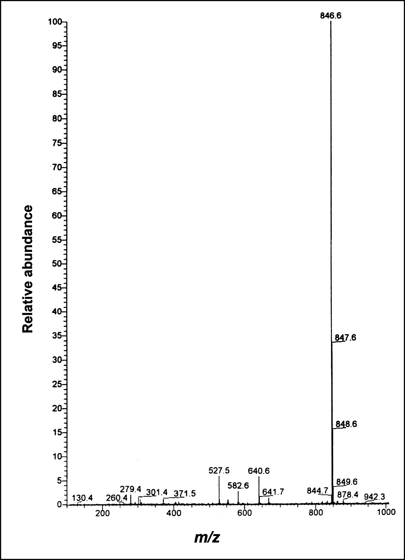
All [99gTc(N)(PNP)(L)]Cl complexes were characterized by elemental analysis and mass spectroscopy. The ESI mass spectrum of the [99gTc(N)(PNP5)(DBODC)]+ complex is shown in Figure 3. The ESI mass spectrum (m/z, percent abundance) was as follows: 847 [M]+, 100; and 640 {M-(CH2)3P[(CH2)2OCH3]2}+, 5.

ESI mass spectrum of [99gTc(N)(PNP5)(DBODC)]Cl complex.
Animal Studies
Biodistribution Studies.
Animal experiments were performed in compliance with the relevant national laws relating to the conduct of animal experimentation and were approved by the Ferrara University hospital ethical committee. Before injection, the complexes were purified by the procedures outlined above. After purification, the collected activity was further diluted with phosphate buffer (0.1 mol dm−3; pH 7.4) to give a final solution with an ethanol content of 10%. When the dithiocarbamate ligand was nonsymmetric (R1 ≠ R2), a racemic mixture of 2 enantiomeric complexes was obtained. These components were not separated, and the whole mixture was used for biologic experiments.
Female Sprague-Dawley rats weighing 200–250 g were anesthetized with an intramuscular injection of a mixture of ketamine (80 mg kg−1) and xylazine (19 mg kg−1). A jugular vein was surgically exposed, and 100 μL (300–370 kBq) of the solution containing the radioactive complex were injected. The animals (n = 3) were sacrificed by cervical dislocation at different times after injection. The blood was withdrawn from the heart through a syringe immediately after the sacrifice, and blood counts were determined. Organs were excised, rinsed in saline, and weighed, and counts were determined with an NaI well counter. Measured activity was corrected for the decay factor. Results, expressed as percent dose per gram of tissue, are reported in Tables 2–5.
Biodistribution in Rats for Selected [99mTc(N)(PNP)(L)]+ Complexes
Biodistribution in Rats of [99mTc(N)(PNP3)(DBODC)]+ Complex
Biodistribution in Rats of [99mTc(N)(PNP5)(DBODC)]+ Complex
Biodistribution in Rats of [99mTc(N)(PNP6)(DBODC)]+ Complex
A similar procedure was applied for determination of the biodistribution in rats of the cardiac agents 99mTc-sestamibi and 99mTc-tetrofosmin prepared with the corresponding commercial kit formulations and injected without further purification.
Small-Animal Scanner Studies.
Tomographic images of the rat heart were obtained by use of a small-animal, hybrid PET-SPECT scanner assembled in our laboratory and described in detail elsewhere (20). Briefly, the YAP-(S)PET scanner has 4 detection heads, each composed of a YAP:Ce planar crystal matrix coupled to a position-sensitive photomultiplier (where YAP is yttrium aluminium Perovskite doped with cerium). The application of high-resolution parallel-hole collimators on 2 opposite detector heads allows this PET device to be used also as a dedicated, small-animal SPECT scanner. The field of view in both PET and SPECT modes is a cylinder with a diameter of 4 cm and a depth of 4 cm. Imaging with 99mTc complexes was performed 1 h after injection of Sprague-Dawley rats (200–250 g) anesthetized as described above by collection of data with 1 detector head working in the SPECT mode. [99mTc(N)(PNP5)(DBODC)]+ complexes, 99mTc-sestamibi, and 99mTc-tetrofosmin were used in imaging studies, and the injected activities were 174, 184, and 159 MBq, respectively. Acquisition and reconstruction parameters were as follows: angular views, 64 over 180°; recorded events, 192 × 104; reconstructed events, ∼64 × 104. Images with dimensions of 40 pixels (radial) × 40 pixels (axial) × 20 pixels (transaxial) were reconstructed by means of an expectation maximization iterative algorithm and by excluding events below 140 keV.
RESULTS
Chemistry
The preparation of a class of monocationic, square-pyramidal, 99mTc-nitride radiopharmaceuticals of the type [99mTc(N)(L)(PNP)]+, where L is the monoanionic form of a dithiocarbamate ligand and PNP is a diphosphine ligand (Fig. 2), was performed with a liquid formulation involving a 2-step reaction. The first step produced the [99mTc≡N]2+ core through the initial reaction of 99mTcO4− with SDH as a donor of nitride nitrogen atoms (N3−) and in the presence of Sn2+ ions as a reductant (5,21). The preparation was completed by adding simultaneously to the same reaction vial the 2 ligands PNP and NaL to afford the final products, [99mTc(N)(PNP)(L)]+. Notably, the asymmetric [99mTc(N)(PNP)(L)]+ heterocomplexes were always obtained without the concomitant formation of the symmetric neutral [99mTc(N)(L)2] complexes, comprising 2 identical bidentate ligands.
A lyophilized, 2-vial kit formulation was specifically developed for the preparation of the compounds [99mTc(N)(PNP3)(DBODC)]+ and [99mTc(N)(PNP5)(DBODC)]+. The first vial was filled with the reagents SDH and SnCl2, required to form the Tc≡N group, while the second vial contained the sodium salt of DBODC, free PNP3 (or PNP5), and γ-cyclodextrin, used as a solubilizing agent for the diphosphine ligand. The presence of EDTA in the first vial of the freeze-dried formulation was required to prevent the precipitation of the neutral disubstituted Sn(DBODC)2 complexes upon the addition of the dithiocarbamate ligand. With the kit formulation, the yields of [99mTc(N)(PNP3)(DBODC)]+ and [99mTc(N)(PNP5)(DBODC)]+ achieved were similar to those obtained with the 2-step liquid formulation (95%–98%).
The [99mTc(N)(PNP)(L)]+ complexes were characterized by chromatographic methods. Their chemical structures were investigated by comparison with the corresponding analogs prepared with the long-lived isotope 99gTc. The ESI mass spectrum of the representative [99mTc(N)(PNP5)(DBODC)]+ complex is shown in Figure 3. The crystal structure of the analogous rhenium complex [Re(N)(PNP)(DEDC)]+, where PNP is Ph2P(CH2)2-N(CH2CH2OCH3)-(CH2)2PPh2 and DEDC is bis-N-ethyl-dithiocarbamato, was reported elsewhere (22).
The [99mTc(N)(PNP)(L)]+ complexes were found to be highly stable in solution and in blood, even after removal of excess free ligands. In particular, all of the complexes were strongly resistant to transchelation by cysteine and GSH. HPLC measurement of log ko′ values showed that these complexes were lipophilic, with log ko′ values ranging from 2.48 to 4.35 (Table 1).
Animal Studies
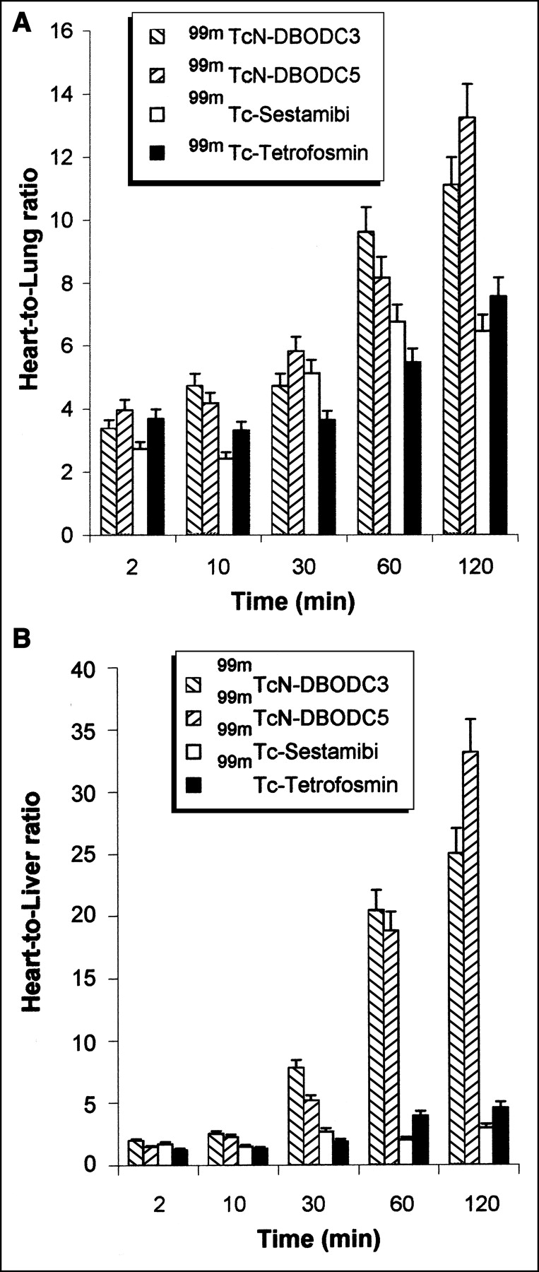
Biodistribution data for the [99mTc(N)(PNP)(L)]+ heterocomplexes in rats are reported in Tables 2–5. Time-activity curves for heart uptake for some selected heterocomplexes and for 99mTc-sestamibi and 99mTc-tetrofosmin are shown in Figures 4 and 5. Variations in heart-to-lung and heart-to-liver uptake ratios for the [99mTc(N)(PNP3)(DBODC)]+ and [99mTc(N)(PNP5)(DBODC)]+ complexes, 99mTc-sestamibi, and 99mTc-tetrofosmin are shown in Figure 6. Tomographic gamma camera images of the rat myocardium that were collected with the YAP-(S)PET small-animal scanner after injection of the [99mTc(N)(PNP5)(DBODC)]+ complex and of the commercial radiopharmaceuticals 99mTc-sestamibi and 99mTc-tetrofosmin are shown in Figure 7.
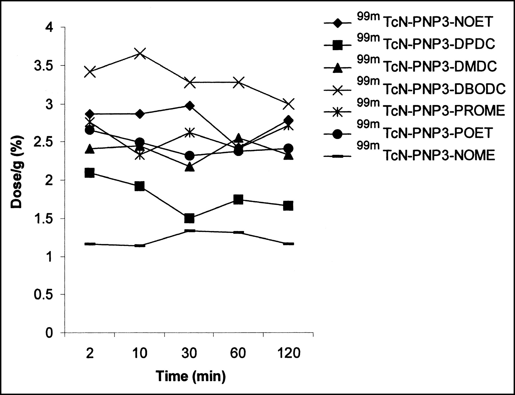
Time-activity curves for heart uptake for some selected [99mTc(N)(PNP)(L)]+ complexes in rats. SDs are omitted for clarity.
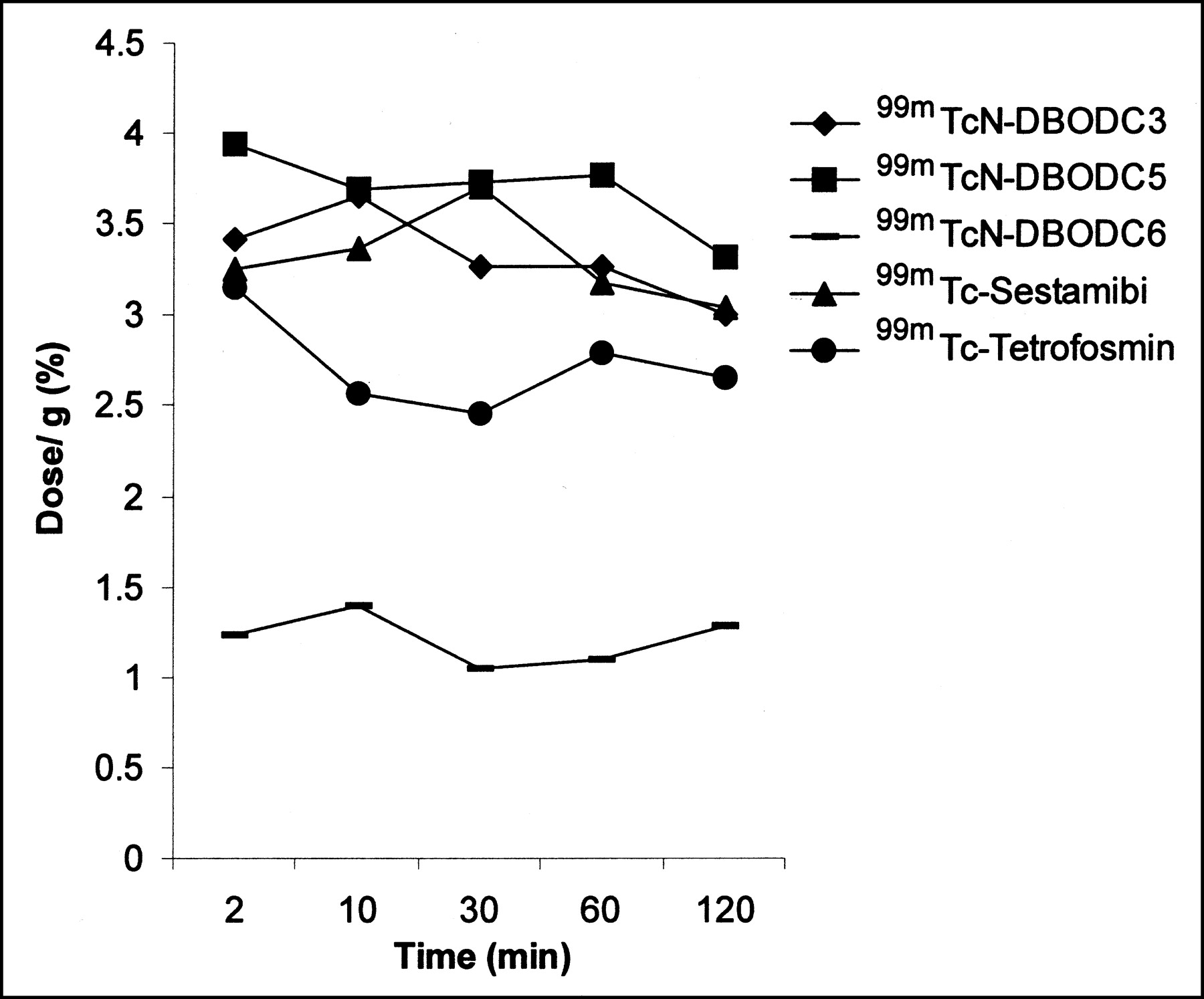
Time-activity curves for heart uptake for [99mTc(N)(PNP3)(DBODC)]+ (99mTcN-DBODC3), [99mTc(N)(PNP5)(DBODC)]+ (9mTcN-DBODC5), and [99mTc(N)(PNP6)(DBODC)]+ (9mTcN-DBODC6) complexes, 99mTc-sestamibi, and 99mTc-tetrofosmin in rats. SDs are omitted for clarity.

Heart-to-lung (A) and heart-to-liver (B) uptake ratios for [99mTc(N)(PNP3)(DBODC)]+ (99mTcN-DBODC3) and [99mTc(N)(PNP5)(DBODC)]+ (9mTcN-DBODC5) complexes, 99mTc-sestamibi, and 99mTc-tetrofosmin.
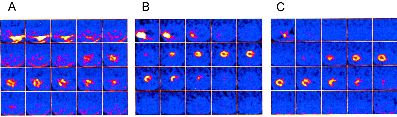
Tomographic transaxial images of rat heart obtained with small-animal SPECT scanner (10) 60 min after injection of 99mTc-sestamibi (A), 99mTc-tetrofosmin (B), and [99mTc(N)(PNP5)(DBODC)]+ complex (C). Slices are ordered from rat tail (top left) to rat head (bottom right). Color intensity is proportional to pixel activity (counts per second).
All of the [99mTc(N)(PNP)(L)]+ complexes accumulated in the rat myocardium, and activity in this region remained almost constant over time. Blood and lung washouts were rapid, elimination occurring mainly through the kidneys and liver. Interestingly, liver uptake was transient, and washout from this organ was fast and quantitative, activity being almost completely eliminated into the intestine at 1 h after injection. Differences in the biologic behaviors of [99mTc(N)(PNP)(L)]+ complexes were found to be dependent on modification of the pendant functional groups attached to both the nitrogen atom of the dithiocarbamate ligand and the phosphorus and nitrogen atoms of the diphosphine ligand (Fig. 2). The most important effects of these variations were changes in accumulation in the heart and elimination from nontarget tissues, such as the lungs, liver, and blood. Replacement of the diphosphine ligands PNP3 and PNP5 with PNP6 caused a significant decrease in accumulation in the heart and a concomitant increase in uptake in nontarget organs.
DISCUSSION
The combination of π-donor and π-acceptor properties associated with 2 different bidentate ligands was the key factor in determining the selective formation of asymmetric 99mTc-nitride heterocomplexes without the concomitant production of the corresponding symmetric compounds containing 2 identical bidentate ligands (12). On the basis of the previous characterization of the molecular structure of the analogous rhenium complex [Re(N)(PNP)(DEDC)]+ (22), the distorted square-pyramidal geometry was attributed to the [99mTc(N)(PNP)(L)]+ complexes. In this arrangement, the Tc≡N multiple bond occupies an apical position and the diphosphine and dithiocarbamate ligands span the residual 4 coordination positions on the basal plane through the 2 phosphorus atoms and the 2 sulfur atoms, respectively (Fig. 1).
The monocationic [99mTc(N)(PNP)(L)]+ heterocomplexes (where PNP is PNP3 or PNP5) localize selectively in the myocardium of rats and exhibit interesting new properties when viewed in terms of the search for an ideal heart perfusion agent. In fact, although the myocardial uptake of these heterocomplexes was comparable to that of 99mTc-sestamibi and 99mTc-tetrofosmin, the [99mTc(N)(PNP)(L)]+ complexes showed much more favorable heart-to-lung and heart-to-liver uptake ratios. Generally, the heart-to-lung and heart-to-liver uptake ratios for the asymmetric complexes increased exponentially over time and became dramatically high in the interval between 60 and 120 min after injection (Fig. 6). In the series of compounds with PNP3 and PNP5, the most promising biodistribution properties were observed for the [99mTc(N)(PNP3)(DBODC)]+ and [99mTc(N)(PNP5)(DBODC)]+ complexes. These findings were interpreted by assuming that the chemical and physical characteristics of these 2 complexes approached a suitable combination of parameters such as size, lipophilicity, and molecular weight, thus resulting in the most favorable biologic behavior. However, substitution of PNP3 (or PNP5) with PNP6, to yield the [99mTc(N)(PNP6)(DBODC)]+ complex, caused a remarkable decrease in heart uptake and a concomitant increase in liver accumulation, results that might be attributable to the excessive growth of the lipophilic character as a result of the presence of longer alkoxy groups on P atoms of PNP6. These characteristics would cause the values to fall outside the most effective range of biologic parameters.
High-quality images for rats were recorded with a small-animal SPECT scanner assembled in our laboratory (20). Specifically, we attempted a comparison among the 99mTc-sestamibi, 99mTc-tetrofosmin, and [99mTc(N)(PNP5)(DBODC)]+ agents. Tomographic images were collected 1 h after injection of the appropriate radiopharmaceutical (Fig. 7). Images were scaled with respect to their own maximum found in the heart region. With the compounds 99mTc-sestamibi and 99mTc-tetrofosmin, peak activity was localized in the abdominal region and was associated mainly with higher liver uptake. In contrast, peak activity with the [99mTc(N)(PNP5)(DBODC)]+ complex was positioned exactly in the myocardial area, and liver activity was found to be almost negligible. As a consequence, with the [99mTc(N)(PNP5)(DBODC)]+ agent, it was possible to obtain a clear visualization of the cardiac region, in close agreement with biodistribution results showing rapid liver clearance. These results support the conclusion that the [99mTc(N)(PNP5)(DBODC)]+ complex appears to possess highly favorable distribution properties in comparison with other cardiac tracers.
CONCLUSION
In this work, a new class of monocationic perfusion heart imaging agents having an unusual asymmetric structure was obtained through the application of improved procedures for the preparation of 99mTc complexes containing a terminal Tc≡N multiple bond. These unprecedented structural properties resulted in more favorable biologic behavior. In particular, the [99mTc(N)(PNP3)(DBODC)]+ and [99mTc(N)(PNP5)(DBODC)]+ complexes displayed the most appropriate molecular properties for application to heart imaging. Heart-to-lung and heart-to-liver uptake ratios for these compounds were observed to be dramatically higher than those for 99mTc-sestamibi and 99mTc-tetrofosmin. These results, in combination with the high heart uptake and fast blood clearance, indicate that the novel complexes possess favorable biologic properties and could be used as improved heart perfusion tracers for collecting high-quality scintigraphic images. Evaluation of the biologic behavior of these new agents in other animal models is currently under way.
Acknowledgments
The authors thank Prof. Alberto Del Guerra and Prof. Adriano Piffanelli for helpful discussions. Financial support provided by Nihon Medi-Physics Co., Ltd., Tokyo, Japan, is gratefully acknowledged.
Footnotes
Received Aug. 21, 2002; revision accepted Jan. 20, 2003.
For correspondence or reprints contact: Adriano Duatti, PhD, Laboratory of Nuclear Medicine, Department of Clinical and Experimental Medicine, University of Ferrara, Via L. Borsari 46, 44100 Ferrara, Italy.
E-mail: dta{at}unife.it
REFERENCES
In this issue


Jump to section
Related Articles
Cited By...
- Whole-Body Biodistribution and Radiation Dosimetry of the New Cardiac Tracer 99mTc-N-DBODC
- Subcellular Distribution and Metabolism Studies of the Potential Myocardial Imaging Agent [99mTc(N)(DBODC)(PNP5)]+
- 99mTc-N-DBODC5, a New Myocardial Perfusion Imaging Agent with Rapid Liver Clearance: Comparison with 99mTc-Sestamibi and 99mTc-Tetrofosmin in Rats