


Abstract
99mTc-[bis (dimethoxypropylphosphinoethyl)-ethoxyethylamine (PNP5)]-[bis (N-ethoxyethyl)-dithiocarbamato (DBODC)] nitride (N-PNP5-DBODC or N-DBODC5) is a new monocationic myocardial perfusion tracer. We sought to compare the myocardial uptake and clearance kinetics and organ biodistribution of 99mTc-N-DBODC5 with 99mTc-sestamibi and 99mTc-tetrofosmin. Methods: Seventy-five anesthetized Sprague–Dawley rats were injected intravenously with 22.2–29.6 MBq 99mTc-N-DBODC5 (n = 25), 99mTc-sestamibi (n = 25), or 99mTc-tetrofosmin (n = 25). Rats were euthanized at either 2, 10, 20, 30, or 60 min after injection and γ-well counting was performed on excised organ (heart, lung, and liver) and blood samples. In 3 additional rats, serial in vivo whole-body γ-camera imaging with each tracer was performed. Results: 99mTc-N-DBODC5 cleared rapidly from the blood pool. At 2 min after injection, 99mTc-N-DBODC5 blood activity was significantly lower than either 99mTc-sestamibi or 99mTc-tetrofosmin (P < 0.01) and remained lower over 60 min. Myocardial 99mTc-N-DBODC5 uptake was rapid (2.9% ± 0.1% injected dose/g at 2 min), and there was no significant clearance over 60 min, similar to 99mTc-sestamibi and 99mTc-tetrofosmin. All 3 tracers exhibited rapid lung clearance. Importantly, 99mTc-N-DBODC5 cleared more rapidly from the liver than either 99mTc-sestamibi or 99mTc-tetrofosmin. As early as 30 min after injection, 99mTc-N-DBODC5 heart-to-liver ratio was 5.7 ± 1.0 versus 1.6 ± 0.1 and 2.9 ± 0.3 for 99mTc-sestamibi and 99mTc-tetrofosmin (P < 0.05). By 60 min, 99mTc-N-DBODC5 heart-to-liver ratio further increased to 18.4 ± 2.0 compared with 2.6 ± 0.2 and 5.8 ± 0.7 for 99mTc-sestamibi and 99mTc-tetrofosmin (P < 0.001). The rapid blood pool, lung, and liver clearance of 99mTc-N-DBODC5 resulted in excellent-quality myocardial images within 30 min after injection. Conclusion: 99mTc-N-DBODC5 is a promising new myocardial perfusion tracer with superior biodistribution properties. The rapid 99mTc-N-DBODC5 liver clearance may shorten the duration of imaging protocols by allowing earlier image acquisition and may markedly reduce the problem of photon scatter from the liver into the inferoapical wall on myocardial images.
Single-photon myocardial perfusion imaging agents labeled with 99mTc have been developed with properties better suited for γ-camera imaging than 201Tl because of the higher photopeak and shorter half-life of 99mTc. Cationic 99mTc complexes, 99mTc-sestamibi (1) and 99mTc-tetrofosmin (2,3), are routinely used for clinical imaging with their favorable myocardial uptake and retention properties (4). Neutrally charged 99mTc complexes, 99mTc-teboroxime (1,5) and 99mTc-N-NOET (6,7), exhibit better flow-extraction properties at high flows compared with these cationic agents and have complete redistribution similar to 201Tl (8). 99mTc-teboroxime is not routinely used because of its very rapid myocardial clearance kinetics (9,10), whereas 99mTc-N-NOET, the first reported compound characterized by the presence of a terminal technetium–nitrogen multiple bond (6), has not yet been approved for clinical imaging.
Although the physical properties of 99mTc are better suited than 201Tl for γ-camera imaging, the organ biodistribution properties of these 99mTc-labeled tracers remain suboptimal for myocardial perfusion imaging. Interfering abdominal activity resulting from intense liver or gastrointestinal uptake is often observed for prolonged periods with these 99mTc-labeled agents because of their prominent hepatobiliary excretion (11,12). In particular, because of its close proximity to the heart, prolonged high liver uptake can make it difficult to accurately assess myocardial perfusion, particularly in the inferior or inferoapical left ventricular wall (13–18). Therefore, it is important to develop new tracers with improved organ biodistribution properties, with less liver uptake.
99mTc-[bis (dimethoxypropylphosphinoethyl)-ethoxyethylamine (PNP5)]-[bis (N-ethoxyethyl)-dithiocarbamato (DBODC)] nitride (N-PNP5-DBODC or N-DBODC5) is a new class nitrido 99mTc agent that is currently under investigation (19,20). The core of this molecule consists of 99mTc triple bonded to nitrogen, and it is highly lipophilic, similar to 99mTc-N-NOET (19). However, unlike 99mTc-N NOET, which is a neutral molecule, 99mTc-N-DBODC5 is monocationic, like 99mTc-sestamibi and 99mTc-tetrofosmin. Accordingly, the goal of this experimental study was to determine the organ biodistribution kinetics of 99mTc-N-DBODC5 in comparison with the existing 99mTc-labeled cationic agents, 99mTc-sestamibi and 99mTc-tetrofosmin.
MATERIALS AND METHODS
All experiments were performed with the approval of the University of Virginia Animal Care and Use Committee in compliance with the position of the American Heart Association on the use of research animals.
Biodistribution Study of 99mTc-Labeled Agents in Rats
The protocol of the present study is shown in Figure 1. Seventy-five Sprague-Dawley rats (200–250 g) were anesthetized with an intraperitoneal injection of either sodium pentobarbital (30 mg/kg) or a mixture of ketamine (80 mg/kg) and xylazine (19 mg/kg). A saphenous vein was exposed and 22.2–29.6 MBq of either 99mTc-N-DBODC5 (n = 25), 99mTc-sestamibi (n = 25), or 99mTc-tetrofosmin (n = 25) were injected. For each of the 3 tracers, the 25 rats were subdivided into 5 groups according to timing of euthanasia after tracer injection. The subgrouped animals (n = 5 for each subgroup) were euthanized at either 2, 10, 20, 30, or 60 min after injection. Samples of blood and organs (excised heart, lung, and liver tissues) were collected in preweighed containers. Tracer activity in each sample was determined using a γ-well scintillation counter (MINAXI 5550; Packard Instruments) with standard window settings for 99mTc (120–160 keV). The tissue counts were corrected for background and decay, and the activity in each organ sample was calculated as a percentage of the total injected dose.
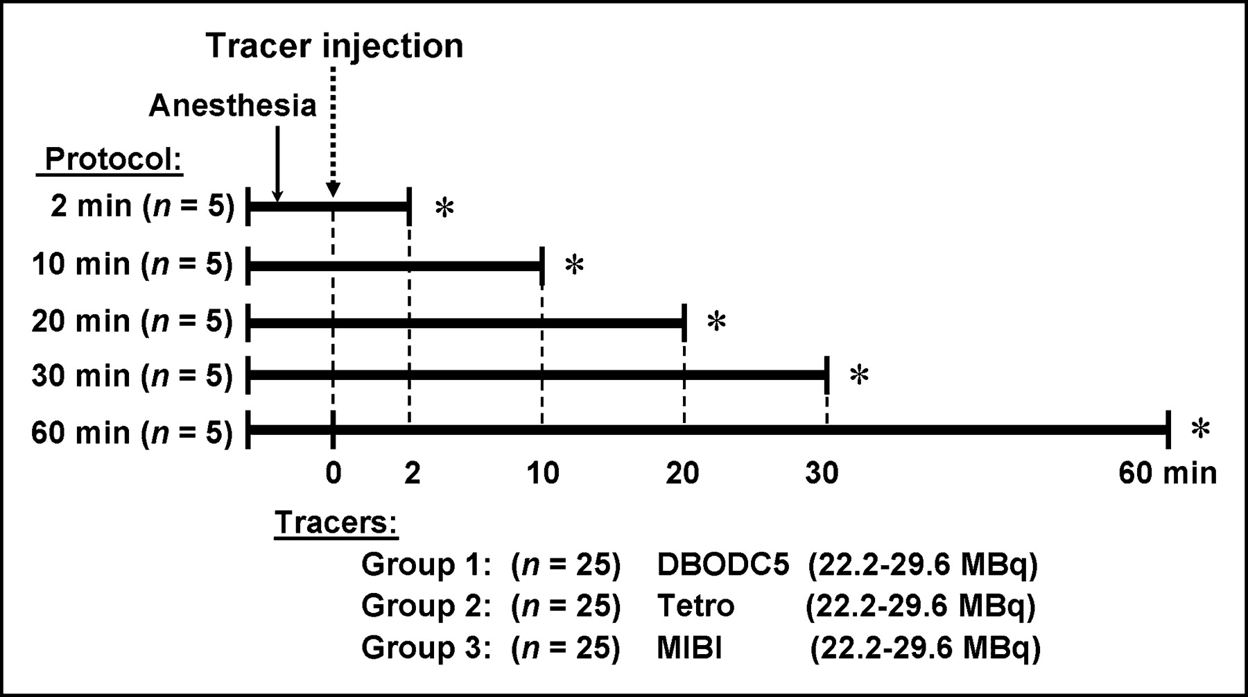
Experimental protocol (n = 75). DBODC5 = 99mTc-N-DBODC5; Tetro = 99mTc-tetrofosmin; MIBI = 99mTc-sestamibi. *Euthanasia; blood and tissue sampling.
In Vivo Whole-Body γ-Camera Imaging
To compare the tracer uptake and washout kinetics from the same animals and the same organs (heart, lung, liver) over time, serial in vivo γ-camera imaging was also performed in 9 additional anesthetized rats. Either 37.7–48.1 MBq of 99mTc-N-DBODC5 (n = 3), 99mTc-sestamibi (n = 3), or 99mTc-tetrofosmin (n = 3) was injected via a saphenous vein, after which the rats were placed supine directly on the surface of the low-energy, high-resolution collimator of a γ-camera (Digirad 20200tc Imager). In vivo whole-body images were acquired at 2, 30, and 60 min after injection using a 15% window centered on the 140-keV 99mTc photopeak. Image acquisition time was 5 min at each time point, resulting in approximately 0.4 × 10⁶ counts in each image. The images were then quantified by regions of interest drawn on the heart, lung, and liver regions on each image.
Preparation of 99mTc-N-DBODC5
A dose of 99mTc-N-DBODC5 was synthesized with a lyophilized kit formulation as previously described by Boschi et al. (20). An aliquot of 1.0 mL of Na-99mTc-O4 (50.0 MBq to 4.5 GBq) was added to a vial containing 5.0 mg of succinate dehydrogenase, 5.0 mg of ethylenediaminetetraacetic acid, 0.1 mg of SnCl2·2H2O, and 1.0 mL of phosphate buffer (0.1 mol/dm3) in a freeze-dried form. The resulting solution was kept at room temperature for 30 min. The contents of a second lyophilized vial consisting of 3.5 mg of PNP5, 3.5 mg of DBODC, and 3.5 mg of γ-cyclodextrin were reconstituted with 1.75 mL of saline. Then, 1.0 mL of the resulting solution was withdrawn from the second vial and added to the first vial, which was heated at 100°C for 15 min. The 99mTc-sestamibi and 99mTc-tetrofosmin doses were obtained from a local commercial radiopharmacy.
Purification and Quality Control of 99mTc-N-DBODC5
A cation exchange C-18 Sep-Pak cartridge (Waters) was activated with 5 mL of ethanol followed by 5 mL of deionized water. Then, the reaction solution containing the final 99mTc complex, 99mTc-N-DBODC5, was diluted with 8.0 mL of deionized water and passed through the activated cartridge. Approximately 60% of the initial activity was retained on the cartridge. After washing the cartridge with 20 mL of deionized water and 3 mL of an 80:20 mixture of ethanol (2.4 mL) and water (0.6 mL), the complex was recovered by passing 1.0 mL of a mixture of ethanol and an aqueous solution of NBu4Br (0.1 mol/L) (90:10). Before injection, the radiochemical purity of all preparations was determined by thin-layer chromatography technique. The labeling efficiency of 99mTc-N-DBODC5 was greater than 96% in each experiment.
Data and Statistical Analysis
All statistical computations were made using SYSTAT software (SYSTAT Inc.). The results were expressed as the mean ± SEM. Differences between means within a group and difference between the 3 groups were assessed using a 1-way ANOVA (biodistribution data from γ-well counting) or a 2-way ANOVA (quantitative data from serial in vivo imaging), with P values < 0.05 considered significant. Nonlinear regression on liver washout kinetic data andcalculation of the T1/2 parameter for all 3 tracers was performed using Prism software (Graphpad Software, Inc.).
RESULTS
Biodistribution in Rats
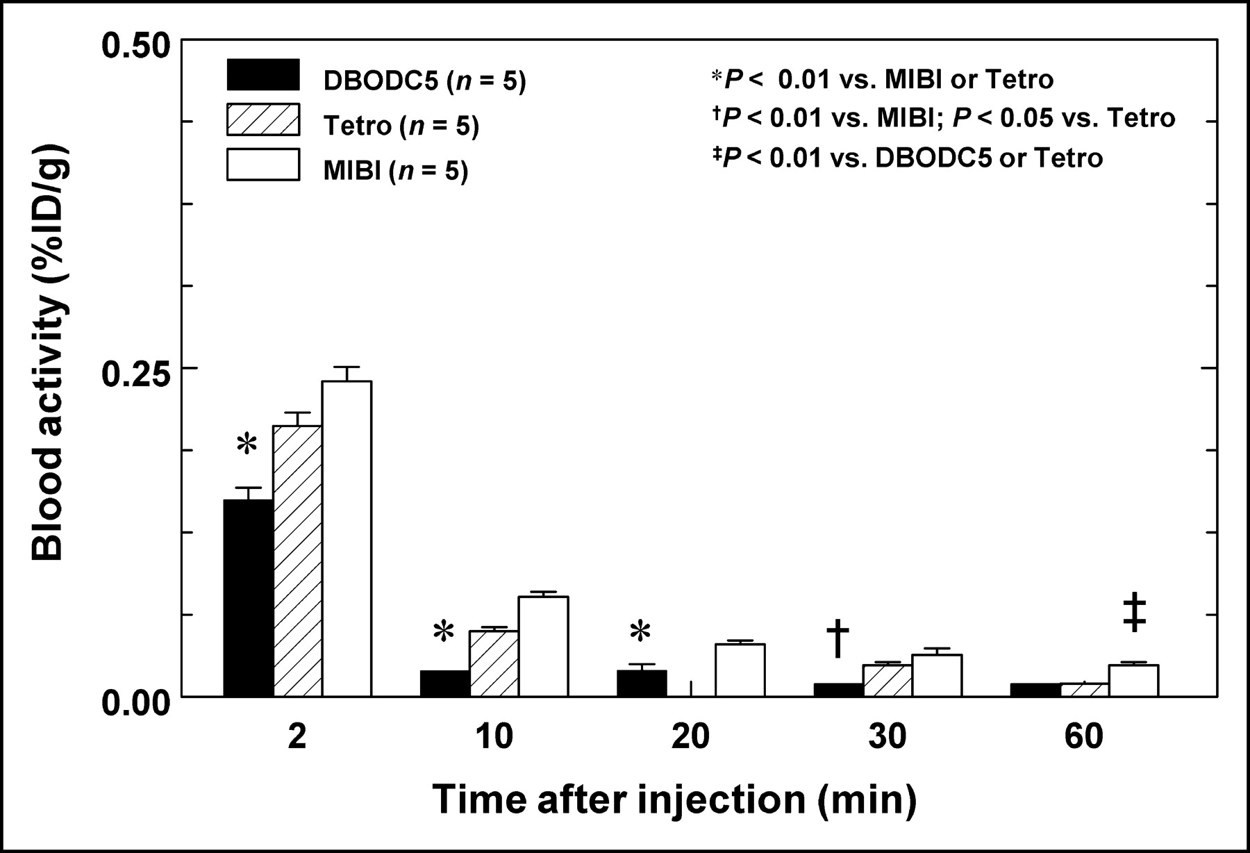
Organ biodistribution data for 99mTc-N-DBODC5 over time are presented in Table 1 and Figures 2–7. No statistically significant differences were observed in the injected doses among the 3 tracers. As shown in Figure 2, 99mTc-N-DBODC5 exhibited rapid clearance from the blood pool. As early as 2 min after injection, the blood activity for 99mTc-N-DBODC5 was less than the other 2 tracers and remained lower for 30 min. At 60 min, 99mTc-N-DBODC5 blood activity was still less than that of 99mTc-sestamibi. The bar graph in Figure 3 compares the amount of myocardial uptake of the 3 tracers at the various time points. As shown, myocardial 99mTc-N-DBODC5 uptake was rapid, and there was no significant clearance over 60 min, similar to 99mTc-tetrofosmin and 99mTc-sestamibi. Figure 4 compares heart-to-lung uptake ratios from γ-well counting. All 3 tracers exhibited rapid lung clearance. At 2 min, however, the mean heart-to-lung ratio for 99mTc-N-DBODC5 was higher than that of 99mTc-sestamibi, and the ratio increased from 2.6 to 7.7 over 60 min (P < 0.001 vs. 2 min). On in vivo images, lung clearance was rapid and comparable for all 3 tracers.

Comparison of blood activity over time. 99mTc-N-DBODC5 exhibited more rapid clearance from blood pool. DBODC5 = 99mTc-N-DBODC5; Tetro = 99mTc-tetrofosmin; MIBI = 99mTc-sestamibi.

Comparison of heart activity over time from γ-well counting. There is no significant clearance of 99mTc-N-DBODC5 from heart over 60 min, similarly to the other cationic 99mTc-labeled tracers. DBODC5 = 99mTc-N-DBODC5; Tetro = 99mTc-tetrofosmin; MIBI = 99mTc-sestamibi.

Comparison of heart-to-lung activity ratios over time from γ-well counting. All 3 tracers exhibited rapid lung clearance; however, at 2 min the mean heart-to-lung ratios for 99mTc-N-DBODC5 were higher than those for 99mTc-sestamibi and increased significantly over 60 min. DBODC5 = 99mTc-N-DBODC5; Tetro = 99mTc-tetrofosmin; MIBI = 99mTc-sestamibi.
99mTc-N-DBODC5 Biodistribution in Rats
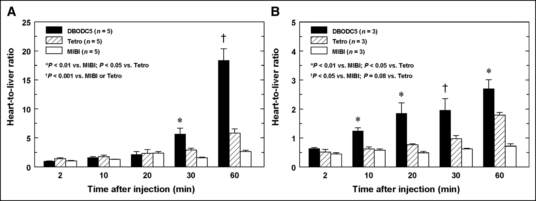
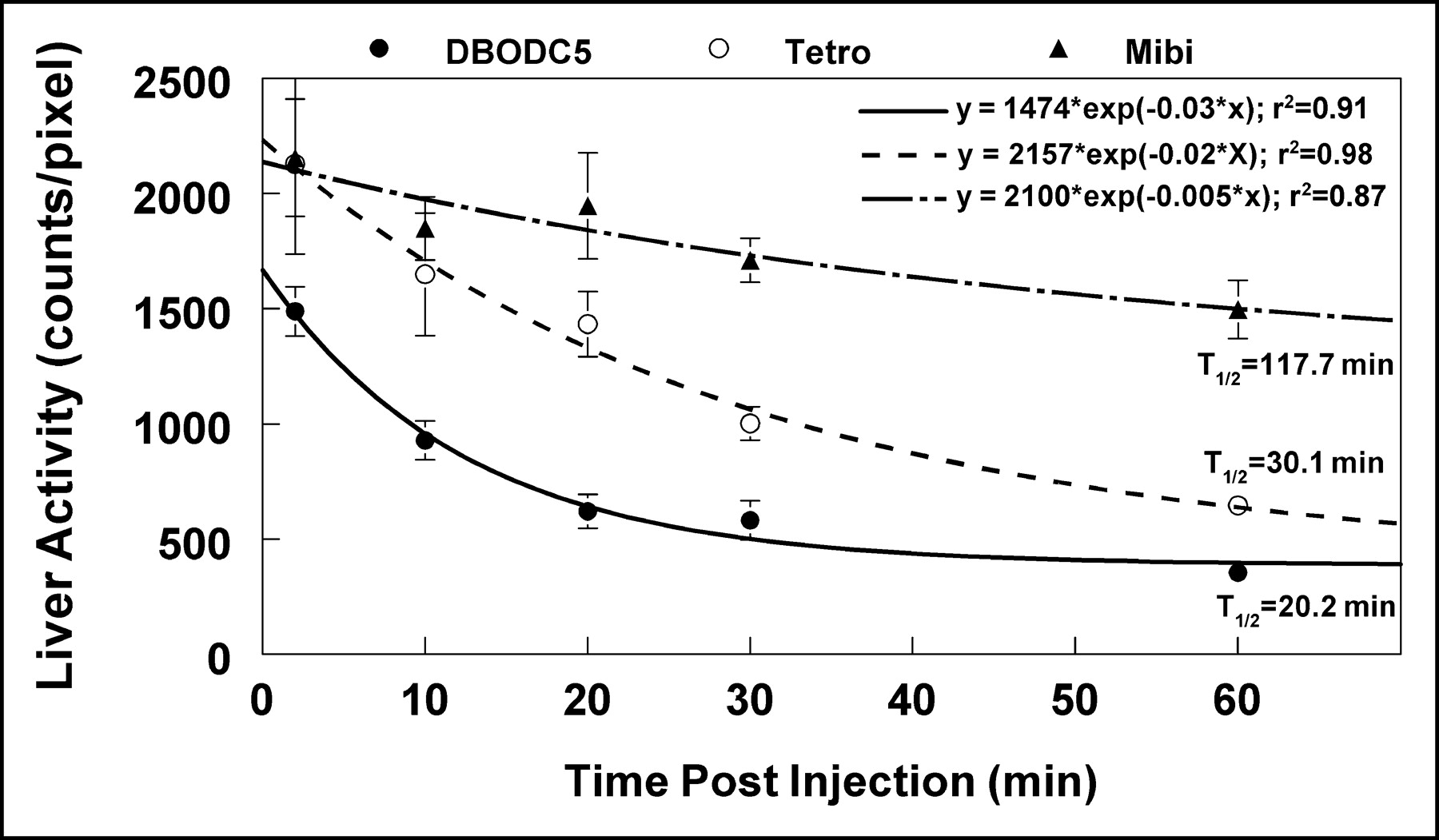
Importantly, 99mTc-N-DBODC5 exhibited more rapid liver clearance than either 99mTc-tetrofosmin or 99mTc-sestamibi. As shown in Figure 5A, as early as 30 min after injection, the mean heart-to-liver ratio for 99mTc-N-DBODC5 from γ-well counting (5.7 ± 1.0) was higher than for either 99mTc-tetrofosmin (2.9 ± 0.3) or 99mTc-sestamibi (1.6 ± 0.1) (P < 0.05 vs. 99mTc-tetrofosmin, and P < 0.01 vs. 99mTc-sestamibi). Furthermore, the mean heart-to-liver ratio for 99mTc-N-DBODC5 increased from 5.7 at 30 min to 18.4 by 60 min, further widening the difference between 99mTc-N-DBODC5 and the other 2 99mTc-agents (5.8 ± 0.7 and 2.6 ± 0.2 for 99mTc-tetrofosmin and 99mTc-sestamibi; P < 0.001, respectively). As shown in Figure 5B, the significantly faster liver clearance of 99mTc-N-DBODC5 was also observed by in vivo image quantification. Figure 6 compares the liver clearance kinetics among these 3 99mTc-labeled tracers. Liver activity was determined from a region of interest placed over the liver on the in vivo images. Nonlinear regression analysis using a monoexponential curve fit revealed that 99mTc-N-DBODC5 cleared from the liver approximately 1.5 times faster than 99mTc-tetrofosmin and 6 times faster than 99mTc-sestamibi. Similar results were obtained from the analysis of the γ-well counting of liver samples from rats euthanized at different time points (P < 0.01, respectively).

Comparison of heart-to-liver activity ratios over time from γ-well counting (A) and in vivo image quantification (B). Faster liver clearance of 99mTc-N-DBODC5, compared with the other 99mTc-labeled agents, was observed with both ex vivo γ-well counting and in vivo whole-body imaging. DBODC5 = 99mTc-N-DBODC5; Tetro = 99mTc-tetrofosmin; MIBI = 99mTc-sestamibi.

Liver clearance kinetics for the 3 99mTc-labeled agents. Tracer activities were determined from a region of interest placed on liver on in vivo images. T1/2 value calculated from monoexponential curve fitting for DBODC5 clearance was approximately 1.5 and 6 times faster than for Tetro and Mibi, respectively. DBODC5 = 99mTc-N-DBODC5; Tetro = 99mTc-tetrofosmin; MIBI = 99mTc-sestamibi.
In Vivo γ-Camera Imaging
Figure 7 displays in vivo whole-body planar images acquired at different time points after tracer injection. On all initial images acquired at 2 min after injection, very high liver uptake can be seen adjacent to the heart. By 30 min after injection, 99mTc-N-DBODC5 liver uptake visibly decreased and nearly disappeared at 60 min after injection, whereas high liver uptake still can be seen on the 99mTc-sestamibi image at this time point. The 99mTc-tetrofosmin liver uptake was intermediate between that of 99mTc-N-DBODC5 and 99mTc-sestamibi at 60 min.
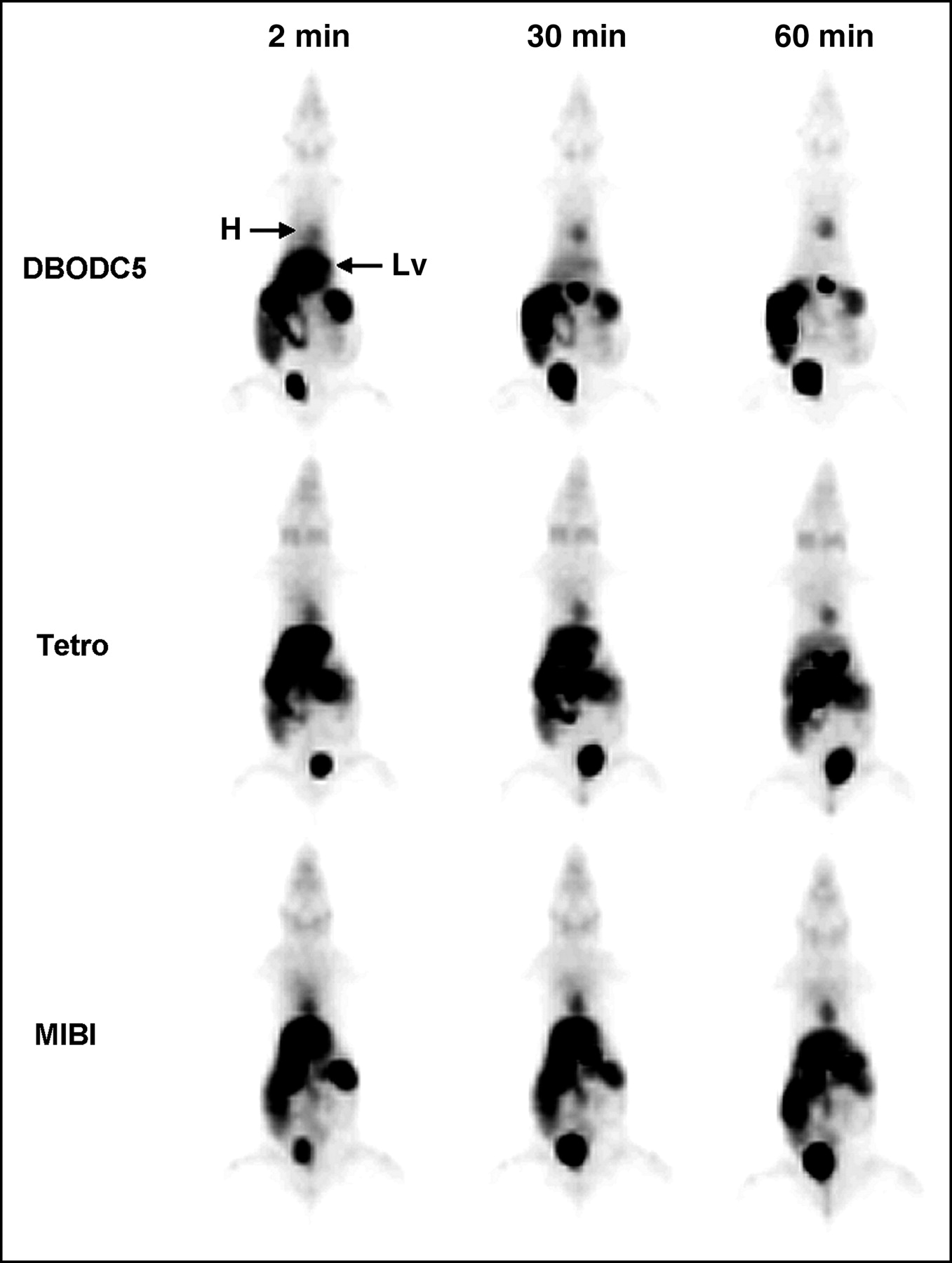
Serial in vivo whole-body images for 99mTc-N-DBODC5 (top), 99mTc-tetrofosmin (middle), and 99mTc-sestaibi (bottom). Persistent heart uptake can be seen with all of the tracers, whereas 99mTc-N-DBODC5 liver uptake cleared more rapidly than the other 2 tracers. DBODC5 = 99mTc-N-DBODC5; Tetro = 99mTc-tetrofosmin; MIBI = 99mTc-sestamibi; H = heart; Lv = liver.
DISCUSSION
Accurate determination of myocardial perfusion status and cellular integrity has major clinical and prognostic importance and may affect critical therapeutic decision-making in patients with ischemic heart disease. Over the past 2 decades, there has been a great deal of effort to develop 99mTc-labeled myocardial perfusion tracers for clinical γ-camera imaging. However, despite the more favorable physical properties of 99mTc compared with 201Tl, at present none of the 99mTc-labeled agents that have been approved for clinical use has ideal biodistribution properties.
In this experimental study, we found that a new nitrido class 99mTc agent, 99mTc-N-DBODC5, exhibits high heart uptake and novel biodistribution kinetics with its rapid liver clearance in anesthetized rat models. The quantitative biodistribution data for 99mTc-N-DBODC5 are consistent with the first published findings of favorable biodistribution kinetics of this new 99mTc-labeled agent (19). Consistent with the results of the quantitative organ biodistribution, the present study compares for the first time serial in vivo whole-body planar images of 99mTc-N-DBODC5 with the existing agents, 99mTc-sestamibi and 99mTc-tetrofosmin. The more rapid liver clearance as well as the relatively high heart uptake yielded high-quality in vivo 99mTc-N-DBODC5 images. These findings suggest that 99mTc-N-DBODC5 is a promising new agent and may allow for more accurate assessment of myocardial perfusion with less photon scatter from the liver. This may provide better accuracy for detection of coronary artery disease and for myocardial viability assessment.
With clinical γ-camera imaging using 99mTc-labeled myocardial perfusion tracers, interfering abdominal activity due to intense liver or gastrointestinal uptake may make it difficult to interpret the heart activity, particularly in the inferior or inferoapical left ventricular wall (13–18). High liver uptake is frequently observed with rest or pharmacologic stress studies (14) and results from the prominent hepatobiliary excretion of the lipophilic 99mTc-labeled tracers (11,12). Prolonged intense abdominal activity adjacent to the heart can lead to a paradoxical decrease of counts in the inferior wall in the absence of perfusion abnormalities (13). Test specificity in detection of coronary artery disease could be affected by a false-positive inferior wall defect on stress and rest images (14). This intense liver activity has been reported for both 99mTc-sestamibi (21) and 99mTc-tetrofosmin (3).
To overcome this problem, several technical attempts have been undertaken in clinical imaging. Early image acquisition after tracer injection has been advised to prevent intestinal artifacts (22), while late image acquisition is recommended to avoid liver artifacts (17,23). These suggestions are based on the fact that, with hepatobiliary clearance, the activity of the excreted tracer moves from the liver and gallbladder to the gastrointestinal area over time (24). Although gastrointestinal activity can be reduced and distanced from the heart by filling the stomach before image acquisition (24–26), it is more important to reduce the liver uptake or to stimulate the liver clearance to achieve high-quality myocardial perfusion image acquisition. Although giving high-lipid foods to stimulate the gallbladder and reduce liver activity has been attempted, Hurwitz et al. demonstrated using milkshake ingestion that this measure was insufficient to reduce intense liver activity (25). The measure was effective only in reduction of interfering intestinal activity on the heart image by increasing the volume of the stomach with fluid (25).
Other means of reducing artifacts caused by high liver uptake have involved modifications to image reconstruction algorithms. Image reconstruction via filtered backprojection is the current standard in clinical myocardial perfusion imaging (27). This process is well known to cause artifactual decreased myocardial wall uptake if the liver activity is greater than the heart with 99mTc myocardial imaging (28). With phantom measurements, Germano et al. suggested higher frequency cutoffs in prereconstruction filters, if count statistics are good and liver uptake is high (15). Nuyts et al. emphasized the importance of accurate attenuation correction in the left ventricular wall counting rate (16). The authors also demonstrated that 360° reconstruction, in comparison with 180° reconstruction, reduces the differences in attenuation between the different projections, therefore reducing the reconstruction artifacts (16). However, despite these efforts in both basic and clinical studies, photon scatter from extremely high liver activity on attenuation-corrected images is still an unresolved problem (29). To date, no technique is commonly available to completely overcome abdominal image artifacts. Thus, developing new 99mTc-labeled myocardial agents that exhibit more favorable organ biodistribution properties, with less liver uptake without reducing myocardial uptake would be a great advance.
Biochemical mechanisms for the markedly rapid liver clearance of 99mTc-N-DBODC5 remain unknown. However, one potential explanation is the lipophilic character and the electronic charge of the tracer. The lower the lipophilicity of a compound the lower the initial uptake in the liver (30,31). Boschi et al. demonstrated that substitution of 99mTc-N-DBODC5 with a similar monocationic nitride 99mTc complex, which has higher lipophilic profile, caused an increased liver accumulation of the tracer (20).
In contrast to the marked difference in liver clearance kinetics, the heart uptake of this new tracer was comparable to that seen with the other cationic 99mTc-labeled tracers, 99mTc-sestamibi and 99mTc-tetrofosmin. The myocardial uptake of 99mTc-sestamibi and 99mTc-tetrofosmin have been reported to be driven by electropotential gradient according to the Nernst equation, and these tracers exhibit prolonged accumulation in mitochondria (32–34). Because 99mTc-N-DBODC5 is also highly lipophilic and monocationic, it is possible that the mechanism for myocardial uptake and washout of this tracer is similar to 99mTc-sestamibi and 99mTc-tetrofosmin. Further studies are necessary to determine the exact mechanism for myocardial uptake of this novel tracer.
The main findings of this rat biodistribution study were, first, high myocardial uptake of 99mTc-N-DBODC5 with slow clearance similar to that of 99mTc-sestamibi and 99mTc-tetrofosmin and, second, rapid blood, lung, and especially liver clearance of 99mTc-N-DBODC5 allowing for excellent-quality in vivo heart images as early as 30 min after injection.
One clinical implication of the present study is that the fast 99mTc-N-DBODC5 liver clearance kinetics may significantly reduce photon scatter from the liver into the inferior and inferoapical walls on myocardial perfusion images, thereby reducing artifacts and potentially improving the diagnostic accuracy for the detection of coronary artery disease compared with the other 99mTc-labeled perfusion agents. Moreover, these novel biodistribution properties might shorten the duration of imaging protocols, allowing for earlier image acquisition.
CONCLUSION
99mTc-N-DBODC5 is a promising new myocardial perfusion imaging agent with superior biodistribution properties. For clinical imaging, the more rapid liver clearance could give the new tracer an advantage over the other 99mTc-labeled tracers.
Acknowledgments
This experimental study was funded by a research grant from Nihon Medi-Physics Co., Ltd. YoshihiroYamamichi is employed by NMP. Dr. Adriano Duatti has a financial interest in 99mTc-N-DBODC5.
Footnotes
Received Aug. 6, 2004; revision accepted Aug. 11, 2004.
For correspondence or reprints contact: David K. Glover, PhD, Cardiovascular Division, Department of Internal Medicine, University of Virginia Health System, P.O. Box 800500, Charlottesville, VA 22908-0500.
E-mail: dglover{at}virginia.edu


