


Abstract
18F-labeled 1-amino-3-fluorocyclobutane-1-carboxylic acid (18F-fluciclovine) is a leucine analog radiotracer that depicts amino acid transport into cells. 18F-fluciclovine PET/CT visualizes malignancy, including prostate cancer, invasive ductal breast cancer, and invasive lobular breast cancer. Whether changes in 18F-fluciclovine avidity reflect changes in tumor burden resulting from treatment has not been shown. In this prospective clinical trial (clinical trials.gov: NCT01864083), changes in 18F-fluciclovine avidity after neoadjuvant therapy were compared to breast cancer therapy response, as determined by residual tumor burden on pathology, were evaluated. Methods: Twenty-four women with a new diagnosis of locally advanced invasive ductal breast cancer (n = 18) or invasive lobular breast cancer (n = 6) underwent 18F-fluciclovine PET/CT before and after the completion of neoadjuvant systemic therapy. SUVmax, SUVmean, metabolic tumor volume, and total lesion avidity were obtained for the primary breast tumor, axillary lymph nodes, and extraaxillary lymph nodes on each examination and corrected for background 18F-fluciclovine avidity. The relationship between changes in 18F-fluciclovine avidity and the percentage of reduction of tumor on pathology was assessed with the Spearman rank correlation. Results: The median decrease in the corrected SUVmax of the primary breast lesions was 99% (range, 33%–100%). The median reduction of tumor on pathology was 92% (range, 10%–100%). Changes in 18F-fluciclovine avidity were strongly correlated with the percentage of reduction of tumor on pathology (Spearman ρ, 0.79; 95% CI, 0.56–0.90; P < 0.001). Conclusion: Changes in 18F-fluciclovine avidity strongly correlated with the tumor response on pathology in this pilot study.
Many tumor cells demonstrate increased amino acid transport; therefore, amino acid metabolism may be a useful target for tumor imaging (1). 18F-labeled 1-amino-3-fluorocyclobutane-1-carboxylic acid (18F-fluciclovine), an amino acid analog labeled with the positron emitter 18F, is under investigation as an imaging agent for several types of malignancy (2–12). 18F-fluciclovine was recently approved by the U.S. Food and Drug Administration for the detection of recurrent prostate cancer. Initial studies of 18F-fluciclovine in breast cancer have provided encouraging results that warrant further investigation (7,8).
Although 18F-fluciclovine has been shown to successfully image several types of malignancy, there is currently no evidence that changes in 18F-fluciclovine avidity correlate with changes in tumor burden. Before 18F-fluciclovine is used for evaluation of the tumor response, the association of 18F-fluciclovine avidity and the tumor response must be demonstrated (13). The purpose of this trial was to assess the ability of 18F-fluciclovine PET to determine the therapeutic response to neoadjuvant chemotherapy in patients with breast cancer. All patients underwent prospectively planned surgical removal of the treated primary breast malignancy, allowing for an analysis of the correlation between therapy-induced changes in 18F-fluciclovine avidity and pathologic tumor burden.
MATERIALS AND METHODS
Study Design and Patients
This prospective clinical trial (clinical trials.gov: NCT01864083) was performed with institutional review board approval and written informed consent from participants. Patients who presented for evaluation at Memorial Sloan Kettering Cancer Center; who had biopsy-proven, locally advanced, nonmetastatic invasive ductal breast cancer (IDC) or invasive lobular breast cancer (ILC); and who were referred for neoadjuvant systemic therapy between August 2013 and August 2015 were invited to participate. ILC is a subtype of breast cancer that is difficult to visualize by all known imaging modalities (14,15), including 18F-FDG PET (16–21). Given the continued need for better metabolic imaging agents for ILC, patients with ILC were actively sought for inclusion in the study, accounting for the higher prevalence of ILC in our cohort than in the overall breast cancer population. The planned sample size of 25 patients was chosen for financial and logistic considerations; the aims of the study were exploratory. In total, 27 patients were invited to participate, but only 24 completed the protocol. Three patients withdrew from the protocol before the final 18F-fluciclovine scan.
Exclusion criteria were an age of less than 18 y, current pregnancy or lactation, prior malignancy other than squamous or basal skin cancers, and unwillingness or inability to consent. There were no restrictions on sex or race. Clinical records were used to document patient age, sex, and race; presence of biopsy-proven known axillary or extraaxillary nodal metastases before 18F-fluciclovine PET/CT; and histology, grade, and receptor status of the primary breast malignancy. Tumors were considered hormone receptor–positive if they showed greater than 1% staining for either the estrogen receptor (ER) or the progesterone receptor. Tumors were considered human epidermal growth factor 2 (HER2)–positive if they had a HER2 immunohistochemistry grade of 3+ or were amplified by fluorescence in situ hybridization at a ratio of greater than or equal to 2.0. Tumors that were negative for the ER, the progesterone receptor, and HER2 were classified as “triple negative.” The initial results of pretherapy 18F-fluciclovine PET/CT examinations from this trial were previously reported (7).
Verification of Malignancy
All patients had a biopsy-proven primary breast malignancy before enrollment in the clinical trial. The results of a pathologic evaluation of axillary nodes were also available for all patients. The results of a pathologic evaluation of extraaxillary nodes were available for the 3 patients with suspicious extraaxillary findings on initial 18F-fluciclovine PET/CT.
18F-Fluciclovine Production
18F-fluciclovine was manufactured in compliance with current good-manufacturing-practice requirements at the Memorial Sloan Kettering Cancer Center Radiochemistry and Molecular Imaging Probes Core Facility. The GE FASTlab automated synthesizer, synthesizer cassettes, reagents, and materials were all supplied by GE Healthcare as previously described (22). The automated synthesis involved nucleophilic incorporation of 18F-fluoride into the 18F-fluciclovine precursor followed by removal of protective groups by hydrolysis and terminal sterilization with a 0.22-μm sterilizing filter. The 18F-fluciclovine final drug product was formulated with a 200 mM citrate buffer solution. All manufactured 18F-fluciclovine drug product batches were quality control tested to ensure conformance with the acceptance specifications for pH, appearance, radiochemical purity, radiochemical identity, radionuclidic identity, endotoxin levels, sterilizing filter integrity, and residual solvent levels before release for patient administration.
18F-Fluciclovine PET/CT Imaging and Image Interpretation
Enrolled patients underwent baseline 18F-fluciclovine PET/CT within 21 d of the initiation of therapy. Repeat 18F-fluciclovine PET/CT was performed within 21 d of the completion of neoadjuvant therapy and before surgical management. There was no patient-specific preparation before 18F-fluciclovine administration, and patients were allowed to eat and drink before the PET/CT examination. Patients were positioned on designated research PET/CT scanners, either GE Healthcare Discovery STE or GE Healthcare Discovery 710, operated in the 3-dimensional mode. 18F-fluciclovine PET studies were performed as hybrid PET/CT examinations for attenuation correction, lesion localization, and availability of additional CT data. A low-milliampere (60–80 mA) CT scan of the chest was acquired first. Next, a bolus of 370 MBq (10 mCi) (±10%) of 18F-fluciclovine was administered intravenously in less than 10 s. Dynamic PET imaging over the chest was performed for 30 min, with data collected for each minute and then summed into 5-min intervals. 18F-fluciclovine avidity was most often greatest during the 5- to 10-min time interval; therefore, quantitative analyses of 18F-fluciclovine avidity were conducted with the data collected during this time interval.
18F-fluciclovine PET/CT images were reconstructed with iterative reconstruction and displayed on a PET/CT workstation (PET VCAR; GE Healthcare) as multiplanar reconstructions. Areas of focal tracer uptake were localized with the companion CT and classified as lesions of the breast, axillary nodes, or extraaxillary nodes. Three-dimensional regions of interest were placed in areas of tracer uptake, and measures of 18F-fluciclovine avidity were recorded; the measures were SUVmax (body weight), metabolic tumor volume (MTV; volume of tumor [cm3] with SUV >42% of SUVmax), SUVmean (within the MTV), and total lesion avidity (TLA; calculated as MTV × SUVmean). A background SUVmax was also measured for each examination.
Calculation of Change in 18F-Fluciclovine Avidity After Therapy
On both pretreatment and posttreatment scans, the lesion SUVmax was corrected for background as follows: corrected SUVmax = lesion SUVmax − background SUVmax. When the corrected SUVmax was less than 0, 0 was recorded. Similar corrections were made for SUVmean. When the corrected SUVmax and the corrected SUVmean were 0 on posttreatment scans, MTV and TLA were also 0. The percentage of change in the 18F-fluciclovine SUVmax after therapy was calculated as follows: [(pretreatment corrected SUVmax − posttreatment corrected SUVmax)/pretreatment corrected SUVmax] × 100. Similar calculations were performed for SUVmean, MTV, and TLA.
Calculation of Tumor Response in Surgical Specimens
All 24 patients proceeded to definitive surgical management of their primary breast malignancy. The entire tumor bed was marked and submitted to a pathologist who specialized in breast malignancy and had 7 y of experience with measurements for estimating the percentage of residual tumor according to published methods (23). In brief, the volume of the tumor bed with posttherapy changes, the volume of residual invasive or in situ tumor, and the percentage of cellularity of the tumor in the tumor bed were measured. From these measurements, the percentage of tumor volume reduction was calculated as described by Symmans et al. (23). A 100% pathologic tumor response, with no remaining invasive or in situ tumor, was defined as a pathologic complete response. Any reduction of less than 100% was defined as a partial response.
Statistical Analysis
Descriptive statistics were calculated for patient characteristics and measures of 18F-fluciclovine avidity. The relationship between the change in 18F-fluciclovine avidity after therapy and the percentage of tumor response in surgical specimens was assessed with the Spearman rank correlation, along with asymptotic 95% confidence limits, and visualized within a scatterplot with a regression line.
The proportion of patients in whom 18F-fluciclovine avidity was reduced to the background level after therapy was calculated on the basis of histology (IDC vs. ILC), receptor status (ER+/HER2− vs. HER2+ vs. triple negative), and tumor grade (1 or 2 vs. 3). Because of the limited sample size and pilot nature of this prospective study, no formal hypothesis tests for group differences were conducted.
The pathologic response of the primary breast malignancy was dichotomized into a complete pathologic response (no residual invasive or in situ malignancy) and a partial pathologic response (any residual invasive or in situ malignancy).
P values of less than 0.05 were considered statistically significant. All analyses were performed with SAS 9.4 (SAS Institute Inc.).
RESULTS
Twenty-four women were prospectively enrolled to receive the institutional review board–approved protocol and underwent baseline and post–neoadjuvant therapy 18F-fluciclovine PET/CT. The median age of the cohort was 53 y (range, 37–79 y). The histology of the primary breast malignancy was IDC in 18 patients (75%) and ILC in the remaining 6 patients (25%). Patient and tumor characteristics are summarized in Table 1.
Patient and Tumor Characteristics
Physiologic 18F-fluciclovine uptake was seen in the liver, pancreas, and skeletal muscle, as previously reported (5). During the dynamic 30-min scan, 18F-fluciclovine uptake was rapid, peaking at 5–10 min; uptake then plateaued or slowly decreased until 30 min. A time–activity curve for the primary breast malignancy of a representative patient is shown in Figure 1. Background uptake in presumably normal breast tissue was low in all patients, with SUVmax ranging from 0.5 to 1.5. 18F-fluciclovine–avid lesions, with an avidity greater than that of the breast background, were observed in the breast (all 24 patients), axillary nodes (19/24 patients), and extraaxillary nodes (3/24 patients) on baseline 18F-fluciclovine scans. On post–neoadjuvant therapy scans, breast lesions in 12 of 24 patients decreased to the background level, whereas breast lesions in the other 12 patients decreased but remained appreciably above the background level. In the 19 patients with 18F-fluciclovine–avid axillary nodes at baseline, 14 lesions decreased to the background level on post–neoadjuvant therapy scans, whereas in the other 5 patients, axillary node lesions remained appreciably above the background level. In the 3 patients with 18F-fluciclovine–avid extraaxillary nodes at baseline, 2 lesions decreased to the background level on post–neoadjuvant therapy scans, whereas 1 lesion remained appreciably above the background level.

Time–activity curve for primary breast malignancy in representative patient. 18F-fluciclovine uptake was rapid, peaking at 5–10 min, and then slowly decreased until 30 min.
The numbers of patients with 18F-fluciclovine–avid breast, axillary node, and extraaxillary node lesions, as well as their median and range for measures of 18F-fluciclovine avidity, are shown in Table 2. An example of a patient with primary breast cancer and axillary node metastases that all decreased to background levels after neoadjuvant therapy is shown in Figure 2.
Numbers of Patients with 18F-Fluciclovine–Avid Breast and Node Lesions and 18F-Fluciclovine Avidity Measurements
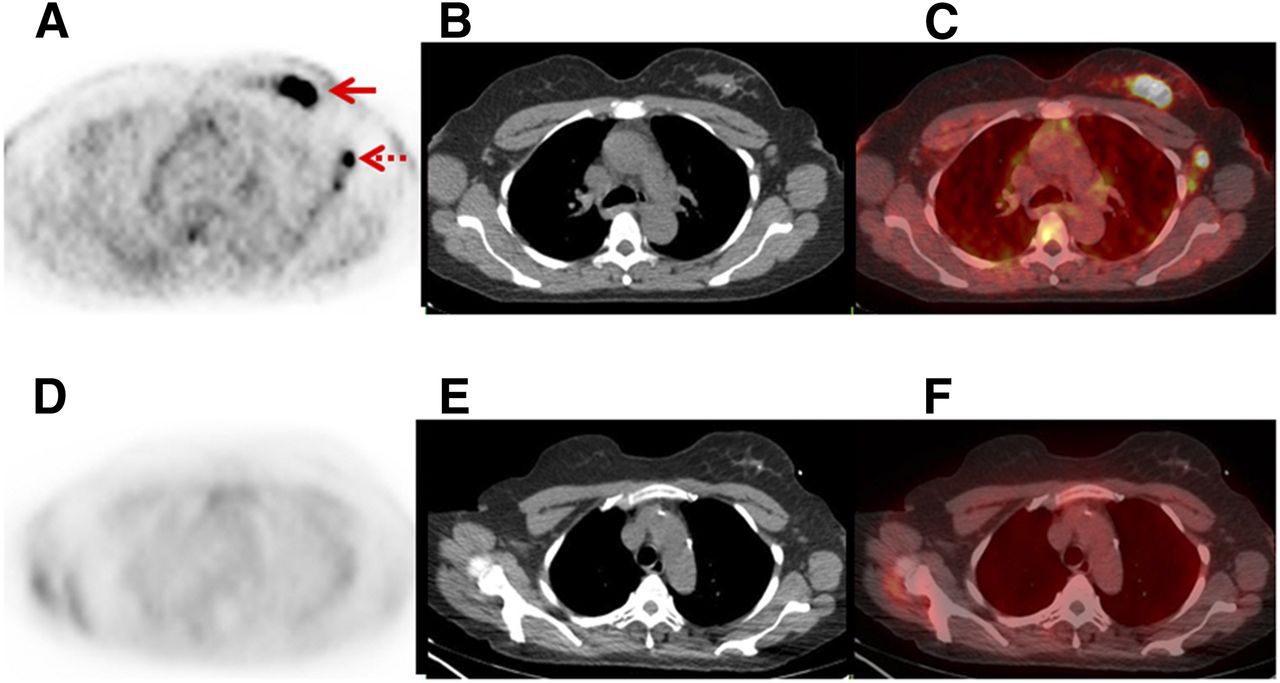
Reduction in 18F-fluciclovine avidity after neoadjuvant therapy in 52-y-old woman with grade 2 ER−/HER2+ IDC. (A–C) Axial 18F-fluciclovine PET (A), axial CT (B), and axial fused (C) images at baseline show 18F-fluciclovine–avid primary breast mass (solid arrow) and 18F-fluciclovine–avid axillary node metastases (broken arrow). (D–F) Axial 18F-fluciclovine PET (D), axial CT (E), and axial fused (F) images after neoadjuvant therapy show decrease in 18F-fluciclovine avidity of all lesions to background levels. Pathology revealed complete pathologic response, with no residual tumor.
The reduction in the corrected SUVmax of the 24 primary malignancies was correlated against the percentage of change in the tumor response on pathology after neoadjuvant therapy. The median decrease in the corrected SUVmax was 99% (range, 33%–100%). The median percentage of reduction in tumor burden on pathology was 92% (range, 10%–100%). The calculated Spearman ρ was 0.79 (95% CI, 0.56–0.90; P < 0.001) (Table 3). A scatterplot of the corrected SUVmax by the tumor response on pathology is shown in Figure 3.
Correlation Between Percentage of Change in Corrected SUVmax of Primary Malignancy and Percentage of Reduction in Tumor Burden on Pathology After Neoadjuvant Therapy
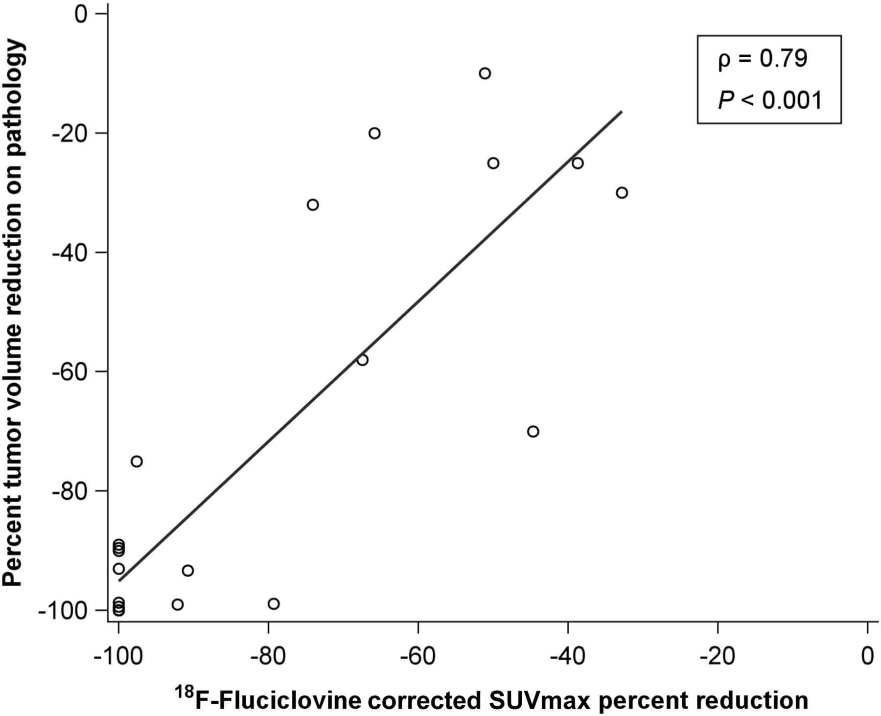
Scatterplot of percentage of change in corrected SUVmax vs. percentage of tumor volume reduction on pathology after neoadjuvant therapy. ρ = Spearman ρ.
The reductions in the corrected SUVmax of the primary malignancies after neoadjuvant therapy were further analyzed by histology, receptor status, and tumor grade (Table 4). By histology, 10 of 18 primary IDC tumors were reduced to the background level (median reduction, 100%; range, 39%–100%), whereas only 2 of 6 primary ILC tumors were reduced to the background level (median reduction, 70%; range, 33%–100%). Analogous comparisons by receptor status and tumor grade are shown in Table 4.
Reduction in Corrected SUVmax of Primary Malignancy After Neoadjuvant Therapy
Only 5 patients had a complete pathologic response in their primary malignancy (no residual invasive or in situ malignancy), compared with 19 patients with a partial pathologic response (any residual invasive or in situ malignancy).
DISCUSSION
18F-fluciclovine is a promising tumor marker recently approved by the U.S. Food and Drug Administration for the detection of recurrent prostate cancer. Recent studies also suggested promise for patients with breast malignancies. However, the detection of malignancy and the evaluation of a tumor response are separate tasks. Changes in 18F-FDG avidity have been extensively proven to correlate with the tumor response in breast cancer and many other malignancies (24–27). This prospective clinical trial provided evidence that changes in 18F-fluciclovine avidity also correlate well with the tumor response in patients with breast cancer (Spearman ρ, 0.79; P < 0.001).
In this prospective trial, we evaluated the treatment response in primary breast malignancies after neoadjuvant therapy. Because the entire primary breast malignancy normally undergoes surgical resection and there are established criteria for measuring the tumor response in the tumor bed from the surgical specimen (23), baseline and posttherapy/presurgery 18F-fluciclovine PET/CT provides an excellent opportunity to correlate changes in 18F-fluciclovine avidity with the tumor response on pathology. Evaluating changes in 18F-fluciclovine avidity as a measure of the tumor response may be most useful for metastatic disease; however, because metastases are not normally resected, the resected primary malignancy serves as a proxy for analysis. In this prospective trial, we evaluated 18F-fluciclovine as a noninvasive biomarker of the tumor response after therapy but not as an early predictor of the tumor response.
ILC, a subtype of breast cancer that accounts for 10%–15% of primary breast malignancies (28), is difficult to visualize by all current imaging modalities (14,15), including 18F-FDG PET (17,18,20,21). Therefore, the development of better metabolic imaging agents for ILC could be clinically valuable. There is preliminary evidence that ILC may be more 18F-fluciclovine–avid than 18F-FDG–avid (7,8). This study included 6 patients with ILC tumors, all of which were 18F-fluciclovine–avid. Changes in the corrected SUVmax appeared to correlate with the ILC tumor response, although the number of patients with ILC was low. Only 2 of 6 primary ILC malignancies demonstrated a 100% reduction in 18F-fluciclovine avidity, and both demonstrated a complete pathologic response. ILC is known to have a lower rate of a pathologic complete response after neoadjuvant therapy than IDC, although this pattern may be secondary to ILC tumors more commonly expressing ER+/HER2− receptor status, a phenotype that has a lower rate of a pathologic complete response (29,30). This pattern appeared in our small dataset.
18F-fluciclovine demonstrated rapid tumor uptake and a gradual decrease in avidity over time. The cause for the gradual decrease in avidity over time was not clear, but this finding may have been due to clearance of the tracer from tumor cells, as 18F-fluciclovine—unlike 18F-FDG—was not modified intracellularly.
No distant metastases were detected in this trial. The detection of distant metastases was limited by the restriction of imaging to include only the chest. 18F-fluciclovine has prominent physiologic hepatic uptake, which may make the detection of liver metastases challenging, although lung, brain, and bone lesions may be readily visible.
The strength of this study was its design as a prospective clinical trial, which decreases study biases and allows follow-up of a well-defined cohort. However, this study was limited in sample size, which particularly limited subgroup analyses for histology, receptor status, and tumor grade.
CONCLUSION
Changes in 18F-fluciclovine avidity were strongly correlated with the tumor response on pathology in this pilot study. Thus, 18F-fluciclovine PET/CT shows promise for monitoring the response to therapy.
DISCLOSURE
This study was supported by Susan G. Komen for the Cure research grant KG110441 (to Gary A. Ulaner), the Memorial Sloan Kettering Cancer Center (MSKCC) Biostatistics Core Facility and Radiochemistry and Molecular Imaging Probes Core Facility (P30 CA008748), the MSKCC Breast Cancer Research Fund, and GE Healthcare. No other potential conflict of interest relevant to this article was reported.
Footnotes
Published online Nov. 17, 2016.
- © 2017 by the Society of Nuclear Medicine and Molecular Imaging.
REFERENCES
- Received for publication August 30, 2016.
- Accepted for publication November 1, 2016.


Jump to section
Related Articles
Cited By...
- Glutamate Transport Proteins and Metabolic Enzymes are Poor Prognostic Factors in Invasive Lobular Carcinoma
- Riluzole suppresses growth and enhances response to endocrine therapy in ER+ breast cancer
- 18F-Fluciclovine PET Imaging of Glutaminase Inhibition in Breast Cancer Models
- Head-to-Head Evaluation of 18F-FES and 18F-FDG PET/CT in Metastatic Invasive Lobular Breast Cancer
- Hypoxia-induced switch in SNAT2/SLC38A2 regulation generates endocrine resistance in breast cancer
- Hypoxia-induced switch in SNAT2/SLC38A2 regulation generates endocrine-resistance in breast cancer
- Imaging Melphalan Therapy Response in Preclinical Extramedullary Multiple Myeloma with 18F-FDOPA and 18F-FDG PET