Article Text

Abstract
Introduction There is increasing evidence that imaging with [123I]FP-CIT SPECT is helpful in differentiating dementia with Lewy bodies (DLB) from Alzheimer's disease (AD) but it is not known how well the scan performs in differentiating DLB from frontotemporal dementia (FTD).
Method We compared the striatal dopamine transporter (DAT) binding in FTD (n=12), DLB (n=10) and AD (n=9) by visually rating the caudate and putamen on [123I]FP-CIT SPECT scans.
Results The majority (9/10) of DLB cases had an abnormal scan and a significant reduction of uptake of DAT binding in the putamen and the caudate. A third (4/12) of the FTD cases also had an abnormal scan and a significant reduction in uptake in the putamen and the caudate. In contrast, only one out of nine AD cases had an abnormal scan. Significant differences were found when comparisons were made between the groups for visual analysis of the entire scan (p=0.001) and the four regions of interest (p=0.001 – 0.013). In contrast to the AD group (specificity of scan 89%), the specificity of [123I]FP-CIT SPECT scans was reduced in the FTD group to 67%. Three quarters of the study population had at least one extrapyramidal motor sign (EPMS), with bradykinesia being the most common EPMS in both FTD (83%) and DLB (70%).
Conclusions This study highlights to clinicians that a positive (abnormal) [123I]FP-CIT SPECT scan, even in a patient with an EPMS, does not exclude the diagnosis of FTD and emphasises the importance of a comprehensive clinical evaluation and a detailed cognitive assessment.
- Frontotemporal dementia
- dementia with Lewy bodies
- [123I]FP-CIT SPECT
- frontotemporal lobar degeneration
- dopamine transporter
Statistics from Altmetric.com
- Frontotemporal dementia
- dementia with Lewy bodies
- [123I]FP-CIT SPECT
- frontotemporal lobar degeneration
- dopamine transporter
Introduction
Frontotemporal lobar degeneration (FTLD) is a clinically, genetically and pathologically1–3 heterogeneous disorder with high rates of misdiagnosis and delays in diagnosis.4 ,5 Clinically, FTLD can be broadly divided into behavioural variant of frontotemporal dementia (FTD), semantic dementia and progressive non-fluent aphasia. There is also a clinical overlap with FTD associated with motor neuron disease and corticobasal and progressive supranuclear palsy syndrome. Patients with FTLD have relatively selective frontotemporal atrophy. The underlying histopathology is characterised by the presence of τ inclusions (FTLD-τ), TAR DNA-binding protein 43 pathology (FTLD-TDP) or fused in sarcoma pathology (FTLD-FUS). Although some of the clinical syndromes are predominantly associated with specific pathology, the underlying histopathology for the behavioural variant of FTD is heterogeneous.3
There is increasing evidence that dopamine transporter (DAT) imaging with [123I]FP-CIT single-photon emission computed tomography (SPECT) is helpful in differentiating dementia with Lewy bodies (DLB) from Alzheimer's disease (AD)6–8 but it is not known how well the scan performs in differentiating DLB from FTD. Due to their overlapping symptoms9 and differing treatment strategies, this is an area of particular clinical importance.
Hallucinations9 ,10 and extrapyramidal motor sign (EPMS), including bradykinesia, rigidity and tremor, are observed in some of the clinical phenotypes of FTLD, namely FTD,10–12 corticobasal degeneration and progressive supranuclear palsy.1 Furthermore, DLB can present with prominent behavioural symptoms,13 a dysexecutive syndrome14 and primitive reflexes,15 making it difficult at times to distinguish it from FTD clinically.
Evidence of basal ganglia dysregulation in FTD includes atrophy on structural imaging1 ,2 ,16–19 and histopathological changes in the striatum and the substantia nigra at postmortem.20–22 Hypometabolism has been shown on positron emission tomography (PET);23 ,24 however, there are limited imaging studies of the striatal DAT binding. Functional imaging with [11C]2β-carbomethoxy-3β-(4-fluorophenyl)tropane (11C-CFT) PET25 and [123I]N-ω-fluoropropyl-2β-carbomethoxy-3β-(4-iodophenyl)nortropane (FP-CIT) SPECT6 ,26 ,27 demonstrates reduced uptake in the basal ganglia in FTD; however, there are no direct comparative studies between DLB and FTD.
Given the evidence of phenotypic and neuropathological heterogeneity in FTD1–3 and the neuropathological changes observed in the basal ganglia,20–22 we hypothesise that a proportion of patients with FTD will have an abnormal (reduced) DAT binding on [123I]FP-CIT SPECT but that this will not be as frequent as in patients with DLB.
Methods
Patients
We compared patients with FTD, DLB and AD. Patients were recruited at two different time points. All patients with DLB and AD as well as two patients with FTD were recruited as part of an earlier study.6 ,8 Ten patients with FTD were recruited later from a neurocognitive and a memory clinic.
AD and DLB
Details of recruitment of DLB and AD patients were previously described.6 ,8 For this study we selected only cases with available brain autopsies. Neuropathological diagnostic criteria were used for the diagnosis of DLB (DLB Consortium, third report) and AD (CERAD, Braak stage and NIA-RIAD diagnosis) and neocortical neurofibrillary tangle pathology was also staged.6
FTD cases
We recruited 10 patients with a clinical diagnosis of FTD from the West Essex Neurocognitive Clinic and the Mascall's Park Memory Clinic. Clinical diagnosis of FTD was made in a multidisciplinary meeting by a consensus of clinicians. Cases were further subclassified into a behavioural and a language variant of FTD.
Patients were eligible for inclusion if they: 1) fulfilled the clinical diagnostic criteria for FTD28 and 2) had either frontotemporal hypoperfusion or hypometabolism on a functional 99mTc-labelled hexamethyl propylene amine oxime SPECT or 2-deoxy-2-[18F] fluoro-D-glucose PET scan or a positive family history of a first-degree relative with confirmed FTD. We also included two neuropathologically confirmed cases of FTD from the autopsy cohort.6
Patients at all stages of illness were considered for inclusion unless they were unable to provide informed consent. Other exclusion criteria included a diagnosis of cerebrovascular disease, based on clinical presentation or structural imaging of significant vascular pathology; English was not a first language; or concomitant use of drugs listed in table 1 within the exclusion period. [123I]FP-CIT SPECT imaging of presynaptic dopaminergic function can be affected by drugs (table 1) that cross the blood brain barrier and act at the synapses, usually by binding and blocking DAT.29 Other drugs, such as selective serotonin reuptake inhibitors (SSRIs), increase the striatal uptake ratio of [123I]FP-CIT by 10% and this needs to be taken into account when performing semiquantitative analysis.30 This, however, is not of a magnitude to alter the outcome of a visual analysis.29 SSRIs are not routinely discontinued in clinical practice prior to a [123I]FP-CIT SPECT scan and as we selected visual analysis as a primary outcome measure, the concomitant use of SSRIs was not an exclusion criterion.
List of drugs that excluded participation in study
Assessment
All patients were assessed by a psychiatrist who took a comprehensive medical and psychiatric history from the patient and an informant and performed a physical and mental state examination, Unified Parkinson's Disease Rating Scale (UPDRS) and Modified Hoehn & Yahr staging. UPDRS was performed by a doctor or a specialist dementia nurse specifically trained to perform the UPDRS. We rated patients as having an EPMS if they had at least one of the following features: bradykinesia, rigidity and/or tremor.
Disease severity was evaluated by the Clinical Dementia Rating (CDR) scale. A psychologist performed a detailed neuropsychological assessment which comprised of Cambridge Cognitive Examination-Revised (CAMCOG-R) that includes Mini Mental State Examination (MMSE) or Repeatable Battery for the Assessment of Neuropsychological Status. Some patients also completed the Logical Memory Test, Wechsler Memory Scale III, National Adult Reading Test, Trail Making Test part A and B, letter fluency (FAS) and ideational fluency.
Attempts were made to collect all clinical and neurocognitive data on the day of the scan. When this was not possible, subsequent appointments were offered and in some cases previous cognitive and physical assessments completed within 12 months of the FP-CIT scan were used.
[123I]FP-CIT SPECT
Medication
For all cases, thyroid metabolism was blocked with potassium iodide to reduce possible irradiation of the thyroid gland. There was no operational policy in place regarding medication listed in table 1 for the original cohort of patients scanned; however, medication use at the time of the scan was recorded. For newly scanned FTD cases, certain prescription medications and recreational drugs listed in table 1 were exclusion criteria. These patients completed a medication checklist prior to their scan.
Autopsy confirmed cases (FTD (n=2), AD and DLB cases)
All subjects underwent scanning with a brain-dedicated scanner, the Strichman Medical Equipment 810, linked to a Macintosh computer. The scans were carried out at the Institute of Nuclear Medicine, University College London Medical School. The Strichman camera consists of 12 individual detectors, each equipped with a focusing collimator. The transaxial resolution of this camera is 7.6 mm full width at half maximum and the axial resolution is 12.5 mm. Usually 8–10 slices were acquired starting at the cerebellum level upwards to include basal ganglia. High dynamic range image files were viewed with MRIcro and saved as JPEG images to improve standardisation of scans between the two groups. Scanning took place between 3 and 4 h after injection of [123I]FP-CIT (150–185 MBq). The overall scanning time for each patient was 30–45 min.
Clinically diagnosed FTD cases
All newly recruited FTD patients underwent scanning with a Siemens SYMBIA T2, SPECT/CT Hybrid scanner at the Princess Alexandra Hospital. This system is dual headed and 360 projections were taken (180 on each head). Each projection took 30 s to complete and all were reconstructed using filtered Backprojection. Slices were taken through the head in three planes and we used transverse slices measuring 3.8 mm for analysis. Therefore, there were only 5–6 slices on which the striata were visible and these key images were saved as a JPEG file directly from the scanner. Scanning took place between 3 and 4 h after injection of FP-CIT (150–185 MBq). The overall scanning time for each patient was 30–45 min.
Visual rating of scans
Data processing
These images were acquired according to standard protocols and following the EANM guidelines.31
In summary, during data processing scatter correction was not applied, as it is not, at the moment, a standard for DAT brain imaging with radiopharmaceuticals, in particular with ioflupane ([123I]FP-CIT). On the other hand, attenuation correction was employed following the standard manufacturers′ algorithm. Although some debate in the literature continues regarding the need for applying attenuation correction, in one of the cameras (Siemens) the Chang method was applied using a coefficient of 0.15/cm. In this camera, the reconstruction method used was filtered back projection. All data acquired in the SME 810 (a dedicated brain SPECT instrument) were processed using iterative reconstruction with attenuation correction adapted for the focused high resolution collimators used during data acquisition.
Blinding of raters
As scan data came from two different cameras we created screen shots saved as JPEG images and containing up to six slices only of the basal ganglia. Figures 1–4 are images used for visual rating and include a positive and a negative [123I]FP-CIT SPECT scan result from the two cameras used. Case 1 (DLB) and case 29 (AD) are images from the Strichman Medical Equipment 810 and case 30 (FTD normal) and case 36 (FTD abnormal) are images from the Siemens camera.
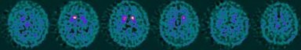
Case 01: abnormal [123I]FP-CIT SPECT result in a patient with autopsy diagnosis of dementia with Lewy bodies.

Case 29: normal [123I]FP-CIT SPECT result in a patient with autopsy diagnosis of Alzheimer's disease.

Case 30: normal [123I]FP-CIT SPECT result in a patient with clinical diagnosis of frontotemporal dementia.

Case 36: abnormal [123I]FP-CIT SPECT result in a patient with clinical diagnosis of frontotemporal dementia.
Independent rating
PK and JB, nuclear medicine experts on DAT imaging, independently rated each scan image, blind to the clinical and autopsy diagnoses. Both raters were trained to read images from different cameras and have done this in other studies.7 They visually assessed each of the four regions of interest (right putamen, left putamen, right caudate and left caudate) separately. Each region was scored as follows: 0, normal uptake; 1, slight reduction; and 2, significant reduction. Scans were then categorised into two groups, that is, abnormal and normal results. The abnormal result group comprised of scans with a score of 2, that is, significant reduction, in at least one of the four regions and the remaining scans, that is, those with scores of 0 or 1 were combined into a normal result group.
Statistical analysis
Data were analysed using SPSS/PC+ V.16 (Statistical Package for Social Sciences).
Descriptive data
Demographic data, presenting complaints, physical symptoms and neurocognitive data were included in the analysis. Raw scores for letter and verbal fluency were converted into age adjusted percentiles32 and into categorical data (impaired, normal). Descriptive statistics (means, medians and percentages) were obtained for each patient group and for the group as a whole where appropriate. Significance testing comprised of χ2, Mann–Whitney and Kruskal–Wallis tests.
[123I]FP-CIT SPECT
Frequencies for abnormal [123I]FP-CIT SPECT scans and abnormal uptake in the four regions of interest were calculated for the entire sample and each patient group. Frequencies of EPMS and impairment on cognitive tests were calculated for patients with abnormal scans. Diagnostic accuracy of [123I]FP-CIT SPECT was assessed by calculation of sensitivity and specificity.
Ethical approval
Ethical approval was obtained from the Essex 1 Research Ethics Committee. The Administration of Radioactive Substances Advisory Committee of the UK granted permission for the radiotracer [123I]FP-CIT to be used for this research project. Written informed consent was obtained from all patients participating in both recruitment phases.
Results
Patients
The autopsy cohort comprised of 10 DLB, nine AD and two FTD cases. We recruited a further 10 cases of FTD who fulfilled the revised Lund Manchester criteria28 and of these nine had frontotemporal hypometabolism or hypoperfusion on functional imaging with PET and SPECT, respectively. The final case of FTD was a male patient who developed his first symptoms at the age of 45 and had a sibling with a confirmed diagnosis of young-onset FTD.
Demographic data
Descriptive data are summarised in table 2. There was a significant difference between the groups for age at the time of FP-CIT scan and onset of dementia. As expected, cases with FTD were significantly more likely to be prescribed an SSRI at the time of their scan compared with DLB and AD (p<0.001; n=30).
Descriptive data for patients with FTD, DLB and AD
Cognitive profiles and physical signs
There was no difference in disease severity between the groups. With the exception of bradykinesia (p=0.017) and visual hallucinations (p=0.007), there were no other significant differences either on physical examination or cognitive testing (table 3).
Physical signs and cognitive rating for FTD, DLB and AD
Three quarters of the study population had at least one EPMS and this was the greatest in the FTD group. Bradykinesia was the most common in FTD (83%) and DLB (70%). Tremors were most frequent in the AD group (55.6%).
Visual rating FP-CIT
Table 4 summarises the consensus visual analysis of the four regions of interest (left caudate, right caudate, left putamen and right putamen) and the overall result of the FP-CIT scan. There was 95% agreement on visual rating of scans between the two raters.
Visual analysis of FP-CIT scan
Comparison of visual analysis for the three groups
The majority of DLB cases (90%) had an abnormal scan and a significant reduction of uptake (score=2) in the left and right putamina (left 80%; right 90%). Overall, 30% also had a significant reduction of uptake in the caudate. A third of the FTD group had an abnormal scan and a significant reduction of uptake in the putamen bilaterally (three cases) and the right caudate (four cases). Only one AD case had an abnormal scan with significant reduction in the right putamen. Significant differences were found when comparisons were made between the groups for visual analysis of the entire scan (p=0.001) and the four regions of interest (p=0.00–0.013).
Comparison of abnormal and normal scan results
For the entire sample, a high score on the modified Hoehn and Yahr staging was associated with an abnormal FP-CIT result (p=0.02). While there were no other significant associations observed between EPMS and scan results, 70% of cases with rigidity had an abnormal scan and more than two-thirds of patients without EPMS had a normal scan.
Diagnostic accuracy of FP-CIT
In comparison with the DLB group, the specificity of FP-CIT scans was reduced in the FTD group (67%) compared with the AD group (88.9%) and the sensitivity remained unchanged (90%) (table 5).
Sensitivity and specificity of FP-CIT in differentiating DLB from AD and FTD
FTD cases
Table 6 summarises the demographic, cognitive and physical assessments for the FTD patients.
Demographic, clinical features and FP-CIT scan results of FTD cases (n=12)
Discussion
There is increasing evidence that FTD is more common in all age groups than previously thought.1 This raises the possibility that some cases are misdiagnosed during life. Most clinicians still consider FTD to be a rare disorder; however, FTLD pathology is found in 18% of autopsy cases that are difficult to clinically classify at baseline.33 When characteristic symptoms of FTD are present at baseline, the specificity (99%) and sensitivity (85%) of clinical diagnosis are fairly good;33 however, in the absence of characteristic behavioural and language impairments, misdiagnosis and delays in diagnosis are more frequent.4 ,5 There are cases of neuropathologically confirmed FTD that fulfilled clinical diagnostic criteria of DLB6 ,9 or had both FTLD and Lewy body pathology at autopsy20 ,34 ,35 and therefore FTD has to be one of the differential diagnoses of DLB and vice versa.
Decreased DAT binding in the striatum is reliably used to differentiate DLB from AD6–8 but there are only a few imaging studies that examine DAT in FTD. Sedaghat et al 26 demonstrated reduced uptake of [123I]FP-CIT of up to 62% and 68% in the right and left striata, respectively, in FTD (n=7) as compared with healthy controls and a negative correlation with UPDRS scores (low uptake correlated with more severe motor signs). Walker et al 6 described one FTD patient in their autopsy study with reduced DAT uptake in the striatum who fulfilled the DLB diagnostic criteria36 at initial presentation and throughout follow-up assessments. Sperfeld et al 27 described a single case with marked reduction of presynaptic DAT striatal binding with N-(3-iodopropen-2-yl)-2β-carbomethoxy-3β-(4-chlorophenyl) tropane SPECT and mild reduction of postsynaptic dopamine D2 receptor binding with 3-iodo-6-methoxybenzamide SPECT. There is one [11C]CFT PET study demonstrating an 18% reduction of DAT binding in the putamen and a 14% reduction of DAT binding in the caudate in 12 FTD cases compared with healthy controls25 and again a negative correlation with severity of EPMS.
We are the first group to compare striatal DAT uptake in FTD with DLB and AD. In keeping with our hypothesis we have shown that DAT uptake, as measured by [123I]FP-CIT SPECT, is abnormal (reduced) in a substantial number of FTD cases. In our four cases with an abnormal [123I]FP-CIT SPECT scan, we found significant reduction (score=2) in uptake of the ligand in the right caudate (n=4) and bilaterally in the putamen (n=3) suggesting that striatal involvement is largely symmetrical and appears to affect both the caudate and the putamen. We are unable to compare this distribution with other DAT imaging studies. Interestingly, and in line with our findings, a recent PET study using a radiotracer for the vesicular monoamine transporter ([11C]dihydrotetrabenazine), which is expressed in the striatum, predominantly in dopaminergic terminals, also showed mildly reduced striatal binding in FTD.37
A significant proportion of our FTD group (83%) had bradykinesia, which is characteristic of DLB and is present in FTD.15 We found higher frequencies of tremor (50%) and lower frequencies of rigidity (25%) than previously reported12 ,26 ,38 in FTD. The overall frequency of EPMS was surprisingly high in FTD and reached similar levels to that observed in DLB. While clinicians do not often think of EPMS as a feature of FTD, previous studies have reported variable prevalence of EPMS in pathological and phenotypical subtypes of FTLD.3 ,10 ,20 ,34 ,38 ,39 Although EPMS was originally included as a supportive feature in the Neary clinical criteria28 they were subsequently excluded from later revisions.40 Overall, studies of FTD that used validated instruments to measure EPMS (eg, motor part of UPDRS)12 report higher rates in comparison with studies that rely purely on routine observations from physical examination.11 ,15 Recruitment of patients from specialist neurology clinics can also inflate the prevalence of EPMS25 and at first glance the high rates in all our patient groups, including AD, may suggest a selection bias. This is unlikely as we recruited patients from a neurocognitive and memory clinic and not from movement disorder clinics. We also excluded cases of corticobasal degeneration and progressive supranuclear palsy which are associated with striatal dysfunction. Our FTD cohort included both behavioural (n=7) and language (n=5) variants.
Other evidence of dopamine dysregulation in FTD includes lower CSF levels of homovanillic acid (a metabolite of dopamine) and dopamine,41 and reduced dopamine D2 receptor ligand uptake in the frontal cortex.42 As yet, it is not known why EPMS occurs in FTD or what the extent of striatal dysfunction is, but the negative correlation observed between an EPMS and dopamine uptake supports a causal relationship.25 Looi et al demonstrated that patterns of striatal atrophy in FTD were consistent with predicted frontostriatal dysfunction and suggested trans-synaptic degeneration, a previously described neuropathological process in which damage to neurons progresses along anatomical and functional pathways, as a model to explain these changes.16 ,17 Frontal cortical atrophy observed in FTD may lead to a reduction of input to the basal ganglia and a disconnection of the striatum from the frontostriatal circuits. Shape analysis of the resultant caudate and putamen atrophy is also consistent with predicted frontostriatal dysfunction associated with FTLD subtypes. This theory of disconnectivity is further supported by worsening hypometabolism of the striatum as the disease progresses.23 There is also growing evidence of basal ganglia atrophy at postmortem18 ,19 ,21 and there appears to be an association with molecular subtypes1 ,22 and disease severity.20 Even though atrophy of the substantia nigra is not described in FTD imaging studies, loss of pigmented neurons and gliosis is well documented at postmortem9 ,21 ,22 ,35 and there is some evidence to suggest an association with certain molecular subtypes and an EPMS but these findings are inconsistent.20
There are some limitations that need to be considered when interpreting our results. Our cohort is relatively small and this may be responsible for some results failing to achieve significance. However, FTD is not as common as other types of dementia and the number of patients recruited is similar to other imaging studies. For 10 FTD cases we have combined two methods, clinical diagnostic criteria and functional imaging or confirmed family history, to increase our diagnostic accuracy; however, this method is less accurate than the neuropathological diagnosis used to identify two FTD cases and all the DLB and AD cases. Future neuropathological confirmation will be sought. In the UK, functional neuroimaging (SPECT and PET) is recommended by the National Institute for Health and Clinical Excellence to help differentiate suspected FTD from AD in the uncertain patient.43 Although we are not aware of any head-to-head trials comparing the accuracies of the two techniques, it would be expected on theoretical grounds that the more expensive technology of PET imaging, allowing greater spatial resolution, would be superior to SPECT.
Comparison of modern [123I]FP-CIT scan images with those obtained 10 years ago in the autopsy study presented a challenge to the blinding of visual raters. Some differences in the images were noticeable, such as the number of slices. Even though we attempted to standardise the images, it is possible that the visual raters may have identified which camera the scans came from and therefore the diagnostic group in the case of the FTD patients. We limited our manipulation of the scans to maintain the overall quality of the images for the visual raters. Most patients were scanned in the early stages of their dementia (median CDR=1) and medians for the groups were comparable. The patterns of cognitive impairment observed on testing were in keeping with the expected disease profiles. AD patients scored poorly on global tests (median: CAMCOG-R 52; MMSE 16) as compared with DLB (median: CAMCOG-R 69; MMSE 18) and FTD (median: CAMCOG-R 72.5; MMSE 24) patients. Assessment of verbal fluency indicated that FTD patients were more impaired on category fluency tasks compared with DLB and AD patients, who were more impaired on letter fluency tasks.
Finally, we only used visual rating to assess the uptake of striatal DAT and we do not report on semiquantitative analysis as most of our FTD patients were prescribed a selective SSRI to treat either comorbid depression or behavioural symptoms. Serotonin transporter blocking antidepressants, such as SSRIs, increase the striatal [123I]FP-CIT uptake ratios by 10%.30 This difference is too small to affect the interpretation of visual assessments; therefore, we used this method as a primary outcome measure. Exclusion of these patients would have significantly reduced our recruitment and we did not want to discontinue these drugs as this could have had clinical implications for patients.
There are different theories postulating the cause and effect of dopamine dysregulation, basal ganglia atrophy and substantia nigra degeneration observed in FTD. Our study has demonstrated that the uptake of striatal DAT is reduced in some cases of FTD. The clinical implications are important as our finding suggests that [123I]FP-CIT SPECT is less useful in differentiating FTD from DLB than it is in differentiating AD from DLB. We have not performed repeated [123I]FP-CIT SPECT imaging and therefore we cannot comment if with more advanced illness the number of abnormal scans will increase. The variation of our results (four abnormal/eight normal) may also reflect the phenotypic and molecular heterogeneity regularly observed in FTLD or the extent of neuropathological findings in the striatum and/or substantia nigra.
In conclusion, this study highlights to clinicians that a positive (abnormal) [123I]FP-CIT SPECT scan, even in a patient with an EPMS, does not exclude the diagnosis of FTD and emphasises the importance of a comprehensive clinical evaluation and detailed cognitive assessment.
Acknowledgments
We are grateful to the patients and their relatives who took part in the study, Paula Sandhu and the Nuclear Medicine Department, Princess Alexandra Hospital, and Livia Bolt and Dr Matt Guy, Department of Medical Physics, Southampton General Hospital.
References
Footnotes
-
Funding Dr Morgan received research support from GE Healthcare (free FP-CIT ligands for FTD cases).
-
Competing interests Dr Walker has received consultancy and speaker fees and research support from GE Healthcare, consultancy fees from Bayer Healthcare and Novartis, and research support from Lundbeck. Professor Booij, Professor Costa and Dr Kemp have received consultancy and speaker fees and research support payments from GE Healthcare. This work was supported by GE Healthcare Ligand order number: DAT-08-02.
-
Ethics approval Ethics approval provided by the Essex 1 Research Ethics Committee. Patients signed consent forms approved by an Ethics Committee and the Caldicott Guardian approval was sought for data sharing.
-
Provenance and peer review Not commissioned; externally peer reviewed.
Linked Articles
- Editorial commentaries