

Abstract
We investigated the feasibility of using 191mIr (half-life, 5 s) to measure rapid dynamic alterations in differential renal blood flow. Methods: A nonobstructive constant renal pelvic pressure model was used. The renal pelves of 6 New Zealand White rabbits were drained by use of bilateral catheters, and increased hydrostatic pressure was achieved by raising 1 catheter to 16, 25, 30, or 35 cm above the level of the renal pelvis. The contralateral kidney served as the control. 191mIr first-pass angiograms were obtained at baseline, after the induction of elevated pressure in the renal pelvis, and after the pressure was returned to normal. A minimum of 3 sequential angiograms were obtained at each point. Results: The differential blood flow values (mean ± SD) were 47.5% ± 7.3% at baseline, decreased to 42.3% ± 2.6% when the renal pelvic pressure was elevated (P = 0.001), and returned to 51.1% ± 4.0% after the pressure was returned to normal (P = 0.0017). There was no significant difference between baseline and postcompression values (P = 0.4807). Conclusion: It is possible to use 191mIr first-pass angiography to evaluate rapid dynamic changes in differential renal blood flow in an experimental animal model.
- 191mIr
- 191Os
- renal scintigraphy
- animal models
Changes in renal blood flow occur in many types of renal disease. Pharmacologic agents such as furosemide and captopril can be used to enhance these changes (1). However, it is not possible to demonstrate these rapid changes scintigraphically with 99mTc-mercaptoacetyltriglycine or 99mTc-dimercaptosuccinic acid because a minimum of several hours is required between studies with these agents, and this time period is significantly longer than the period of action of furosemide and captopril (minutes). Data about minute-to-minute changes in renal blood flow may reveal important diagnostic information that is currently unavailable because of the limitations of these radiopharmaceuticals. This study was undertaken to determine whether it is possible to measure rapid changes in relative renal blood flow by first-pass 191mIr (half-life, 5 s) angiography.
MATERIALS AND METHODS
Renal blood flow in New Zealand White rabbits was measured with 191mIr. The first objective was to determine whether it is possible to reproducibly measure differential renal blood flow with first-pass 191mIr angiography. The second objective was to determine whether it is possible to use 191mIr to detect changes in renal blood flow that occur in response to acute changes in hydrostatic pressure in the pelvicalyceal system.
Animal Model
All animal experiments were performed under a protocol approved by the Children’s Hospital Animal Care and Use Committee.
All experiments were performed with conditioned New Zealand White rabbits (mean weight, 3.87 kg; range, 3.25–4.4 kg) under general anesthesia by use of a mixture of acepromazine (0.75 mg/kg), ketamine (40 mg/kg), and xylazine (10 mg/kg). Intravenous access for the injection of 191mIr was established by use of a 20-gauge internal jugular central venous catheter inserted via surgical cutdown and sutured in place. The catheter was filled with heparin to maintain patency, and the skin wounds were closed. Ureteral catheters, one in each side, also were inserted via a midline abdominal incision.
Changes in renal blood flow were induced by use of a nonocclusive renal obstruction model. This model simulates the effects of acute renal obstruction by inducing acute elevations in renal pelvic pressure (2). On the experimental side, elevations in renal pelvic pressure were induced by raising the tip of the ureteral catheter to the desired level above the renal pelvis (2). This model allows rapid and reliable reductions in renal blood flow in response to increases in renal pelvic pressure without the risk of renal artery injury. On the contralateral side, the ureteral catheter was maintained at the level of the renal pelvis, without changes in pelvic pressure, to serve as an internal control.
Radiopharmaceutical and Imaging
191mIr was produced by use of 37 GBq (1 Ci) of 191Os/191mIr generators of the oxalate type (3). These generators provide an injected dose of 3.7–7.4 GBq (100–200 mCi) of 191mIr, depending on the day of use and injection volume (3).
Radionuclide angiograms were obtained by use of a Siemens E-Cam equipped with ultra-high-resolution collimators and with 20% windows centered on the 65- and 129-keV photopeaks of 191mIr. Data were collected at 1 frame per second for 30 s. The animals were placed supine above the camera with all 4 extremities secured. The generator was placed as close as possible to the animal but outside the field of view of the camera. The generator outlet was connected to the internal jugular catheter by use of a 3-way connector with the saline flush on the third arm of the 3-way connector. 191mIr was injected directly into the animal from the generator as a rapid (<2-s) bolus and was followed immediately by a saline flush (4). An elution volume of 2 mL followed by a 3-mL saline flush was found to produce optimal renal images with no evidence of detector saturation. Differential renal blood flow was calculated by comparing the peak 191mIr counts in regions of interest drawn around each kidney.
Reproducibility of Differential Renal Blood Flow Measurements
The reproducibility of the differential renal blood flow measurements was evaluated with 5 consecutive intravenous injections of 191mIr into each of 4 rabbits. No other manipulations were performed on these animals. Differential renal blood flow was calculated for each study as described above.
Scintigraphic Measurements of Acute Changes in Differential Renal Blood Flow
Multiple differential renal blood flow measurements in 6 rabbits were obtained as described above. These animals included the 4 used for the initial reproducibility measurements as well as 2 additional animals, and these measurements were obtained several weeks after the initial reproducibility measurements. A total of 3–5 baseline 191mIr studies were obtained, the renal pelvic pressure was increased by raising the outlet of the ureteral catheter above the level of the renal pelvis, and 3 or more consecutive measurements were obtained at the higher pressure. Renal pelvic pressures of 16, 25, 30, and 35 cm of water were evaluated in each of the animals. The outlet of the ureteral catheter was returned to the level of the pelvis, and a final set of 3 or more measurements was obtained.
RESULTS
Baseline Renal Blood Flow Measurements
The results of the study of the reproducibility of 191mIr measurements of differential renal blood flow are summarized in Table 1. The mean differential renal blood flow ranged from 44.4% to 46.4%, with SDs of 0.5–1.1. The mean differential renal blood flow for all 4 animals was 45.8%, and the SD was 0.9; these values were similar to the intraanimal values.
Baseline Differential Renal Blood Flow Values
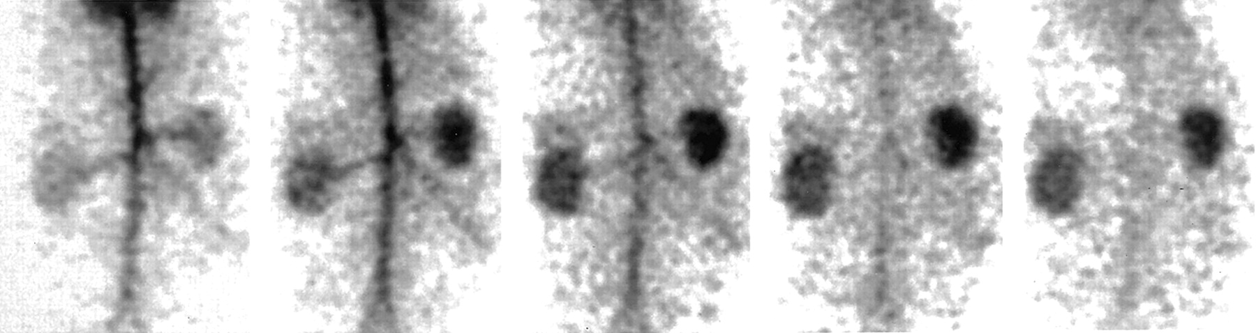
An example of the images obtained with this technique is shown in Figure 1. These images, obtained at 2-s intervals, clearly show the 191mIr bolus passing down the aorta, through the renal arteries, and into the kidneys. The left kidney, with elevated pelvic pressure, shows visibly lower uptake of the tracer than the right kidney. Because the half-life of 191mIr is only 5 s, it is possible to obtain serial images of similar quality at approximately 5-min intervals.
191mIr radionuclide angiography in a rabbit, demonstrating reduced renal blood flow to the left kidney as a result of increased hydrostatic pressure in the pelvicalyceal system.
Detection of Changes in Differential Renal Blood Flow
Studies were performed at elevated renal pelvic pressures in 6 animals (Table 2). For these animals, the mean baseline differential renal blood flow ranged from 41.6% to 61.7% (mean, 47.5%; SD, 7.3%). At an elevated pressure, a decrease in differential renal blood flow was observed in all 6 animals. This decrease ranged from 4.2% to 8.0% of the baseline value and was found to be statistically significant by a 2-tailed paired Student t test (P = 0.001; n = 6). When the pressure was reduced to normal, differential renal blood flow returned to the baseline value within 5 min. The postcompression change in differential renal blood flow also was found to be statistically significant (P = 0.0017; n = 4). There was no significant difference between baseline and postcompression values (P = 0.4807).
Effect of Pelvic Pressure on Renal Blood Flow
The magnitude of the decrease in renal blood flow as a function of renal pelvic pressure was evaluated by comparing mean differential blood flow and renal pelvic pressure, but no statistically significant correlation was observed (r = 0.145). Therefore, although there was a significant decrease in differential renal blood flow with increased intrarenal pressure, the magnitude of the decrease in renal blood flow did not correlate directly with the magnitude of the change in renal pelvic pressure.
DISCUSSION
Many types of renal disease are characterized by changes in renal blood flow, and these changes can be enhanced by pharmacologic agents such as furosemide and captopril (1). Observing the rapid changes induced by these drugs is difficult, however, because although the time frame of action of the drugs is on the order of minutes, a minimum of several hours is required between studies with 99mTc-mercaptoacetyltriglycine or 99mTc-dimercaptosuccinic acid. Data about short-term variations in renal blood flow might reveal clinically significant information that is currently unavailable because of the limitations of these radiopharmaceuticals. This limitation might be circumvented by use of a radiopharmaceutical with a much shorter half-life than 99mTc. Accordingly, we investigated the possibility of using 191mIr (half-life, 5 s) to obtain these data.
This study confirmed that the differential blood flow values obtained with 191mIr are reproducible, with repeated measurements in the same animal showing variations on the order of 1% (Table 1). It was also possible to measure rapid changes in differential renal blood flow with 191mIr. Changes in renal pelvic pressure ranging from 16 to 35 cm of water caused changes in differential renal blood flow ranging from 2.2% to 8.0%; the measurements reverted to their original values after the pressure was reduced to baseline (Table 2). The magnitude of these changes was not correlated with changes in renal pelvic pressure, but the changes were reproducible. Furthermore, the images obtained with 191mIr are of high quality.
We previously demonstrated that it is possible to obtain high-quality SPECT images of the kidney in rabbits with 191mIr (5). In this earlier study, the estimated steady-state 191mIr dose was 170 MBq (4.5 mCi) at an infusion rate of 3 mL/min, and a 5-min acquisition was required to obtain acceptable images. In the present study, the injected dose was 3.7–7.4 GBq (100–200 mCi), and the radionuclide angiograms were obtained in 30 s. The images from the 2 studies were of similar quality. The bolus injection method, however, allows images to be obtained somewhat faster and, perhaps more important, without the potential complications associated with a continuous infusion study—which, for an ultra-short-lived radionuclide such as 191mIr, means that the generator and the infusion pump are connected directly to the animal.
The primary limitation of extending this technique to humans is the significantly longer circulation transit time of an 191mIr bolus in humans than in rabbits. However, Heller et al. previously showed that it is possible to measure both left and right ventricular ejection fractions in adults with 191mIr (6), and the vascular transit time from the left ventricle to the kidney is relatively short. Loss of tracer to decay during transit also can be offset by injection of a larger dose. It is also worth noting that a significant fraction of patients with obstructive renal disease are children, in whom the transit time is much shorter than in adults. Therefore, although the vascular transit time in these patients is not as fast as in a 4-kg rabbit, it may nevertheless be possible to perform this type of study in these patients with only a modest increase in radiopharmaceutical dose. If it is demonstrated that this technique is feasible in patients, the potential advantages include shorter imaging times, the ability to perform serial measurements to detect acute alterations in differential renal blood flow, and lower patient radiation dose, even when serial studies are performed. It also may be possible to perform renal stress tests by measuring differential renal blood flow before and after a pharmacologic or physical challenge.
CONCLUSION
We have demonstrated that 191mIr can be used to measure differential renal blood flow in an animal model and that the measurements obtained are reproducible. The very short half-life of 191mIr means that repeated studies can be performed within 2–3 min, thereby making it possible to observe acute dynamic alterations in differential renal blood flow—which may in turn be valuable in the assessment of the effects of pharmacologic or physical interventions. The very short half-life of 191mIr also means that, although similar studies may be a challenge in adult patients, such studies may be possible in smaller, pediatric patients.
Acknowledgments
Technical support was provided by Nancie Keane, Diane Itrato, Pamela Day, and Royal Davis. Karl Mitchell assisted with image processing. This work was partially supported by Department of Health and Human Services Grant FD-R-000719-06-1.
Footnotes
Received Jun. 25, 2003; revision accepted Oct. 23, 2003.
For correspondence or reprints contact: S. Ted Treves, MD, Division of Nuclear Medicine, Children’s Hospital, 300 Longwood Ave., Boston, MA 02115.
E-mail: ted.treves{at}tch.harvard.edu