
Main menu

  • Home
  • Content
    • Current
    • Ahead of print
    • Past Issues
    • JNM Supplement
    • SNMMI Annual Meeting Abstracts
  • Subscriptions
    • Subscribers
    • Institutional and Non-member
    • Rates
    • Corporate & Special Sales
    • Journal Claims
  • Authors
    • Submit to JNM
    • Information for Authors
    • Assignment of Copyright
    • AQARA requirements
  • Info
    • Permissions
    • Advertisers
    • Continuing Education
  • About
    • About Us
    • Editorial Board
    • Contact Information
  • More
    • Alerts
    • Feedback
    • Help
    • SNMMI Journals
  • SNMMI
    • JNM
    • JNMT
    • SNMMI Journals
    • SNMMI

User menu

  • Subscribe
  • My alerts
  • Log in
  • My Cart

Search

  • Advanced search
Journal of Nuclear Medicine
  • SNMMI
    • JNM
    • JNMT
    • SNMMI Journals
    • SNMMI
  • Subscribe
  • My alerts
  • Log in
  • My Cart
Journal of Nuclear Medicine

Advanced Search

  • Home
  • Content
    • Current
    • Ahead of print
    • Past Issues
    • JNM Supplement
    • SNMMI Annual Meeting Abstracts
  • Subscriptions
    • Subscribers
    • Institutional and Non-member
    • Rates
    • Corporate & Special Sales
    • Journal Claims
  • Authors
    • Submit to JNM
    • Information for Authors
    • Assignment of Copyright
    • AQARA requirements
  • Info
    • Permissions
    • Advertisers
    • Continuing Education
  • About
    • About Us
    • Editorial Board
    • Contact Information
  • More
    • Alerts
    • Feedback
    • Help
    • SNMMI Journals
  • Follow JNM on Twitter
  • Visit JNM on Facebook
  • Join JNM on LinkedIn
  • Subscribe to our RSS feeds
Meeting ReportNeurosciences

Multiparametric magnetic resonance imaging and positron emission tomography findings in the diagnosis of primary progressive aphasia – A case based approach and review of literature

Neetu Soni, Savita Puri, PRERANA RANA and Steven Meyers
Journal of Nuclear Medicine June 2022, 63 (supplement 2) 2648;
Neetu Soni
1University of Rochester URMC
  • Find this author on Google Scholar
  • Find this author on PubMed
  • Search for this author on this site
Savita Puri
2University of Rochester -URMC
  • Find this author on Google Scholar
  • Find this author on PubMed
  • Search for this author on this site
PRERANA RANA
3PET Suite: Indraprastha Apollo Hospital & House of Diagnostics, New Delhi) Depar
  • Find this author on Google Scholar
  • Find this author on PubMed
  • Search for this author on this site
Steven Meyers
4URMC
  • Find this author on Google Scholar
  • Find this author on PubMed
  • Search for this author on this site
  • Article
  • Figures & Data
  • Info & Metrics
Loading

Abstract

2648

Introduction: Primary progressive aphasia (PPA) is considered a heterogeneous syndrome, with different clinical subtypes and neuropathological causes. Based on clinical, neuroimaging, and neuropathology, PPA is classified into 3 subtypes: non-fluent (nfvPPA) with agrammatism or apraxia of speech, semantic (svPPA) with a progressive and selective loss of lexical semantics, and logopenic (lvPPA) with frequent word-finding pauses and phonemic paraphasias. However, classification is complicated, and some individuals do not fit into one subtype. The svPPA shows association with FTLD-TDP-43, nfvPPA with FTLD-tau pathologies and lvPPA develops into AD.

Neuroimaging is an essential tool for the early diagnosis of PPA in both clinical and research settings. Structural, functional, and metabolic imaging modalities, including magnetic resonance imaging (MRI) and positron emission tomography (PET), are widely available.

This educational exhibit will focus on the complementary role of various imaging modalities, the qualitative and quantitative utility of newer MRI techniques, novel radiopharmaceuticals, and integrated PET/MRI in the setting of PPA.

Methods: To provide neuroimaging characteristics of PPA to understand their pathophysiology and provide an accurate diagnosis.

Help familiarize neuroradiologists with integrated PET/MR imaging in evaluating and diagnosing PPA and its subtypes.

Results: The clinical diagnosis of a specific variant has been associated with a relatively distinct neuroimaging pattern.

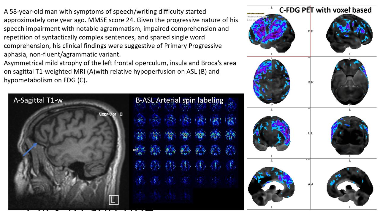
MRI- In svPPA, asymmetrical atrophy in the left anterior temporal lobe and hippocampus progresses to the posterosuperior temporal lobe, orbitofrontal, and cingulate cortices. In nfvPPA, atrophy primarily involves the left frontal operculum, insula, Broca's area, basal ganglia, and thalamus. In lvPPA, atrophy initially includes left posterior-perisylvian and later involves ipsilateral parietal-frontal and contralateral temporal lobes. FDG-PET has excellent diagnostic performance (sensitivity 87.8%, specificity 89.9%) in differentiating PPA patients from controls. In svPPA, hypometabolism involves the temporal lobe, entorhinal, perirhinal cortex, inferior temporal poles, and amygdala. In nfaPPA, hypometabolism involves the left inferior frontal and superior temporal regions. lvPPA shows left-sided hypometabolism involving lateral frontal, posterior-lateral temporal lobe, caudate, PCG, and precuneus. Amyloid-PET may be especially useful in lvPPA due to its frequent association with amyloid pathology. Tau-PET correlates with disease severity, atrophy, and FDG hypometabolism.

Conclusions: Multiparametric magnetic resonance imaging and positron emission tomography with FDG, amyloid and tau tracers help in the diagnosis of primary progressive aphasia and their subtypes.

Figure
  • Download figure
  • Open in new tab
  • Download powerpoint
Previous
Back to top

In this issue

Journal of Nuclear Medicine
Vol. 63, Issue supplement 2
June 1, 2022
  • Table of Contents
  • Index by author
Article Alerts
Sign In to Email Alerts with your Email Address
Email Article

Thank you for your interest in spreading the word on Journal of Nuclear Medicine.

NOTE: We only request your email address so that the person you are recommending the page to knows that you wanted them to see it, and that it is not junk mail. We do not capture any email address.

Enter multiple addresses on separate lines or separate them with commas.
Multiparametric magnetic resonance imaging and positron emission tomography findings in the diagnosis of primary progressive aphasia – A case based approach and review of literature
(Your Name) has sent you a message from Journal of Nuclear Medicine
(Your Name) thought you would like to see the Journal of Nuclear Medicine web site.
Citation Tools
Multiparametric magnetic resonance imaging and positron emission tomography findings in the diagnosis of primary progressive aphasia – A case based approach and review of literature
Neetu Soni, Savita Puri, PRERANA RANA, Steven Meyers
Journal of Nuclear Medicine Jun 2022, 63 (supplement 2) 2648;

Citation Manager Formats

  • BibTeX
  • Bookends
  • EasyBib
  • EndNote (tagged)
  • EndNote 8 (xml)
  • Medlars
  • Mendeley
  • Papers
  • RefWorks Tagged
  • Ref Manager
  • RIS
  • Zotero
Share
Multiparametric magnetic resonance imaging and positron emission tomography findings in the diagnosis of primary progressive aphasia – A case based approach and review of literature
Neetu Soni, Savita Puri, PRERANA RANA, Steven Meyers
Journal of Nuclear Medicine Jun 2022, 63 (supplement 2) 2648;
del.icio.us logo Digg logo Reddit logo Twitter logo CiteULike logo Facebook logo Google logo Mendeley logo
  • Tweet Widget
  • Facebook Like
  • Google Plus One
Bookmark this article

Jump to section

  • Article
  • Figures & Data
  • Info & Metrics

Related Articles

  • No related articles found.
  • Google Scholar

Cited By...

  • No citing articles found.
  • Google Scholar

More in this TOC Section

  • PET quantifies target engagement of MAGL therapeutics for Alzheimer’s disease
  • CSF Shunt Scintigraphy - spills and stops, review of protocols via challenging cases
  • A Critical Assessment of PET Radiotracers in the Imaging of Astrocytic Tumors
Show more Neurosciences

Similar Articles

SNMMI

© 2023 Journal of Nuclear Medicine

Powered by HighWire