
Main menu

  • Home
  • Content
    • Current
    • Ahead of print
    • Past Issues
    • JNM Supplement
    • SNMMI Annual Meeting Abstracts
  • Subscriptions
    • Subscribers
    • Institutional and Non-member
    • Rates
    • Corporate & Special Sales
    • Journal Claims
  • Authors
    • Submit to JNM
    • Information for Authors
    • Assignment of Copyright
    • AQARA requirements
  • Info
    • Permissions
    • Advertisers
    • Continuing Education
  • About
    • About Us
    • Editorial Board
    • Contact Information
  • More
    • Alerts
    • Feedback
    • Help
    • SNMMI Journals
  • SNMMI
    • JNM
    • JNMT
    • SNMMI Journals
    • SNMMI

User menu

  • Subscribe
  • My alerts
  • Log in
  • My Cart

Search

  • Advanced search
Journal of Nuclear Medicine
  • SNMMI
    • JNM
    • JNMT
    • SNMMI Journals
    • SNMMI
  • Subscribe
  • My alerts
  • Log in
  • My Cart
Journal of Nuclear Medicine

Advanced Search

  • Home
  • Content
    • Current
    • Ahead of print
    • Past Issues
    • JNM Supplement
    • SNMMI Annual Meeting Abstracts
  • Subscriptions
    • Subscribers
    • Institutional and Non-member
    • Rates
    • Corporate & Special Sales
    • Journal Claims
  • Authors
    • Submit to JNM
    • Information for Authors
    • Assignment of Copyright
    • AQARA requirements
  • Info
    • Permissions
    • Advertisers
    • Continuing Education
  • About
    • About Us
    • Editorial Board
    • Contact Information
  • More
    • Alerts
    • Feedback
    • Help
    • SNMMI Journals
  • Follow JNM on Twitter
  • Visit JNM on Facebook
  • Join JNM on LinkedIn
  • Subscribe to our RSS feeds
Meeting ReportClinical Science

Low-count Rb-82 cardiac PET/CT image denoising via deep learning for clinical dose reduction with preserved quantification accuracy.

Michelle Fridlund, Martin Lassen, Philip Hasbak, Liselotte Højgaard, Ole Munk, Flemming Andersen and Claes Ladefoged
Journal of Nuclear Medicine June 2022, 63 (supplement 2) 2263;
Michelle Fridlund
1Rigshospitalet, Copenhagen University Hospital, Denmark
  • Find this author on Google Scholar
  • Find this author on PubMed
  • Search for this author on this site
Martin Lassen
2Rigshospitalet, Copenhagen University Hospital
  • Find this author on Google Scholar
  • Find this author on PubMed
  • Search for this author on this site
Philip Hasbak
2Rigshospitalet, Copenhagen University Hospital
  • Find this author on Google Scholar
  • Find this author on PubMed
  • Search for this author on this site
Liselotte Højgaard
3Rigshospitalet, Copenhagen University
  • Find this author on Google Scholar
  • Find this author on PubMed
  • Search for this author on this site
Ole Munk
4Aarhus University Hospital
  • Find this author on Google Scholar
  • Find this author on PubMed
  • Search for this author on this site
Flemming Andersen
3Rigshospitalet, Copenhagen University
  • Find this author on Google Scholar
  • Find this author on PubMed
  • Search for this author on this site
Claes Ladefoged
5Rigshospitalet
  • Find this author on Google Scholar
  • Find this author on PubMed
  • Search for this author on this site
  • Article
  • Figures & Data
  • Info & Metrics
Loading

Abstract

2263

Introduction: Myocardial perfusion imaging (MPI) with PET allows for evaluation of perfusion defect sizes. Current guidelines recommend injections of 1100-1500 MBq (30-40 mCi) Rubidium-82 (82Rb) for MPI. Such thresholds might saturate the PET/CT systems; hence reductions in the administered doses are desired. However, directly limiting the amount of tracer leads to impaired image quality in the form of increased noise. The purpose of this study was to develop a robust AI deep learning (DL) approach to obtain standard-dose image quality from quarter-dose reconstructions without compromising the clinically relevant quantification accuracy and allowing for dose reduction in future studies.

Methods: This retrospective study comprised 82Rb MPI from 111 patients with known or suspected cardiac disease, imaged on a Siemens Biograph 128 mCT/PET scanner at rest and adenosine-induced stress states from June 2016 to July 2020. Only patients who received between 1000 MBq and 1200 MBq of 82Rb were included in this study. The resulting clinical listmode data were decimated with the Siemens e7 tools to simulate their respective quarter-dose counterparts. Images at both dose levels were reconstructed with the PSF-TOF 3D OSEM algorithm, using 4 iterations, 21 subsets, matrix size of 128×128×111 and a 2 mm Gaussian postfiltering. The high-/low-dose image pairs were then used to train a DL algorithm. 84 patients were reserved for training, 17 for validation and 10 for clinical metric testing. The model was validated by comparison of quantitative image metrics peak signal-to-noise ratio (PSNR), structural similarity index (SSIM) and normalized root-mean-squared-error (NRMSE). A paired t-test was used to verify the noise reduction outcomes. Total perfusion deficit (defect extent and severity) values at rest and stress (rTPD/sTPD) in the left ventricle (LV) were extracted with the Cedars QPS software. Correlation between TPD values before and after denoising was compared with Pearson’s R coefficient.

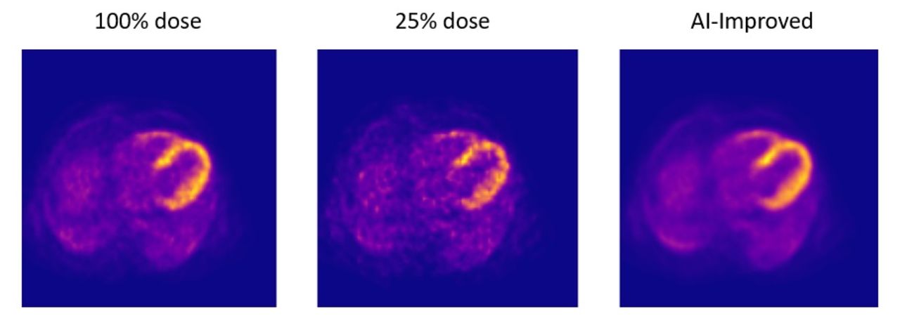
Results: The AI-denoising yielded quantitative improvement across all images (PSNR: 37.53 ± 0.40/41.52 ± 0.30, SSIM: 0.89 ± 0.01/0.95 ± 0.00, NRMSE: 1.12 ± 0.04/0.70 ± 0.02 in input and output respectively; expressed as mean ± SD). All relative differences between the means were statistically significant (p < 0.0001). Substantial noise reduction was observed in DL-enhanced quarter-dose images (Figure 1 shows a single slice example from one of the test patients). Out of the 10 patients selected for testing 5 exhibited normal TPD (< 5% of LV), 2 showed moderate (10-15%) and 3 severe (> 20%) abnormalities during the stress acquisition. Only the 3 patients with severe abnormalities had fixed perfusion defects, present during rest test (between 5 and 15% of LV), while the remainder had little to none. The denoising method did not introduce any incongruencies to neither rest nor stress TPDs. Their values pre- and post-denoising resembled those of the reference dose, with strong correlation in both (RsTPD: 0.99/0.99; p < 0.0001 and RrTPD: 0.98/0.98; p < 0.0001). The regression slope was 1.01 for sTPD before and after denoising, while a slight underestimation was observed in denoised rTPD values (0.99 and 0.85 respectively).

Conclusions: The proposed AI method demonstrated excellent denoising ability without significant loss of diagnostic accuracy for low-dose 82Rb cardiac PET/CT images. Dose reduction to 25% with quantitative clinical metrics comparable to reference dose was possible.

Figure
  • Download figure
  • Open in new tab
  • Download powerpoint
Previous
Back to top

In this issue

Journal of Nuclear Medicine
Vol. 63, Issue supplement 2
June 1, 2022
  • Table of Contents
  • Index by author
Article Alerts
Sign In to Email Alerts with your Email Address
Email Article

Thank you for your interest in spreading the word on Journal of Nuclear Medicine.

NOTE: We only request your email address so that the person you are recommending the page to knows that you wanted them to see it, and that it is not junk mail. We do not capture any email address.

Enter multiple addresses on separate lines or separate them with commas.
Low-count Rb-82 cardiac PET/CT image denoising via deep learning for clinical dose reduction with preserved quantification accuracy.
(Your Name) has sent you a message from Journal of Nuclear Medicine
(Your Name) thought you would like to see the Journal of Nuclear Medicine web site.
Citation Tools
Low-count Rb-82 cardiac PET/CT image denoising via deep learning for clinical dose reduction with preserved quantification accuracy.
Michelle Fridlund, Martin Lassen, Philip Hasbak, Liselotte Højgaard, Ole Munk, Flemming Andersen, Claes Ladefoged
Journal of Nuclear Medicine Jun 2022, 63 (supplement 2) 2263;

Citation Manager Formats

  • BibTeX
  • Bookends
  • EasyBib
  • EndNote (tagged)
  • EndNote 8 (xml)
  • Medlars
  • Mendeley
  • Papers
  • RefWorks Tagged
  • Ref Manager
  • RIS
  • Zotero
Share
Low-count Rb-82 cardiac PET/CT image denoising via deep learning for clinical dose reduction with preserved quantification accuracy.
Michelle Fridlund, Martin Lassen, Philip Hasbak, Liselotte Højgaard, Ole Munk, Flemming Andersen, Claes Ladefoged
Journal of Nuclear Medicine Jun 2022, 63 (supplement 2) 2263;
Twitter logo Facebook logo LinkedIn logo Mendeley logo
  • Tweet Widget
  • Facebook Like
  • Google Plus One
Bookmark this article

Jump to section

  • Article
  • Figures & Data
  • Info & Metrics

Related Articles

  • No related articles found.
  • Google Scholar

Cited By...

  • No citing articles found.
  • Google Scholar

More in this TOC Section

  • Role of FDG PET scan in evaluating the extent of viability using quantitative method in addition to routine visual method, and its impact on management and clinical outcome.
  • xSPECT-based myocardial perfusion scintigraphy: feasibility of myocardial uptake quantitation in patients with suspected coronary artery disease – a pilot study
  • Non-Invasive Measurement of Increased Vessel Wall Inflammation by [18F]FDG PET/CT in Patients with Multiple Myeloma upon Allogeneic Hematopoietic Cell Transplantation
Show more Clinical Science

Similar Articles

SNMMI

© 2023 Journal of Nuclear Medicine

Powered by HighWire