


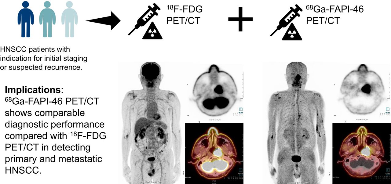
Visual Abstract

Abstract
68Ga-conjugated fibroblast activation protein inhibitor (68Ga-FAPI) has become an attractive agent for PET. This study aimed to compare 68Ga-FAPI-46 PET/CT with 18F-FDG PET/CT for detecting primary cancer and metastatic lesions in patients with head and neck squamous cell carcinoma (HNSCC). Methods: Twelve patients and 28 patients with HNSCC underwent 68Ga-FAPI-46 and 18F-FDG PET/CT for initial staging and recurrence detection, respectively. The concordance and diagnostic accuracy of both tracers were analyzed. Semiquantitative parameters, including SUVmax, SUVmean, and tumor-to-background ratio, were compared. Fibroblast activation protein (FAP) expression tumor volume and total lesion FAP expression of 68Ga-FAPI-46 were compared with metabolic tumor volume and total lesion glycolysis of 18F-FDG, respectively. Differences between semiquantitative parameters were analyzed using paired t testing. Results: 68Ga-FAPI-46 PET/CT was 83.3% and 96.4% concordant with 18F-FDG PET/CT for initial staging and recurrence detection, respectively. Eighteen lesions had histopathologic validation, and both tracers displayed 100% sensitivity, 50% specificity, and 94.4% accuracy for lesion-based analysis. FAP expression tumor volume was greater than metabolic tumor volume (P < 0.05), but no significant differences were observed for the other parameters. Conclusion: 68Ga-FAPI-46 PET/CT showed good concordance with, and comparable diagnostic performance to, 18F-FDG PET/CT for initial staging and recurrence detection in HNSCC patients.
Head and neck squamous cell carcinoma (HNSCC) is the sixth most common carcinoma worldwide, with 890,000 new cases and 450,000 deaths reported in 2018 (1). The treatment of HNSCC depends on the anatomic site, tumor stage, and functional outcome. Early-stage cancers are treated with a single modality, such as surgery or radiotherapy alone, whereas locally advanced cancers require multimodal treatment, which is often a combination of surgery, radiotherapy, and chemotherapy. Therefore, accurate tumor staging is crucial for planning treatment strategies. 18F-FDG PET/CT is a widely accepted tool for imaging various cancers. However, 18F-FDG PET/CT has some limitations when used for HNSCC. High glucose uptake is observed in several normal tissues, such as salivary glands, lymphoid tissues, and lymph nodes. Furthermore, false-positive uptake may occur in areas of peritumoral inflammation or after surgery and radiotherapy (2).
The tumor microenvironment in HNSCC is a mix of tumor and stromal cells, including endothelial cells, immune cells, and cancer-associated fibroblasts. Cancer-associated fibroblasts secrete a broad range of growth factors, cytokines, and chemokines that promote tumor growth, angiogenesis, and recruitment of immunosuppressive immune cells and thus have a role in HNSCC invasion and progression (3). Fibroblast activation protein (FAP) is overexpressed by cancer-associated fibroblasts in several types of cancer, including HNSCC, with relatively low expression in normal tissue. 68Ga-conjugated FAP inhibitor (FAPI), has been developed for targeting FAP and tumor stromal visualization (4). In previous studies, 68Ga-FAPI-04 PET/CT showed a higher sensitivity than 18F-FDG PET/CT in various types of cancers (5), and FAPI PET precisely delineated HNSCC for radiotherapy planning (6). The aim of this study was to conduct a head-to-head comparison of 68Ga-FAPI-46 PET/CT and standard 18F-FDG PET/CT imaging for detecting primary cancer and metastatic lesions in patients with HNSCC.
MATERIALS AND METHODS
Study Design
This was a single-center exploratory comparative-imaging study. The study was approved by the Human Research Ethics Committee of Chulabhorn Research Institute (study registration number, TCTR20210902003 [https://www.thaiclinicaltrials.org/show/TCTR20210902003]), and all subjects provided written informed consent. There was no external source of funding. The study protocol is provided as a supplemental file (supplemental materials are available at http://jnm.snmjournals.org).
Study Population
Potentially eligible HNSCC patients were recruited for enrollment in this study from August 2020 through May 2021. The inclusion criteria were pathologically confirmed HNSCC, an age of more than 18 y, and scheduled PET/CT for initial staging or suspected recurrence. Exclusion criteria included a fasting blood sugar of more than 150 mg/dL, pregnancy or breast feeding, and unwillingness to participate. The flowchart of the study design is presented in Figure 1.

Flowchart of study design.
Preparation of 68Ga-FAPI-46
68Ga-FAPI-46 was synthesized using an iTG 68Ge/68Ga generator and automated module (iQS-TS; ITM Medical Isotopes) and a good-manufacturing-practice–compliant process, as previously described (7,8) with some modifications.
PET/CT Imaging
18F-FDG and 68Ga-FAPI-46 PET/CT were performed on separate days within a 2-wk period. The patients fasted for 6 h before undergoing 18F-FDG PET/CT, whereas no specific preparation was required for 68Ga-FAPI-46 PET/CT. Before the 18F-FDG PET/CT scan, the plasma glucose level was determined to ensure it was no more than 150 mg/dL. The tracer dose was calculated according to the patient’s weight in kilograms (2.59 MBq/kg for 18F-FDG; 2.0 MBq/kg for 68Ga-FAPI-46). Sixty minutes after intravenous administration, scanning was performed from the vertex to the proximal thigh using a 64-slice Biograph Vision PET/CT scanner (Siemens Healthcare GmbH) in 3-dimensional mode with continuous bed motion, at a speed of 1.6–1.8 mm/s. The matrix was 440 × 440, and the reconstruction methods were True X (Siemens) and time of flight. The CT parameters were a tube voltage of 120 kV, a current of 25 mAs, and a slice thickness of 3.0 mm. 68Ga-FAPI-46 PET/CT was performed for comparative purposes without impacting the final patient management.
PET/CT Imaging Analysis
18F-FDG PET/CT scans were interpreted separately from 68Ga-FAPI-46 PET/CT scans within 2 wk of each other by board-certified nuclear medicine physicians working in consensus. One team interpreted the 18F-FDG PET/CT scans, and a second team interpreted the 68Ga-FAPI-46 PET/CT scans; one of the interpreters was on both teams (5 interpreters total). The interpreters were unaware of the clinical data at the time of review. PET, CT, and PET/CT images were viewed using a Syngo.via workstation (Siemens Healthcare GmbH).
An area of focal uptake visually higher than that of the surrounding background was considered positive. The lesion was categorized as a primary tumor, nodal metastasis, or distant metastasis. Nodal metastasis was classified according to location: neck, supraclavicular, mediastinal, axillary, or intraabdominal. Involvement of the brain, visceral organs in the chest and abdomen, bone, and soft tissues was classified as individual sites. Synchronous and second primary tumors were also analyzed. For initial staging, the clinical TNM stage of HNSCC was based on the eighth edition of the American Joint Committee on Cancer staging system (9).
The 3 designated physicians of each team drew 3-dimensional voxels of interest around the lesions and performed semiquantitative analysis, making adjustments to avoid false-positive results in regions of normal physiologic uptake. The tumor region was delineated automatically using an SUV that was 40% of the SUVmax. The SUVmax, SUVmean, and tumor-to-background ratio (T/B) of the primary tumor and distant metastases were recorded. T/B was determined by dividing SUVmax by the SUVmean of contralateral normal tissue. Metabolic tumor volume (MTV) and total lesion glycolysis assessed by 18F-FDG were compared with the equivalent values assessed by 68Ga-FAPI-46 (FAP expression tumor volume [FTV] and total lesion FAP expression, respectively). The T/B was determined by dividing tumor SUVmax by the SUVmean of contralateral normal tissue. MTV and FTV were calculated by multiplying the number of voxels in the tumor region by voxel size. Total lesion glycolysis and total lesion FAP expression were calculated by multiplying MTV or FTV, respectively, by the corresponding SUVmean for each tumor volume. If multiple positive lesions occurred at a single metastatic site, the lesion with the highest activity was analyzed. For nodal metastasis, the SUVmax was calculated for each site.
Reference Standard
Histopathology served as the gold standard for analysis of diagnostic accuracy. The reference standard for nonbiopsied lesions was the anatomic abnormality observed on CT or MRI. An anatomic criterion for nodal metastasis was either a cluster of at least 3 size-independent nodes at 1 site or fewer than 3 lymph nodes, at least 1 of which measured at least 1 cm along the short axis or had a spheric form or central necrosis. The anatomic criteria for lung metastasis included solid pulmonary nodules, a reticulonodular pattern, cavitating nodules, or lymphangitis carcinomatosis. The anatomic criteria for bone metastasis were lytic or sclerotic lesions with cortical breakthrough, a periosteal reaction, an expansile appearance, pathologic fracture on CT, or an abnormal marrow signal intensity on MRI. The anatomic criterion for distant metastasis was a nodule or mass lesion at another site. Lesions showing focally increased uptake above the background level and with corresponding anatomic criteria were defined as true-positives. Patients with negative PET/CT findings were followed up clinically for at least 3 mo to confirm a true-negative result.
Statistical Analysis
The primary outcome was concordance of 8F-FDG and 68Ga-FAPI-46 PET/CT results for initial staging and recurrence detection. The secondary outcome was the diagnostic accuracy of both tracers. Comparison of semiquantitative parameters was the tertiary outcome.
The visually interpreted PET/CT images were compared with the reference standards. Concordance rates between the 2 tracers for initial staging and recurrence detection were calculated. The diagnostic accuracy of both tracers defined by sensitivity, specificity, positive predictive value, negative predictive value, and accuracy was calculated for lesions with histopathologic validation. Differences in semiquantitative parameters between 18F-FDG and 68Ga-FAPI-46 PET/CT were analyzed using paired t tests. Data are presented as number or as mean ± SD. A P value of less than 0.05 was considered statistically significant. STATA software, version 11 (StataCorp LLC), was applied for all analyses.
RESULTS
The characteristics of each patient are shown in Table 1.
Characteristics of Study Patients
Twenty-five primary tumors were detected in 25 patients using both tracers. The mean size of the primary tumors was 3.5 ± 1.4 cm, with a minimum and maximum of 1.5 and 7.4 cm, respectively.
18F-FDG and 68Ga-FAPI-46 identified 128 and 94 lymph nodes, respectively. Overall, there were 33 sites (17 neck, 5 supraclavicular, 1 axillary, 7 mediastinal, and 3 intraabdominal) of nodal involvement in 24 patients detected by both tracers. 18F-FDG PET/CT detected more lymph nodes than did 68Ga-FAPI-46 PET/CT; however, the numbers of sites involved did not differ between the 2 tracers. The sizes of detected nodes ranged from 0.4 to 4.2 cm. Patient with lower nodal detection by 68Ga-FAPI-46 are shown in Figure 2.

18F-FDG PET/CT (A) and 68Ga-FAPI-46 PET/CT (B) images of 69-y-old woman with left oropharyngeal cancer, stage IVA, who underwent PET/CT for initial staging. 18F-FDG PET/CT detected involved lymph nodes on left side at levels IIA and IIB, whereas 68Ga-FAPI-46 PET/CT did not detect level IIB node (arrows). Left IIB node was confirmed as nodal metastasis by anatomic abnormality criteria.
Ten of 40 patients presented with distant metastases involving 15 sites (5 pulmonary, 5 bone, 1 pleural, 1 thyroidal, 1 adrenal, 1 hepatic, and 1 muscle). Synchronous tumors were noted in 4 patients (supraglottis in 1 and esophagus in 3). Two patients had a second primary thyroid cancer with histopathologic confirmation. The lesions at each site were detected with both tracers, except for 2 bone lesions in 2 patients that were observed only on 68Ga-FAPI-46 PET/CT. Both bone lesions were confirmed by anatomic criteria for bone metastasis.
Concordance of 18F-FDG and 68Ga-FAPI-46 PET/CT
There was no difference in the assessment of TNM staging between the 2 tracers in 10 of 12 patients, with 83.3% concordance. 68Ga-FAPI-46 PET/CT upstaged 1 patient (patient 28). The upstaged lesion was confirmed by MRI after a marrow change at the right scapula and suspected bone metastasis. In patient 31, 68Ga-FAPI-46 PET/CT detected a lower number of nodal metastases than did 18F-FDG; the multiple nodal metastases were confirmed by anatomic criteria (the size of the 1 discordant node was 1.0 cm in the short axis and 1.2 cm in the long axis). The PET/CT results for initial staging are detailed in Table 2.
Comparative 18F-FDG and 68Ga-FAPI-46 PET/CT Results for Initial Staging
A difference in recurrence detection between the 2 tracers was observed in only 1 of 28 patients, with 96.4% concordance. In this patient, 68Ga-FAPI-46 PET/CT showed focal uptake without corresponding 18F-FDG uptake in a sclerotic lesion at the ninth right rib, which was a suspected bone metastasis according to our criteria. Images of the discordant cases are presented in Figure 3. The false-positive results in patient 1 may be explained by postoperative inflammation due to primary-tumor excision about 1 mo before the PET studies. False-positive results with biopsy validation in patient 14 may be explained by postradiation fibrotic changes at 5 mo after radiation. The PET/CT results for recurrence detection are detailed in Table 3.

18F-FDG PET/CT (A) and 68Ga-FAPI-46 PET/CT (B) images of 49-y-old woman with nasopharyngeal cancer after concurrent chemoradiation who underwent PET/CT for recurrence detection. 68Ga-FAPI-46 PET/CT showed focal uptake, without corresponding 18F-FDG uptake, in small sclerotic lesion at ninth right lateral rib, which was suspected of being bone metastasis (arrows).
Comparative 18F-FDG and 68Ga-FAPI-46 PET/CT Results for Recurrence Detection
Diagnostic Accuracy of 18F-FDG and 68Ga-FAPI-46 PET/CT
Eighteen lesions had histopathologic results. Both tracers detected 16 true-positive, 1 true-negative, and 1 false-positive lesions. No false-negative results were found. The diagnostic accuracy of 68Ga-FAPI-46 and 18F-FDG PET/CT is shown in Table 4.
Comparative Diagnostic Accuracy of 18F-FDG and 68Ga-FAPI-46 PET/CT
Comparison of Semiquantitative Parameters
There were no significant differences in semiquantitative parameters, except for the FTV of the primary tumor, which was significantly higher than MTV (P = 0.03). An example of a patient with a higher FTV than MTV is shown in Figure 4. The semiquantitative comparisons of primary tumor and distant metastasis are shown in Table 5. The semiquantitative parameters for nodal metastasis are compared in Table 6.

18F-FDG PET/CT (A) and 68Ga-FAPI-46 PET/CT (B) images of 60-y-old man with glottic cancer who underwent PET/CT for recurrence detection. Recurrent tumor avid for 18F-FDG and 68Ga-FAPI-46 was seen at right side of oropharynx (arrows). MTV was 19.37 cm3. FTV was 33.75 cm3.
Comparisons of Semiquantitative Parameters Between 18F-FDG and 68Ga-FAPI-46 PET/CT of Primary Tumor and Distant Metastasis
Semiquantitative Comparisons Between 18F-FDG and 68Ga-FAPI-46 PET/CT for Nodal Metastasis
DISCUSSION
To our knowledge, this study was the first head-to-head comparison of diagnostic performance and semiquantitative parameters—such as uptake, image contrast, and tumor volume—between 68Ga-FAPI-46 and 18F-FDG PET/CT in HNSCC patients. 68Ga-FAPI-46 PET/CT had an 83.3% and 96.4% concordance with 18F-FDG PET/CT for initial staging and recurrence detection, respectively. Lesion-based analysis showed comparable diagnostic accuracy. All primary tumors were detected by both tracers. The number of avid nodes detected by 68Ga-FAPI-46 was less than that detected by 18F-FDG. Our findings corresponded with those of Serfling et al. (10), who reported that 18F-FDG PET/CT had a higher detection rate for cervical nodal metastases than did 68Ga-FAPI PET/CT, if the metastatic nodes were smaller than 0.7 cm; smaller nodes resulted in weaker FAP expression and delayed conversion of normal fibroblasts to cancer-associated fibroblasts. However, 68Ga-FAPI-46 may have higher tumor specificity than 18F-FDG, potentially resulting in fewer instances of false-positive uptake in inflamed or otherwise reactive lymph nodes. Differences in detection of avid nodes will require further verification. Our findings were discordant with those of Chen et al. (5), who observed higher sensitivity and lower specificity for nodal metastatic detection by 68Ga-FAPI-04 than by 18F-FDG PET/CT. However, Chen et al. compared 68Ga-FAPI with 18F-FDG PET/CT in various types of cancer, with only 6 HNSCC patients. In our study, 68Ga-FAPI-46 and 18F-FDG PET/CT showed consistency for detection of distant metastases in most cases. However, we observed detection differences in 2 bone lesions that showed 68Ga-FAPI-46 avidity but no 18F-FDG uptake. Chen et al. reported a false-positive 68Ga-FAPI–avid bone lesion because of myelofibrosis, which was not observed with 18F-FDG. When histopathologic confirmation is not practical, multimodal imaging is required to obtain morphologic details on metastasis.
Compared with 18F-FDG PET/CT, 68Ga-FAPI-46 PET/CT showed higher contrast images with lower physiologic background in the brain, salivary glands, and Waldeyer ring. However, we found no significant differences between the 2 tracers for SUVmax, SUVmean, T/B, or total lesion glycolysis versus total lesion FAP expression in the primary tumors. No significant differences in these semiquantitative parameters were observed between the tracers for nodal and distant metastases. Although there may be some variation in methods of SUV measurement, our results were consistent with those of Ballal et al. (11), who demonstrated that patients with head and neck cancer had comparably high uptake of 68Ga-DOTA.squaramide-FAPI and 18F-FDG. Our findings did not agree with those of Pang et al. (12), who observed higher uptake of 68Ga-FAPI than of 18F-FDG in primary and metastatic lesions of patients with gastric, duodenal, and colorectal cancers. This difference may be explained by differences in glucose metabolism among various tumor cell types. Pang et al. studied patients with adenocarcinoma or signet-ring cell carcinoma. More than 50% of their subjects were gastric cancer patients who showed low-to-moderate 18F-FDG avidity, whereas our study recruited HNSCC patients, who usually demonstrate high 18F-FDG uptake.
Interestingly, we observed a significantly higher FTV than MTV for the primary HNSCC tumor. Syed et al. (6) used 68Ga-FAPI PET/CT to contour head and neck cancer and found that 68Ga-FAPI–based gross tumor volume was significantly different from CT-based gross tumor volume. When 68Ga-FAPI– and CT-based gross tumor volumes were merged using SUVmax thresholds of 3-fold (20%–25% SUVmax) and 5-fold (40%–50% SUVmax), the derived tumor volumes were significantly larger than CT-based volumes. We suggest that 68Ga-FAPI-46–derived FTV may be an important semiquantitative parameter for HNSCC, but this possibility will require further standardization and validation.
The sensitivity, specificity, positive predictive value, negative predictive value, and accuracy of both tracers were 100%, 50%, 94.1%, 100%, and 94.4%, respectively. The 8 patients with negative 68Ga-FAPI-46 and 18F-FDG PET/CT findings had no histopathologic confirmation, resulting in low true-negative results and poor specificity in our study.
Our study was limited by the lack of histopathologic confirmation. Use of a fixed 40% threshold of SUVmax was dependent on the signal-to-noise ratio, T/B, and tumor size. An adaptive threshold–based method or taking the background into consideration may be more suitable for tumor delineation. A precise definition of FTV is needed for further study. Although there were some limitations in our study, the diagnostic performance of 68Ga-FAPI-46 PET/CT agreed well with that of standard 18F-FDG PET/CT molecular imaging.
CONCLUSION
68Ga-FAPI-46 PET/CT has good concordance with, and comparable diagnostic performance to, 18F-FDG PET/CT for initial staging and recurrence detection in HNSCC patients. Most semiquantitative parameters were comparable between the 2 tracers. However, the 68Ga-FAPI-46–derived FTV was higher than the MTV of 18F-FDG. Therefore, FTV may be a potential semiquantitative parameter for tumor volume of primary HNSCC, but further standardization and validation are required.
DISCLOSURE
Sofie iTheranostics Inc. provided the 68Ga-FAPI-46 precursor. No other potential conflict of interest relevant to this article was reported.
KEY POINTS
QUESTION: Does 68Ga-FAPI-46 PET/CT compare favorably with 18F-FDG PET/CT in HNSCC patients?
PERTINENT FINDINGS: 68Ga-FAPI-46 PET/CT was 83.3% and 96.4% concordant with 18F-FDG PET/CT for initial staging and recurrence detection, respectively. The diagnostic accuracy of 68Ga-FAPI-46 PET/CT was equivalent to that of 18F-FDG PET/CT. 68Ga-FAPI-46–derived FTV was higher than MTV assessed by 18F-FDG, but the other semiquantitative parameters were comparable.
IMPLICATIONS FOR PATIENT CARE: 68Ga-FAPI-46 PET/CT shows comparable diagnostic performance to 18F-FDG PET/CT in detecting primary and metastatic HNSCC.
ACKNOWLEDGMENTS
We thank Supanida Mayurasakorn, MD, for collecting the clinical data and Sunattee Kessung for editing the draft of the manuscript.
Footnotes
Published online Dec. 2, 2021.
- © 2022 by the Society of Nuclear Medicine and Molecular Imaging.
REFERENCES
- Received for publication July 1, 2021.
- Accepted for publication November 16, 2021.


