
Main menu

  • Home
  • Content
    • Current
    • Ahead of print
    • Past Issues
    • JNM Supplement
    • SNMMI Annual Meeting Abstracts
  • Subscriptions
    • Subscribers
    • Institutional and Non-member
    • Rates
    • Corporate & Special Sales
    • Journal Claims
  • Authors
    • Submit to JNM
    • Information for Authors
    • Assignment of Copyright
    • AQARA requirements
  • Info
    • Permissions
    • Advertisers
    • Continuing Education
  • About
    • About Us
    • Editorial Board
    • Contact Information
  • More
    • Alerts
    • Feedback
    • Help
    • SNMMI Journals
  • SNMMI
    • JNM
    • JNMT
    • SNMMI Journals
    • SNMMI

User menu

  • Subscribe
  • My alerts
  • Log in
  • My Cart

Search

  • Advanced search
Journal of Nuclear Medicine
  • SNMMI
    • JNM
    • JNMT
    • SNMMI Journals
    • SNMMI
  • Subscribe
  • My alerts
  • Log in
  • My Cart
Journal of Nuclear Medicine

Advanced Search

  • Home
  • Content
    • Current
    • Ahead of print
    • Past Issues
    • JNM Supplement
    • SNMMI Annual Meeting Abstracts
  • Subscriptions
    • Subscribers
    • Institutional and Non-member
    • Rates
    • Corporate & Special Sales
    • Journal Claims
  • Authors
    • Submit to JNM
    • Information for Authors
    • Assignment of Copyright
    • AQARA requirements
  • Info
    • Permissions
    • Advertisers
    • Continuing Education
  • About
    • About Us
    • Editorial Board
    • Contact Information
  • More
    • Alerts
    • Feedback
    • Help
    • SNMMI Journals
  • Follow JNM on Twitter
  • Visit JNM on Facebook
  • Join JNM on LinkedIn
  • Subscribe to our RSS feeds
Meeting ReportOral - PhysicianPharm

68Ga-Pentixafor PET/CT in vivo CXCR4 imaging for the detection of primary and recurrent glioma and its utility for response assessment to image-based radiation dose escalation

Abdul Waheed, Baljinder Singh, Renu Madan, Ankit Watts, Harmandeep Singh, Chirag Ahuja, Amanjit Bal and Apinder Singh
Journal of Nuclear Medicine May 2021, 62 (supplement 1) 47;
Abdul Waheed
1Nuclear Medicine Postgraduate Institute of Medical Education & Research (PGIMER) Chandigarh India
  • Find this author on Google Scholar
  • Find this author on PubMed
  • Search for this author on this site
Baljinder Singh
1Nuclear Medicine Postgraduate Institute of Medical Education & Research (PGIMER) Chandigarh India
  • Find this author on Google Scholar
  • Find this author on PubMed
  • Search for this author on this site
Renu Madan
2Radiotherapy Postgraduate Institute of Medical Education & Research (PGIMER) Chandigarh India
  • Find this author on Google Scholar
  • Find this author on PubMed
  • Search for this author on this site
Ankit Watts
1Nuclear Medicine Postgraduate Institute of Medical Education & Research (PGIMER) Chandigarh India
  • Find this author on Google Scholar
  • Find this author on PubMed
  • Search for this author on this site
Harmandeep Singh
1Nuclear Medicine Postgraduate Institute of Medical Education & Research (PGIMER) Chandigarh India
  • Find this author on Google Scholar
  • Find this author on PubMed
  • Search for this author on this site
Chirag Ahuja
3Radiodiagnosis and Imaging Postgraduate Institute of Medical Education & Research (PGIMER) Chandigarh India
  • Find this author on Google Scholar
  • Find this author on PubMed
  • Search for this author on this site
Amanjit Bal
4Histopathology Postgraduate Institute of Medical Education & Research (PGIMER) Chandigarh India
  • Find this author on Google Scholar
  • Find this author on PubMed
  • Search for this author on this site
Apinder Singh
5Neurosurgery Postgraduate Institute of Medical Education & Research (PGIMER) Chandigarh India
  • Find this author on Google Scholar
  • Find this author on PubMed
  • Search for this author on this site
  • Article
  • Figures & Data
  • Info & Metrics
Loading

Abstract

47

Objectives: Glioma constitutes approximately 30% of the CNS tumours and approximately 80% of all the malignant brain tumours. Magnetic resonance imaging (MRI) is the most commonly used procedure in the follow-up of GBM patients. However, molecular imaging (SPECT, PET) is often required for the tumour characterization and detection of active recurrent/residual disease. The currently available PET tracers i.e. 18F-FET and 11C-methionine have proven high diagnostic efficacy in GBM. A strong association has been reported between CXCR4 chemokine receptors, with the development of an invasive phenotype in malignant GBM and eventually resulting in poor prognosis. The aim of this study was to evaluate the patient outcome with escalated radiation dose derived from the accurate residual/ recurrent glioma disease mapping using CXCR4 targeting 68 Ga-Pentixafor PET imaging.

Methods: 68Ga-Pentixafor PET/CT imaging was performed in 9 patients (7M; 2F; mean age =54.3±8.9 years) at a single point pre-operatively. Tissue diagnosis in 7/9 patients was made on fixed tumor tissue and CXCR4 receptors’ expression was documented by IHC using anti-CXCR4 antibody and visual quantitative scoring. In the 11 (6M:5F; mean age = 44.3±11.7 years) patients, who presented for the clinical/radiological suspicion of recurrence/residual disease, 68Ga-Pentixafor PET/CT imaging was done at presentation (baseline) and at 3-mo post radiation treatment. About 110-150 MBq radioactivity of freshly prepared 68Ga-Pentixafor was administered intravenously. After the scan evidence of recurrent/residual disease, all these 11 patients received radical radiotherapy (67.0 Gy - escalation of 7.0 Gy) with or without concurrent temozolomide as indicated. Regional Brain CT (140Kv, 200mAs, Pitch 0.625:1, Slice thickness 2.5 mm) followed by PET acquisition (single bed position, 8min) at 1-hour post injection. Data was reconstructed using iterative method (2 iterations, 20 sub-sets) and interpreted both visually and semi-quantitatively. All the patients (n=20) underwent contrast enhanced MRI (ceMRI) and MR spectroscopy (MRS) and the data was analysed for visual scan interpretation and quantitative tumour metabolites (choline/NAA; choline/creatine) ratios.

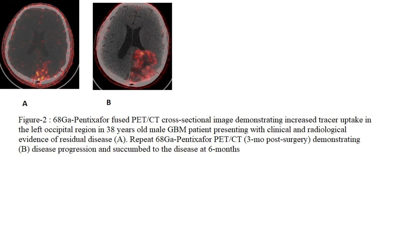
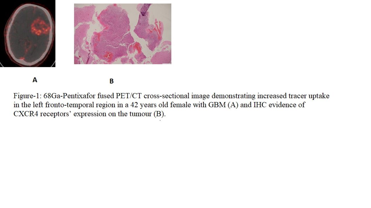
Results: 68Ga-Pentixafor PET/CT findings with focally increased uptake of the radiotracer were interpreted as positive (Figure-1) for primary tumour in 9/9 patients in the pre-surgery group with histopathological/IHC disease evidence in 7/9 patients. The CeMRI findings were positive in 9/9 patients. The metabolites’ ratios of i.e. choline/NAA; choline/creatine on MRS were 4.82±2.5 and 3.0±2.6 respectively.The mean values (n=7) for 68Ga-Pentixafor PET/CT quantitative parameters i.e. SUVmax, SUVmean, SUVpeak, T/B ratio were 4.5±1.6, 0.6±0.26, 1.95±0.8, 6.8 ±4.5 respectively. In the post-surgery group of patients, all the patients (n=11) showed scan evidence of disease recurrence with focally increased tracer uptake. However, no significant difference was observed in the mean values of quantitative parameters (SUVmax, SUVmean, SUVpeak) between the baseline and the 3-mo post RT follow-up PET imaging (Figure-2). Likewise, no significant difference was observed for Cho/NAA; Choline/creatine ratios on MRS imaging. Conclusion: 68Ga-Pentixafor concentrated preferentially in the primary GBM tumours and this preliminary study thus indicates that the specific binding affinity of this tracer may help in the development of CXCR4 targeting radiotherapeutics (alpha/beta emitters). The diagnostic utility of this tracer for response assessment to chemo/radiotherapy and for overall prognostication however, needs to be established in a larger cohort of patients with clinical follow-up and repeat CXCR4 based imaging beyond 3-months.

Figure
  • Download figure
  • Open in new tab
  • Download powerpoint
Figure
  • Download figure
  • Open in new tab
  • Download powerpoint
Previous
Back to top

In this issue

Journal of Nuclear Medicine
Vol. 62, Issue supplement 1
May 1, 2021
  • Table of Contents
  • Index by author
Article Alerts
Sign In to Email Alerts with your Email Address
Email Article

Thank you for your interest in spreading the word on Journal of Nuclear Medicine.

NOTE: We only request your email address so that the person you are recommending the page to knows that you wanted them to see it, and that it is not junk mail. We do not capture any email address.

Enter multiple addresses on separate lines or separate them with commas.
68Ga-Pentixafor PET/CT in vivo CXCR4 imaging for the detection of primary and recurrent glioma and its utility for response assessment to image-based radiation dose escalation
(Your Name) has sent you a message from Journal of Nuclear Medicine
(Your Name) thought you would like to see the Journal of Nuclear Medicine web site.
Citation Tools
68Ga-Pentixafor PET/CT in vivo CXCR4 imaging for the detection of primary and recurrent glioma and its utility for response assessment to image-based radiation dose escalation
Abdul Waheed, Baljinder Singh, Renu Madan, Ankit Watts, Harmandeep Singh, Chirag Ahuja, Amanjit Bal, Apinder Singh
Journal of Nuclear Medicine May 2021, 62 (supplement 1) 47;

Citation Manager Formats

  • BibTeX
  • Bookends
  • EasyBib
  • EndNote (tagged)
  • EndNote 8 (xml)
  • Medlars
  • Mendeley
  • Papers
  • RefWorks Tagged
  • Ref Manager
  • RIS
  • Zotero
Share
68Ga-Pentixafor PET/CT in vivo CXCR4 imaging for the detection of primary and recurrent glioma and its utility for response assessment to image-based radiation dose escalation
Abdul Waheed, Baljinder Singh, Renu Madan, Ankit Watts, Harmandeep Singh, Chirag Ahuja, Amanjit Bal, Apinder Singh
Journal of Nuclear Medicine May 2021, 62 (supplement 1) 47;
Twitter logo Facebook logo LinkedIn logo Mendeley logo
  • Tweet Widget
  • Facebook Like
  • Google Plus One
Bookmark this article

Jump to section

  • Article
  • Figures & Data
  • Info & Metrics

Related Articles

  • No related articles found.
  • Google Scholar

Cited By...

  • No citing articles found.
  • Google Scholar

More in this TOC Section

Oral - PhysicianPharm

  • Spleen targeting H2S donating liposome as theranostic immune modulator in dextran sodium sulfate induced colitis model
  • Imaging non-coding microRNA upregulation in pressure overload heart failure using the complementary anti-miR-21 locked nucleic acid
  • Human Research Studies on the PennPET Explorer
Show more Oral - PhysicianPharm

Neurosciences Young Investigator Award Session

  • Evaluation of [18F]SynVesT-2 for imaging SV2A in the human brain: kinetics, test-retest reproducibility, and binding specificity
  • Similarity and dissimilarity between synaptic density, blood flow and glucose metabolism in Alzheimer’s disease
Show more Neurosciences Young Investigator Award Session

Similar Articles

SNMMI

© 2023 Journal of Nuclear Medicine

Powered by HighWire