
Main menu

  • Home
  • Content
    • Current
    • Ahead of print
    • Past Issues
    • JNM Supplement
    • SNMMI Annual Meeting Abstracts
    • Continuing Education
    • JNM Podcasts
  • Subscriptions
    • Subscribers
    • Institutional and Non-member
    • Rates
    • Journal Claims
    • Corporate & Special Sales
  • Authors
    • Submit to JNM
    • Information for Authors
    • Assignment of Copyright
    • AQARA requirements
  • Info
    • Reviewers
    • Permissions
    • Advertisers
  • About
    • About Us
    • Editorial Board
    • Contact Information
  • More
    • Alerts
    • Feedback
    • Help
    • SNMMI Journals
  • SNMMI
    • JNM
    • JNMT
    • SNMMI Journals
    • SNMMI

User menu

  • Subscribe
  • My alerts
  • Log in
  • My Cart

Search

  • Advanced search
Journal of Nuclear Medicine
  • SNMMI
    • JNM
    • JNMT
    • SNMMI Journals
    • SNMMI
  • Subscribe
  • My alerts
  • Log in
  • My Cart
Journal of Nuclear Medicine

Advanced Search

  • Home
  • Content
    • Current
    • Ahead of print
    • Past Issues
    • JNM Supplement
    • SNMMI Annual Meeting Abstracts
    • Continuing Education
    • JNM Podcasts
  • Subscriptions
    • Subscribers
    • Institutional and Non-member
    • Rates
    • Journal Claims
    • Corporate & Special Sales
  • Authors
    • Submit to JNM
    • Information for Authors
    • Assignment of Copyright
    • AQARA requirements
  • Info
    • Reviewers
    • Permissions
    • Advertisers
  • About
    • About Us
    • Editorial Board
    • Contact Information
  • More
    • Alerts
    • Feedback
    • Help
    • SNMMI Journals
  • View or Listen to JNM Podcast
  • Visit JNM on Facebook
  • Join JNM on LinkedIn
  • Follow JNM on Twitter
  • Subscribe to our RSS feeds
Meeting ReportPoster - PhysicianPharm

Experimental evaluation of a prototype combined PET-Compton imaging system based on 3-D position-sensitive CZT detectors for dedicated breast cancer imaging

Yifei Jin, Phillip Tanton, Michael Streicher, Hao Yang, Steven Brown, Zhong He and Ling-Jian Meng
Journal of Nuclear Medicine May 2021, 62 (supplement 1) 1129;
Yifei Jin
1Nuclear, Plasma and Radiological Engineering University of Illinois At Urbana and Champaign Urbana IL United States
  • Find this author on Google Scholar
  • Find this author on PubMed
  • Search for this author on this site
Phillip Tanton
1Nuclear, Plasma and Radiological Engineering University of Illinois At Urbana and Champaign Urbana IL United States
  • Find this author on Google Scholar
  • Find this author on PubMed
  • Search for this author on this site
Michael Streicher
2H3D, Inc. Ann Arbor MI United States
  • Find this author on Google Scholar
  • Find this author on PubMed
  • Search for this author on this site
Hao Yang
2H3D, Inc. Ann Arbor MI United States
  • Find this author on Google Scholar
  • Find this author on PubMed
  • Search for this author on this site
Steven Brown
2H3D, Inc. Ann Arbor MI United States
  • Find this author on Google Scholar
  • Find this author on PubMed
  • Search for this author on this site
Zhong He
3H3D, Inc. Ann Arobor MI United States
4The University of Michigan Ann Arbor MI United States
  • Find this author on Google Scholar
  • Find this author on PubMed
  • Search for this author on this site
Ling-Jian Meng
5University of Illinois At Urbana-Champaign Champaign IL United States
  • Find this author on Google Scholar
  • Find this author on PubMed
  • Search for this author on this site
  • Article
  • Figures & Data
  • Info & Metrics
Loading

Abstract

1129

Objectives: Breast cancer is the most common cancer in women worldwide, with 1.3 million cases diagnosed per year. The current standard of care in breast cancer management has challenges. Mammograms are well known for having low specificity and often being inconclusive for patients with dense breasts, leading to unnecessary surgical procedures and patient trauma. The conventional PET systems have neither the photon sensitivity nor the spatial resolution required to affect earlier stages of breast cancer management. In recent years, there have been several groups and companies that developed high-performance dedicated positron emission mammography (PEM) systems that show promise for more sensitive cancer detection than standard clinical cameras while also providing better specificity than traditional anatomic imaging modalities such as x-ray mammography [1-5]. In this study, we used a prototype PET setup based on 3-D position-sensitive CZT detectors to demonstrate the combined PET-Compton data acquisition and its application in breast cancer imaging. The CZT detector used in the system not only provides an excellent intrinsic spatial resolution of around 0.5 mm in X-, Y-, and Z- directions, but also offers an excellent energy resolution of around 1% at 511 keV and the ability to detect and localize multiple interactions. This makes it capable of forming images with single photons through Compton scattering, and also use the Compton kinematics on coincidence photons to help reject scattered or chance coincidence in PET data. The objective of this study is to use PET and Compton data to jointly reconstruct the distribution of positron emitters in the breast and to enhance the image quality in regions close to the chest wall and even inside the chest wall through Compton imaging.

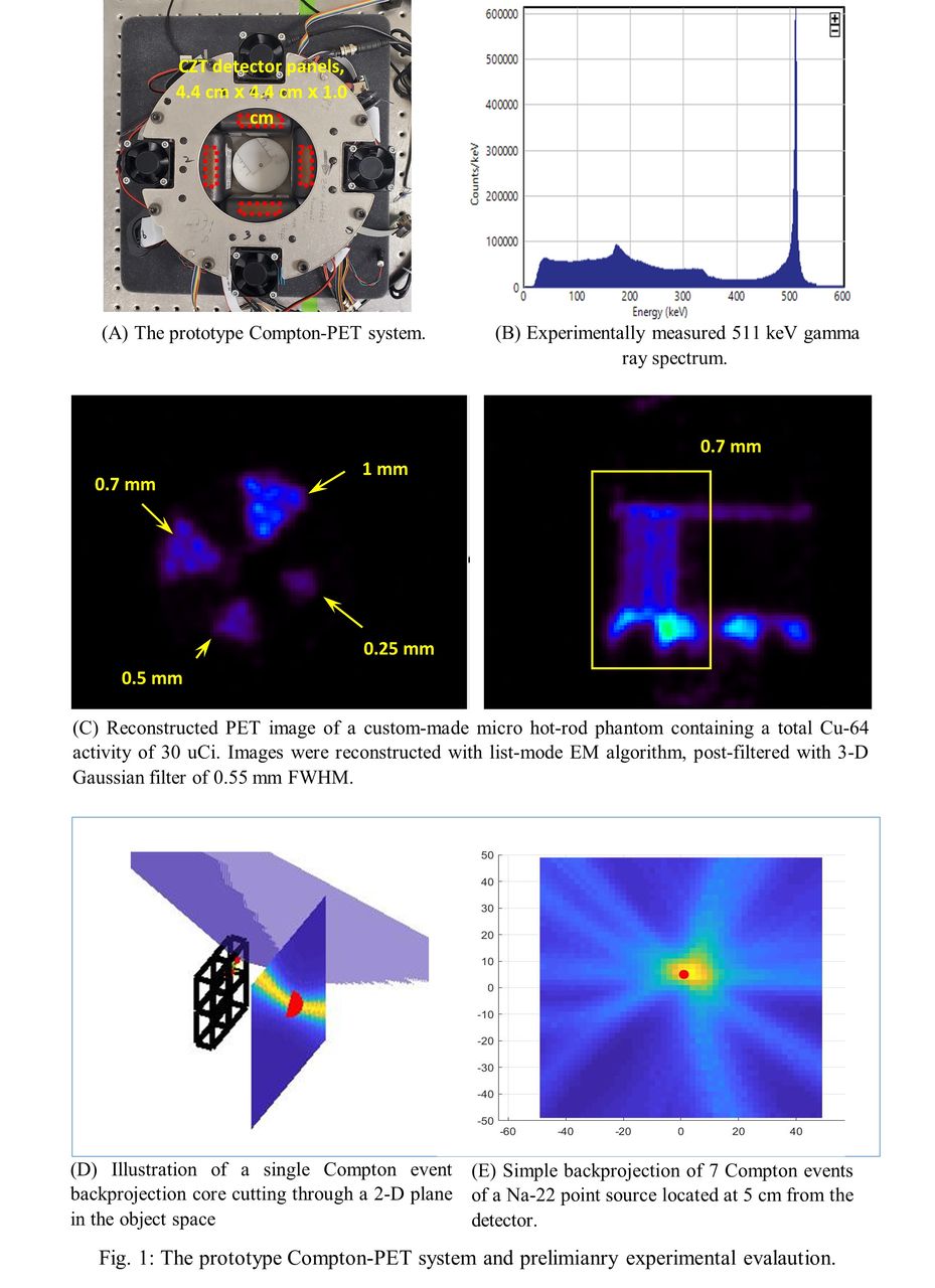
Methods: The proof-of-concept Compton-PET scanner is shown in Fig. 1(A). The system consists of four CZT detector panels, and each panel consists of four (2×2 ) CZT detectors of 2.2×2.2×1.0 cm3 in size. The detector offers a subpixel resolution of <0.5 mm FWHM in all three dimensions with CZT crystal of up to 1.5 cm thickness, and an energy resolution of 5.4 keV FWHM at 511 keV [6]. In the prototype PET system, the distance between opposite PET panels is designed to be 10 cm. The output of all CZT detectors are saved in list-mode with the position, energy and timing information of each individual gamma ray interaction. The coincidence pairs and or the single-photon Compton events are determined in post-processing through the time stamp of individual detected events. We computed a list-mode system response function (SRF) that represents the probability of each source voxel contributing to the given event, and the SRF is used by a list-mode penalized maximum likelihood algorithm for reconstructing the joint-PET-Compton images.

Results: Fig. 1(B) shows the energy spectrum measured with the CZT detector in this Compton-PET prototype. Fig. 1(C) shows the PET image of a resolution phantom acquired with the Compton-PET prototype, where the hot-rods of 0.75 mm diameter can be resolved. Fig. 1(D&E) illustrates a simple back-projection of seven single photons detected on the CZT detector through Compton interactions from a Na-22 point source placed at 5 cm from the CZT detector.

Conclusions: The joint-PET-Compton imaging reported in this paper not only offers very high sensitivity and an ultrahigh-resolution in conventional PET acquisition, but it also allows simultaneous detection of single (or coincidence) photons through Compton interactions. This offers the unique ability to enhance the PET images in regions close to the chest wall and provide imaging information, using Compton data alone, for areas inside the chest wall. The joint-PET-Compton image acquisition would, therefore, help to address one of the major limitations in current positron emission mammography (PEM) instrumentations.

Figure
  • Download figure
  • Open in new tab
  • Download powerpoint
Previous
Back to top

In this issue

Journal of Nuclear Medicine
Vol. 62, Issue supplement 1
May 1, 2021
  • Table of Contents
  • Index by author
Article Alerts
Sign In to Email Alerts with your Email Address
Email Article

Thank you for your interest in spreading the word on Journal of Nuclear Medicine.

NOTE: We only request your email address so that the person you are recommending the page to knows that you wanted them to see it, and that it is not junk mail. We do not capture any email address.

Enter multiple addresses on separate lines or separate them with commas.
Experimental evaluation of a prototype combined PET-Compton imaging system based on 3-D position-sensitive CZT detectors for dedicated breast cancer imaging
(Your Name) has sent you a message from Journal of Nuclear Medicine
(Your Name) thought you would like to see the Journal of Nuclear Medicine web site.
Citation Tools
Experimental evaluation of a prototype combined PET-Compton imaging system based on 3-D position-sensitive CZT detectors for dedicated breast cancer imaging
Yifei Jin, Phillip Tanton, Michael Streicher, Hao Yang, Steven Brown, Zhong He, Ling-Jian Meng
Journal of Nuclear Medicine May 2021, 62 (supplement 1) 1129;

Citation Manager Formats

  • BibTeX
  • Bookends
  • EasyBib
  • EndNote (tagged)
  • EndNote 8 (xml)
  • Medlars
  • Mendeley
  • Papers
  • RefWorks Tagged
  • Ref Manager
  • RIS
  • Zotero
Share
Experimental evaluation of a prototype combined PET-Compton imaging system based on 3-D position-sensitive CZT detectors for dedicated breast cancer imaging
Yifei Jin, Phillip Tanton, Michael Streicher, Hao Yang, Steven Brown, Zhong He, Ling-Jian Meng
Journal of Nuclear Medicine May 2021, 62 (supplement 1) 1129;
Twitter logo Facebook logo LinkedIn logo Mendeley logo
  • Tweet Widget
  • Facebook Like
  • Google Plus One
Bookmark this article

Jump to section

  • Article
  • Figures & Data
  • Info & Metrics

Related Articles

  • No related articles found.
  • Google Scholar

Cited By...

  • No citing articles found.
  • Google Scholar

More in this TOC Section

Poster - PhysicianPharm

  • Preliminary result of Texture Analysis on prediction of overall outcome of neuroendocrine tumors based on pre-therapy heterogeneity of somatostatin receptors on 68Ga Dotatate PET/CT scans.
  • Diagnostic and prognostic value of chronotropic incompetence on exercise gated 99mTc-MIBI SPECT in patients with suspected CAD
  • Comparison of regadenoson and dypiridamole myocardial blood flow and coronary flow reserve hyperemic response by non-invasive quantitative N-13 ammonia PET.
Show more Poster - PhysicianPharm

PIDS-Instrumentation

  • Design and Development of an MRI Compatible Human Brain PET Scanner by Using Dual-ended Readout Detectors
  • First prototype of a novel dual round-edge detector arrangement for breast PET imaging
  • A variable-aperture full-ring pixelated CZT-SPECT system for whole-body study: A simulation study combined with a finite element method
Show more PIDS-Instrumentation

Similar Articles

SNMMI

© 2025 SNMMI

Powered by HighWire