


Visual Abstract

Abstract
Radionuclide therapy targeting prostate-specific membrane antigen (PSMA) is promising for prostate cancer. We previously reported a ligand, 64Cu-CuSarbisPSMA, featuring 2 lysine-ureido-glutamate groups. Here, we report the therapeutic potential of 67Cu-CuSarbisPSMA. Methods: Growth of PSMA-positive xenografts was evaluated after treatment with 67Cu-CuSarbisPSMA or 177Lu-LuPSMA imaging and therapy (I&T). Results: At 13 d after injection, tumor growth was similarly inhibited by the 2 tracers in a dose-dependent manner. Survival was comparable after single (30 MBq) or fractionated (2 × 15 MBq, 2 wk apart) administrations. Conclusion: 67Cu-CuSarbisPSMA is efficacious in a PSMA-expressing model of prostate cancer.
Prostate-specific membrane antigen (PSMA) is a membrane-bound enzyme that can act as a glutamate carboxypeptidase or folate hydrolase. In prostate cancer cells, it becomes membrane-bound and overexpressed with androgen independence and metastasis (1), making it a promising target for both imaging and therapy (2). Radiolabeled peptidomimetic inhibitors of PSMA containing a lysine-ureido-glutamate functional group are effective tracers for imaging prostate cancer using PET (2–4). A theranostic paradigm involves PET with Glu-NH-CO-NH-Lys-(Ahx)-N,N′-bis-[2-hydroxy-5-(carboxyethyl)benzyl]ethylenediamine-N,N′-diacetic acid labeled with 68Ga (half-life, 68 min; mean β+ energy, 0.89 MeV) (68Ga-GaPSMA-11) to guide therapy with 177Lu-LuPSMA-617 (half-life, 6.65 d; β−, 100%; mean β− energy, 134 keV) (5–7). This approach has allowed personalized treatment of advanced prostate cancer, but the short half-life of 68Ga limits the ability to do prospective dosimetry (8), as does the use of differing ligands for diagnosis and therapy. Use of the DOTAGA-containing urea-based PSMA inhibitor called PSMA imaging and therapy (I&T), which can be labeled with either 68Ga or 177Lu, or use of 68Ga-Ga-PSMA-617 in place of 68Ga-GaPSMA-11 (5,9,10) can overcome the latter limitation, but both approaches remain constrained for prospective dosimetry. Both limitations could be potentially addressed by using the positron-emitting (β+) 64Cu (half-life, 12.7 h; β+, 17.4%; mean β+ energy, 278 keV) for diagnosis and β−-emitting 67Cu (half-life, 61.9 h; β−, 100%; mean β− energy, 141 keV) for therapy. The β− emissions of 67Cu have a mean range of 0.2 mm and are appropriate for the treatment of small tumors down to 5 mm in diameter (11). The γ emission of 67Cu (185 keV, 49%; 93 keV, 16%) may be beneficial for quantitative SPECT to verify the radiation dose to the tumor and critical organs (12). The efficacy of targeted therapy with 67Cu has been demonstrated previously in non-Hodgkin lymphoma and neuroendocrine tumors (11,13,14).
The potential advantages of copper radiopharmaceuticals are dependent on high retention in tumors and clearance from normal tissues. The use of chelators that form copper complexes susceptible to release of copper in vivo can lead to high liver uptake at late time-points (15). Importantly, copper(II) complexes of sarcophagine (Sar = 3,6,10,13,16,19-hexa-azabicyclo[6.6.6]icosane)-based ligands are stable in vivo (16). A Sar derivative conjugated to a somatostatin receptor-targeting peptide, 64Cu-CuSarTATE, allows acquisition of high-quality images at 24 h after injection in patients with neuroendocrine tumors (16), and its therapeutic partner, 67Cu-CuSarTATE, is highly efficacious in a neuroendocrine tumor model (14). We recently reported high tumor uptake and retention of a 64Cu-labeled sarcophagine ligand tethered to 2 lysine-ureido-glutamate functional groups in a PSMA-positive model (17). In the current study, we evaluated the therapeutic efficacy of its 67Cu-labeled partner, 67Cu-CuSarbisPSMA (Fig. 1), in the same PSMA-positive tumor model.

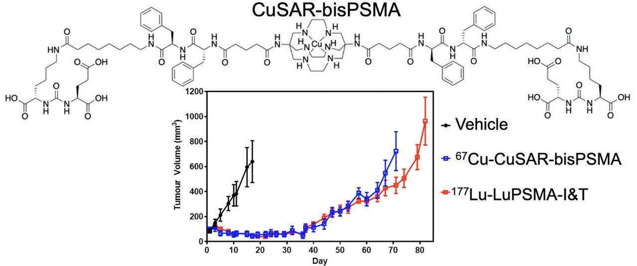
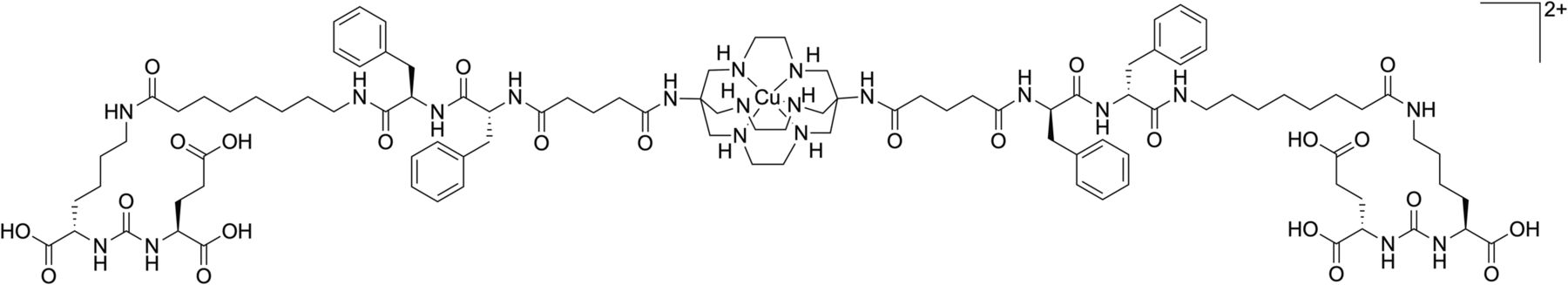
Chemical structure of 64/67Cu-CuSarbisPSMA.
MATERIALS AND METHODS
Radiochemistry
To synthesize 67Cu-SarbisPSMA, 67Cu-CuCl2 (756 MBq, 70 μL, 0.05 M HCl; Idaho State University) was added to a mixture of SarbisPSMA (AusPep) (17) (20 μg, 10 nmol, in 10 μL of 50:50 ethanol:water) and sodium phosphate buffer (0.1 M, pH 6.2, 350 μL). After 10 min at room temperature, analysis by high-performance liquid chromatography indicated at least 95% radiochemical purity (72 GBq/μmol; retention time, 10.9 min; precursor retention time, 11.0 min). The high-performance liquid chromatography was performed on a Shimadzu SPD-10ATvP chromatograph using a Phenomenex Luna C18 100Å column (4.6 × 150 mm, 5 μm) at a rate of 1 mL/min (5%–100% acetonitrile; 0.05% trifluoroacetic acid) over 15 min.
177Lu-LuPSMA I&T was prepared according to published procedures in at least 95% radiochemical yield using PSMA I&T (200 μg, 0.13 μmol) (ABX) and 177LuCl3 (8 GBq) (ANSTO) (58 GBq/μmol) (5).
In Vivo Comparative Experiment
All animal experiments were performed with the approval of the institutional animal ethics committee. Eight-week old male NSG mice (Australian BioResources) were implanted with LNCaP (human prostate adenocarcinoma) cells as described previously (17). Mice (n = 5) bearing subcutaneous LNCaP xenografts (mean tumor volume, ∼90 mm3) were randomized into 5 groups and injected intravenously with vehicle (saline), 67Cu-CuSarbisPSMA (5 or 30 MBq), or 177Lu-LuPSMA I&T (5 or 30 MBq) on day 1 of the experiment. Tumor size and health were monitored twice weekly, with mice being euthanized when tumor volume (calculated as length × width × height [mm] × π/6) exceeded 1,200 mm3.
In Vivo Dose-Dependency Experiment
Male NSG mice (n = 8 per group) with subcutaneous LNCaP xenografts (mean tumor volume, ∼240 mm3) were injected with either saline or 67Cu-CuSarbisPSMA (7.5, 15, or 30 MBq) on day 1 of the experiment. An additional cohort was injected with 67Cu-CuSarbisPSMA (15 MBq) on days 1 and 15 of the experiment (n = 8) and monitored as above.
Data Analysis
Percentage tumor growth inhibition was calculated as 100 × (1 – ΔT/ΔC), where ΔC and ΔT were determined by subtracting the mean tumor volume (in the vehicle control and treated groups, respectively) on day 1 of treatment from the mean tumor volume on either day 17 for the comparative experiment or day 13 for the dose-dependency experiment. Statistical analysis was performed using Prism, version 8.0 (GraphPad). Statistical comparisons between the vehicle control and treated cohorts were done by a 1-way ANOVA followed by a Dunnett post hoc test. Toxicity was assessed by body-weight loss and by physical and behavioral observation. The experiment was ended on day 82 or 85, with the remaining mice being censored for survival. Survival curves (tumor volume ≥ 1,200 mm3) were analyzed using the Mantel–Cox log-rank test.
RESULTS
67Cu-CuSarbisPSMA and 177Lu-LuPSMA I&T Are Efficacious Against LNCaP Tumor Xenograft Model
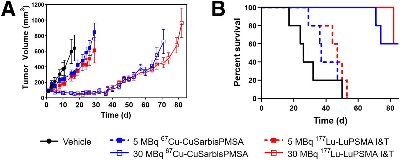
67Cu-CuSarbisPSMA was prepared in high radiochemical purity (>95%) in sodium phosphate buffer without the need for further purification before injection. Mice bearing subcutaneous LNCaP tumors were randomized into groups of 5 animals (mean tumor volume, 90 mm3) and then injected with saline, 67Cu-CuSarbisPSMA (5 MBq, 0.06 nmol, or 30 MBq, 0.36 nmol), or 177LuPSMA I&T (5 MBq, 0.08 nmol, or 30 MBq, 0.48 nmol). The inhibition of tumor growth was similar for both agents after administration of both 5 and 30 MBq, but demonstrated dose dependency (Table 1; Fig. 2). Survival was increased significantly in the 30-MBq dose cohorts when compared with the cohorts treated with 5 MBq (P = 0.002 for both agents) (Fig. 2B).
Percentage Growth Inhibition of LNCaP Tumors Treated with 67Cu-CuSarbisPSMA and 177Lu-LuPSMA I&T as Compared with Vehicle Control

(Left) Inhibition of LNCaP tumor growth after treatment with either 67Cu-CuSarbisPSMA or 177Lu-LuPSMA I&T, expressed as mean tumor volume (±SEM) (n = 5). (Right) Kaplan–Meier curve of percentage survival data; endpoint represents day on which tumor size was at least 1,200 mm3 or censoring occurred (day 82).
Inhibition of LNCaP Tumor Growth Is Dependent on the Activity of 67Cu-CuSarbisPSMA Administered
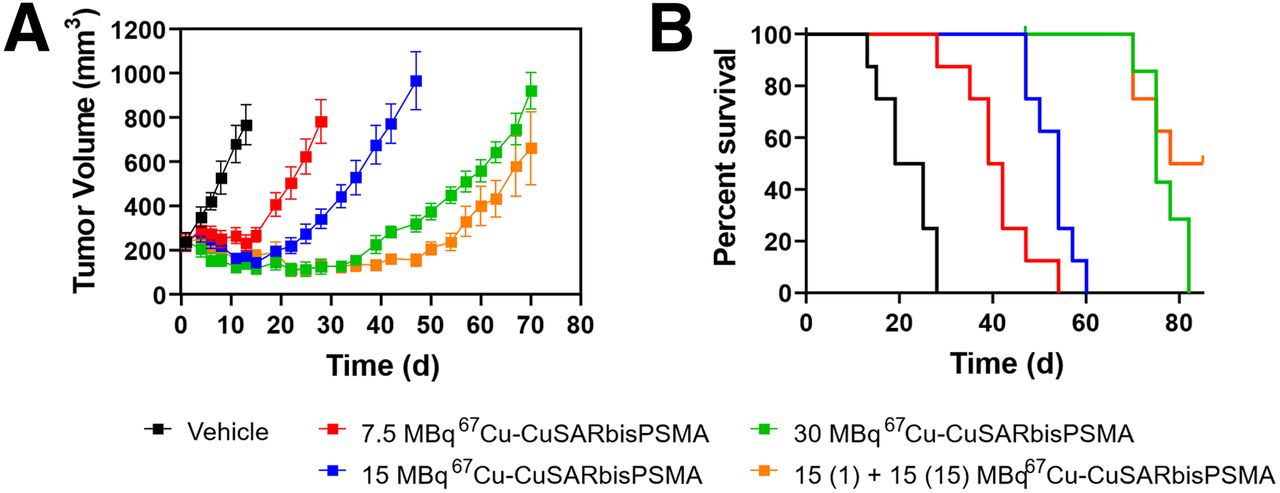
Mice were inoculated with LNCaP cells and, once tumors were established, were randomized into 5 groups (mean tumor volume, 240 mm3) and intravenously injected with either saline or increasing doses of 67Cu-CuSarbisPSMA (7.5 MBq, 0.21 μg, 0.1 nmol; 15 MBq, 0.45 μg, 0.22 nmol; or 30 MBq, 0.89 μg, 0.44 nmol) on day 1 of the experiment. An additional group was injected with 67Cu-CuSarbisPSMA (15 MBq. 0.45 μg, 0.22 nmol) on days 1 and 15 to investigate the potential of fractionated dose protocols. On day 13, tumor growth in each treatment group was suppressed versus the control group (Table 2). All treatments increased survival significantly compared with the vehicle control (7.5 MBq, P <0.001; 15 MBq, P <0.001; 30 MBq, P <0.001). There was a trend for prolonged tumor growth inhibition and survival in the fractionated dose group (2 × 15 MBq) compared with the single dose (30 MBq), although this was not statistically significant (Fig. 3).
Percentage Growth Inhibition of LNCaP Tumors Treated with 67Cu-CuSarbisPSMA as Compared with Vehicle Control

(A) Antitumor efficacy of 67Cu-CuSarbisPSMA against LNCaP tumor xenografts, expressed as average tumor size (±SEM) (n = 8). (B) Kaplan–Meier curve of percentage survival data; endpoint represents day on which tumor size was at least 1,200 mm3 or censoring occurred (day 85).
DISCUSSION
64Cu-CuSarbisPSMA has excellent uptake in LNCaP tumors in male NSG mice and, importantly, showed excellent retention in the tumor up to 24 h after injection (17), suggesting that the 67Cu variant may be suited to PSMA-targeted radiotherapy. In this work, we demonstrated that 67Cu-CuSarbisPSMA and 177Lu-LuPSMA I&T provide similar tumor inhibition and survival extension at equivalent administered activities. This finding is not surprising, as the energy from the β− emissions from 67Cu and 177Lu are similar. The shorter half-life of 67Cu than of 177Lu (61.9 h vs. 6.7 d) means that repeated dosing might be feasible over a shorter time frame, potentially providing better control of rapidly repopulating tumors. Administration of 2 cycles of 15 MBq of activity inhibited tumor growth similarly to a single 30 MBq administration, and although there was a trend for prolonged tumor growth inhibition in the fractionated dose group, the difference was not statistically significant. Further studies could investigate the efficacy of administering 4 cycles of 7.5 MBq of activity. The half-life of 67Cu is similar to that of 90Y (64.6 h), but with a particulate energy similar to that of 177Lu. How these physical characteristics might influence therapeutic efficacy in lesions of differing size and biology remains to be determined. Future work will include comparative biodistribution studies quantifying tumor uptake and retention to allow estimates of dose to the tumor and normal tissue.
CONCLUSION
67Cu-CuSarbisPSMA is efficacious in the PSMA-expressing LNCaP model of prostate cancer, and further evaluation of the combination of 64Cu-CuSarbisPSMA and 67Cu-CuSarbisPSMA as a theranostic approach to prostate cancer is warranted.
DISCLOSURE
This work was partially funded by Clarity Pharmaceuticals. Paul Donnelly and Carleen Cullinane received funding from the Victorian Cancer Council. Rodney Hicks is a NHMRC Practitioner Fellow (APP1108050). Ellen van Dam and Matthew Harris are employed by Clarity Pharmaceuticals, the licensee of relevant intellectual property. Paul Donnelly and Nicholas Zia are inventors of intellectual property, licensed from the University of Melbourne to Clarity. Paul Donnelly serves on the scientific advisory board of Clarity and has a financial interest. Unrelated to this project, Rodney Hicks has shares in Telix Radiopharmaceuticals, with proceeds donated to his institution. No other potential conflict of interest relevant to this article was reported.
KEY POINTS
QUESTION: Is 67Cu-CuSarbisPSMA therapeutically efficacious.
PERTINENT FINDINGS: 67Cu-CuSarbisPSMA appears as efficacious as an agent already used in clinical practice.
IMPLICATIONS FOR PATIENT CARE: Theoretic advantages of the 64/67Cu-CuSarbisPSMA theranostic pair are the ability to use a chemically identical radiopharmaceutical for treatment selection, dosimetry, and therapy, as well as the shorter half-life of 67Cu than of 177Lu, which may allow closer cycles.
Acknowledgments
We thank Dr. Peter Eu for synthesis of 177Lu-LuPSMA I&T.
Footnotes
Published online Oct. 16, 2020.
- © 2021 by the Society of Nuclear Medicine and Molecular Imaging.
REFERENCES
- Received for publication June 15, 2020.
- Accepted for publication September 17, 2020.


Jump to section
Related Articles
Cited By...
- No citing articles found.