


Abstract
64Cu-DOTATATE PET/CT imaging 1 h after injection is excellent for lesion detection in patients with neuroendocrine neoplasms (NENs). We hypothesized that the imaging time window can be extended up to 3 h after injection without significant differences in the number of lesions detected. Methods: From a prospective study, we compared, on a head-to-head basis, sets of 64Cu-DOTATATE PET/CT images from 35 patients with NENs scanned 1 and 3 h after injection of 200 MBq of 64Cu-DOTATATE. The number of lesions on both PET scans was counted and grouped according to organs or regions and compared with negative binomial regression. Discordant lesions (visible on only the 1-h images or only the 3-h 64Cu-DOTATATE PET images) were considered true if found on simultaneous CT or later MR, CT, or somatostatin receptor imaging. We measured lesion SUVmax, reference normal-organ or -tissue SUVmean, and tumor–to–normal-tissue ratios calculated from SUVmax and SUVmean. Results: We found 822 concordant lesions (visible on both 1-h and 3-h 64Cu-DOTATATE PET) and 5 discordant lesions, of which 4 were considered true. One discordant case in 1 patient involved a discordant organ system (lymph node) detected on 3-h but not 1-h 64Cu-DOTATATE PET that did not alter the patient’s disease stage (stage IV) because the patient had 11 additional concordant liver lesions. We found no significant differences between the number of lesions detected on 1-h and 3-h 64Cu-DOTATATE PET. Throughout the 1- to 3-h imaging window, the mean tumor–to–normal-tissue ratio remained high in all key organs: liver (1 h: 12.6 [95% confidence interval (CI), 10.2–14.9]; 3 h: 11.0 [95%CI, 8.7–13.4]), intestines (1 h: 24.2 [95%CI, 14.9–33.4]; 3 h: 28.2 [95%CI, 16.5–40.0]), pancreas (1 h: 42.4 [95%CI, 12.3–72.5]; 3 h: 41.1 [95%CI, 8.7–73.4]), and bone (1 h: 103.0 [95%CI, 38.6–167.4]; 3 h: 124.2 [95%CI, 57.1–191.2]). Conclusion: The imaging time window of 64Cu-DOTATATE PET/CT for patients with NENs can be expanded from 1 h to 1–3 h without significant differences in the number of lesions detected.
Neuroendocrine neoplasms (NENs) represent a heterogeneous class of diseases with large variability in aggressiveness and prognosis. NENs most frequently originate from the pancreas, the gastrointestinal tract, or the lung (1). A common feature of most NENs is the overexpression of somatostatin receptors (SSTRs) (2,3), and SSTR imaging with radioactively labeled SSTR-specific peptides plays an important role in the staging, follow-up, and treatment guidance of patients with NENs (4–7).
We have previously demonstrated that SSTR-targeting PET imaging 1 h after injection of 200 MBq of 64Cu-DOTATATE is excellent in lesion detection in patients with NENs, compared with other clinically available 68Ga-based PET and 111In-based SPECT SSTR-imaging modalities (8,9). Accordingly, in 2018 64Cu-DOTATATE PET was introduced at Rigshospitalet, Copenhagen, Denmark, as routine SSTR imaging for patients with NENs.
64Cu has a low maximal positron energy (0.653 MeV) resulting in a short mean positron range (0.6 mm) that leads to excellent image resolution (10). For comparison, the mean positron range of 68Ga is 2.9 mm (11). In addition, 64Cu has the advantage of a longer half-life than 68Ga (12.7 h vs. 68 min), which prolongs the postsynthesis use time of 64Cu-based tracers compared with 68Ga-based tracers. This advantage allows for central production of the tracer at a 64Cu-cyclotron site, and the tracer can then be distributed to other nonproduction sites with sufficient activity left at time of injection. The shelf life of 64Cu-DOTATATE is 24 h (12), making it possible to prepare the tracer on one day and inject and image patients on the following day.
The long 64Cu half-life also makes it possible to perform imaging in a broader time window and up to several hours after injection with sufficient count statistics on later scans. An obvious benefit of the flexibility in acquisition timing is the possibility to complete a scan of an already injected patient if delays occur such that the scan cannot be completed at the target time (1 h after injection). Initial analysis from our first-in-humans study with 64Cu-DOTATATE suggested that the imaging window could be extended up to at least 3 h after injection without loss of image quality or tumor detection ability (12). However, the study included a relatively small number of patients, and no formal quantitative analysis on the lesion numbers was performed.
The aim of the current study was therefore to compare quantitatively, on a head-to-head basis, 64Cu-DOTATATE PET/CT scans for tumor lesion detection in patients with NENs scanned at 1 and 3 h after injection in a larger group of patients. We hypothesized that the imaging time window can be expanded from 1 h to 3 h without significant differences in the number of lesions detected.
MATERIALS AND METHODS
Patients and Study Design
The study was part of our previously published prospective study of patients with NENs scanned with 64Cu-DOTATATE PET/CT at Rigshospitalet, Copenhagen, Denmark, from 2009 to 2013 (8). In that study, the first 35 patients were scanned using 64Cu-DOTATATE PET/CT both at 1 h and at 3 h after injection, and these 35 sets of scans were included in the current head-to-head analysis. Patients were characterized by sex, age, primary tumor location, tumor functional status, tumor grade, and previous NEN-related treatments. All patients had provided written informed consent before inclusion. The study was approved by the Regional Scientific Ethical Committee (reference no. H-D-2008-045).
Radiotracer Synthesis and Image Acquisition
The 64Cu-DOTATATE production and PET/CT imaging have previously been described in detail (8,12). In brief, 64Cu-DOTATATE was produced in-house, and the injected activity was approximately 200 MBq. Subsequent whole-body PET imaging (from skull to mid-thigh) was performed at 1 and 3 h after injection. The axial and transaxial fields of view were 216 and 205 mm, respectively, and the acquisition time was 3 min per bed position. All patients were scanned on a Biograph 64 TruePoint PET/CT scanner (Siemens Medical Solutions), and the PET data were reconstructed with the TrueX algorithm (Siemens Medical Solutions) using 3 iterations and 21 subsets and smoothed by a gaussian filter (2 mm in full width at half maximum). Before the 1-h PET scan, a diagnostic CT scan was acquired with the following settings: 3-mm slice thickness, 120 kV, and a quality reference of 225 mA modulated by the Care Dose 4D automatic exposure control system (Siemens Medical Solutions). Unless contraindicated, patients received 75 mL of iodine-containing contrast agent administered by an automatic infusion system (Optiray 300; Covidien) with scan delays of 60 s (flow rate, 1.5 mL/s) followed by an infusion of 100 mL of NaCl (flow rate, 2.5 mL/s). The diagnostic CT scan was used for attenuation correction of the 1-h PET scan. Before the subsequent 3-h PET scan, a low-dose CT scan (20 mA, 140 kV) without a contrast agent was acquired for attenuation correction.
Imaging Analysis
The 1- and 3-h 64Cu-DOTATATE PET/CT scans were reviewed side by side from June to August 2019 by the same SSTR reader in cooperation with a radiologist masked to the previous imaging analysis. All foci were identified on PET, and the CT scan was used mainly to confirm the anatomic location of the PET foci. Lesion sites were divided into organs or regions: liver, pancreas, intestines, lung/pleura, bones, lymph nodes, intraabdominal carcinomatosis, and other (soft tissue, heart, stomach, adrenal, and other less common regions), and all PET-positive lesions (defined as a clearly detectable lesion distinguishable from the surrounding tissue or organ) in each organ or region were counted. The concordance of the lesions on 1- and 3-h 64Cu-DOTATATE PET was assessed for each organ or region. Discordant findings were followed up until October 2019 (≤10 y of follow-up) using available MR, CT, or SSTR images to determine whether the discordant finding was a true lesion. A discordant finding was considered true if the lesion had a visible CT correlate on 1- or 3-h 64Cu-DOTATATE PET/CT or could be found on a later SSTR-imaging modality or other imaging methods on follow-up.
To establish a set of reference uptake values for the 64Cu-DOTATATE PET images in patients with NENs, we also quantified uptake in lesions and normal organs and tissues on both the 1-h and the 3-h 64Cu-DOTATATE PET images. The SUVmax of the hottest concordant lesion, if any, in each organ or region was measured from a region of interest covering the entire lesion. The SUVmean of the reference organs or tissues was measured from spheric regions of interest on a nondiseased area of the right liver lobe, the lung adjacent to the right hilus, the cauda and uncinate process of the pancreas, the spleen, the intestines (ileum), the gluteal muscle, and an area 5 cm below the trochanter of the right femur for a bone reference. Reference values for the pituitary gland and the adrenal glands were measured with the SyngoVIA (version VB30A-HF04; Siemens Medical Solutions) isocontour region-of-interest tool (default threshold, 40% of maximum) for a region of interest covering the entire organ. Tumor–to–normal-tissue ratios (TTN) for lesions in the liver, pancreas, and lung were calculated by dividing the lesion SUVmax by the SUVmean of the nondiseased area in the corresponding organ. Similarly, the femur SUVmean was used for bone TTN, intestinal SUVmean for intestinal and intraabdominal carcinomatosis TTN, and gluteal muscle SUVmean for lymph node TTN. All images were analyzed using SyngoVIA.
Statistical Methods
Differences in the number of lesions between 1- and 3-h 64Cu-DOTATATE PET were analyzed with negative binomial regression. The 1- and 3-h 64Cu-DOTATATE PET lesion SUVmax, reference organ and tissue SUVmean, and TTN are shown as mean with 95% confidence intervals and were analyzed with paired t tests adjusted for multiple comparisons by Bonferroni adjustments (13). Two-sided P values of less than 0.05 were considered statistically significant. All statistical analyses were performed using R statistical software (version 3.6.1; R Foundation for Statistical Computing).
RESULTS
Patient and Imaging Characteristics
The mean injected dose of 64Cu-DOTATATE was 205 MBq (range, 183–232 MBq). Tracer uptake time was 52.9 min (range, 43–80 min) and 180.0 min (range, 167–194 min) for 1- and 3-h 64Cu-DOTATATE PET, respectively. The characteristics of the patients are shown in Table 1.
Patient Characteristics
Lesion Numbers
The 1- and 3-h 64Cu-DOTATATE PET images were of excellent quality, with detection of most lesions on both scans. Figure 1 shows representative examples of patients with detectable lesions on both 1-h and 3-h 64Cu-DOTATATE PET in different organs: liver (Fig. 1A), bone (Fig. 1B), lymph nodes (Fig. 1C), and pancreas (Fig. 1D). Lesions divided into organs and regions are shown in Table 2. In total, 822 lesions (99.4% of all lesions) were identified on both 1-h and 3-h 64Cu-DOTATATE PET (concordant lesions), whereas 5 lesions (0.6% of all lesions) were identified on one of the PET scans but not the other (discordant lesions). No significant differences in the number of lesions per organ or region or in the total number of lesions were found. Table 3 shows discordant findings on a per-patient basis. The 5 discordant lesions were detected in 5 different patients. In 4 of these cases, the discordant lesion was present on 3-h but not 1-h 64Cu-DOTATATE PET. Two of the discordant lesions (lymph node in patient 1 and bone in patient 4) had visible CT correlates on 64Cu-DOTATATE PET/CT and were thus considered true-positive (Figs. 2 and 3). Two additional discordant findings (liver in patients 3 and 5) were considered true-positive since they could be identified on later 68Ga-DOTATOC PET (Figs. 4 and 5). The remaining discordant lesion (bone in patient 2) was considered false-positive since the lesion could not be identified on later imaging modalities (Fig. 6). One of the discordant lesions (lymph node in patient 1) revealed, on the 3-h 64Cu-DOTATATE PET, involvement of an additional organ system not detected on the 1-h PET. This patient had 11 additional concordant metastatic liver lesions.
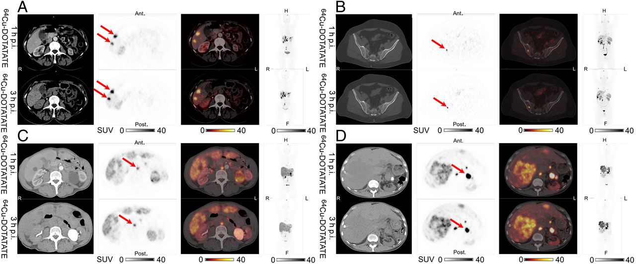
Representative examples of liver lesions (A), bone lesions (B), lymph node lesions (C), and pancreatic lesions (D) in same patients scanned at 1 and 3 h after injection. From left to right are shown CT, 64Cu-DOTATATE PET, 64Cu-DOTATATE PET/CT, and maximum-intensity projection, with corresponding SUV color bars below. All lesions were identified at both 1 h and 3 h on 64Cu-DOTATATE PET (arrows). Ant = anterior; F = feet; H = head; p.i. = after injection; post = posterior.
Lesions per Organ or Region
Discordant Lesions per Patient

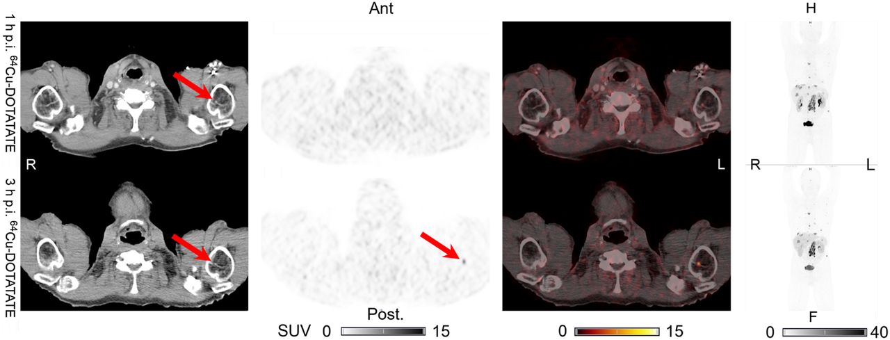
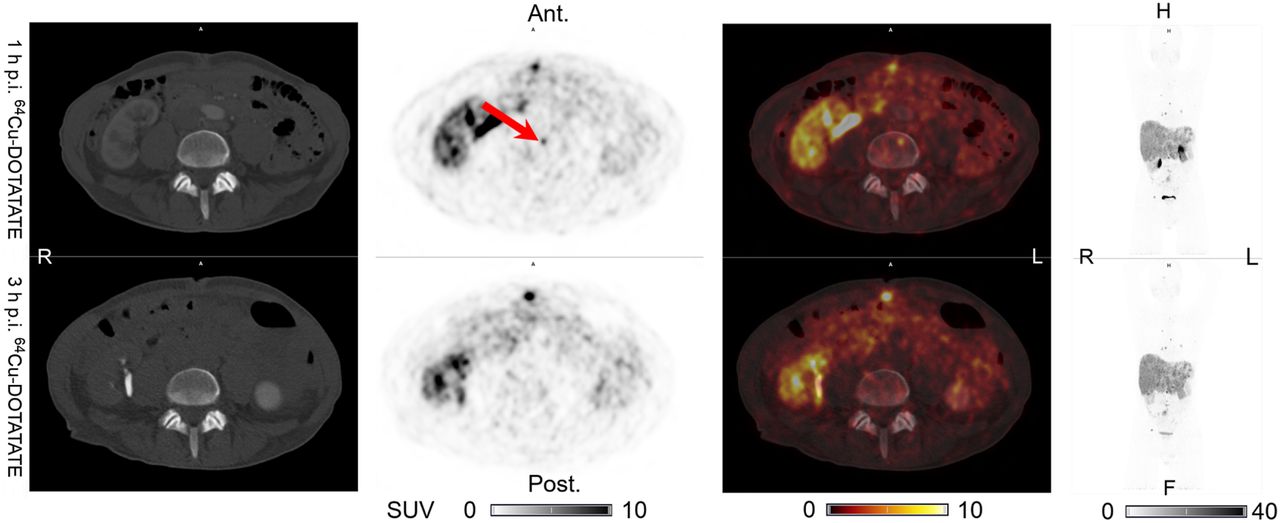
True-positive finding in patient 1: one additional lymph node lesion (arrows) visible on 64Cu-DOTATATE PET at 3 h after injection but not at 1 h, with visible CT correlate. Lesion was also visible on PET and CT at 1 h after injection on 64Cu-DOTATATE PET/CT performed 9 mo later. From left to right are shown CT, 64Cu-DOTATATE PET, 64Cu-DOTATATE PET/CT, and maximum-intensity projection, with corresponding SUV color bars below. Ant = anterior; F = feet; H = head; p.i. = after injection; post = posterior.

True-positive finding in patient 4: one additional bone lesion (arrows) visible on 64Cu-DOTATATE PET at 3 h after injection but not at 1 h, with visible CT correlate. No later SSTR PET images were available. From left to right are shown CT, 64Cu-DOTATATE PET, 64Cu-DOTATATE PET/CT, and maximum-intensity projection, with corresponding SUV color bars below. Ant = anterior; F = feet; H = head; p.i. = after injection; post = posterior.

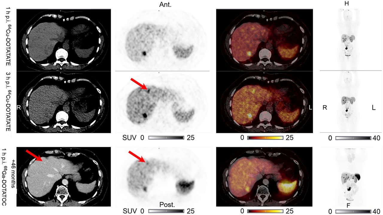
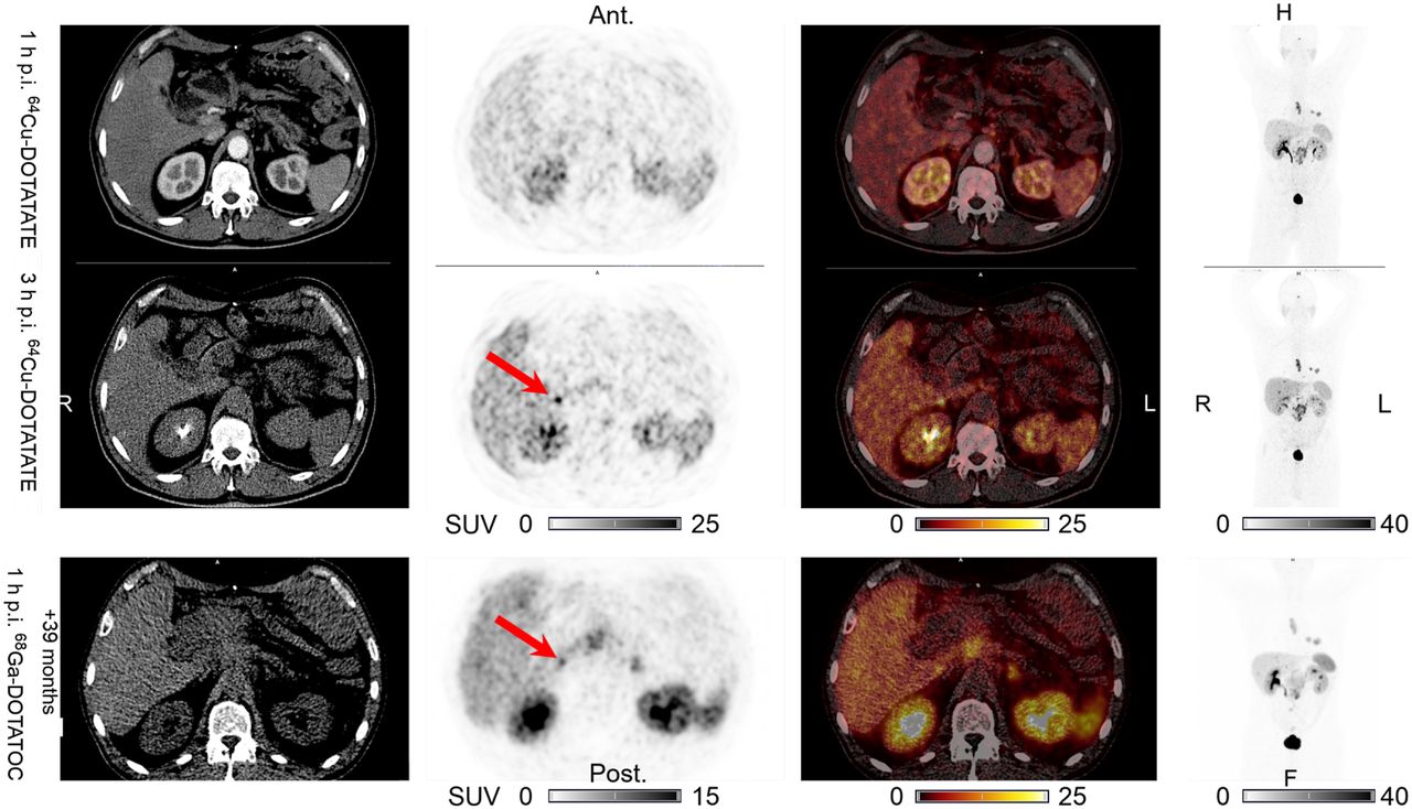
True-positive finding in patient 3: one additional liver lesion (arrows) visible on 64Cu-DOTATATE PET at 3 h after injection but not at 1 h, without CT correlate. This discordant lesion was also visible on PET and CT at 1 h after injection on 68Ga-DOTATOC PET performed 46 mo later. From left to right are shown CT, 64Cu-DOTATATE PET (68Ga-DOTATOC PET), 64Cu-DOTATATE PET/CT (68Ga-DOTATOC PET/CT), and maximum-intensity projection, with corresponding SUV color bars below. Ant = anterior; F = feet; H = head; p.i. = after injection; post = posterior.

True-positive finding in patient 5: one additional liver lesion (arrows) visible on 64Cu-DOTATATE PET at 3 h after injection but not at 1 h, with no visible CT correlate. This discordant lesion was also visible on PET without CT correlate at 1 h after injection on 68Ga-DOTATOC PET/CT performed 39 mo later. From left to right are shown CT, 64Cu-DOTATATE PET (68Ga-DOTATOC PET), 64Cu-DOTATATE PET/CT(68Ga-DOTATOC PET/CT), and maximum-intensity projection, with corresponding SUV color bars below. Ant = anterior; F = feet; H = head; p.i. = after injection; post = posterior.

False-positive finding in patient 2: one additional bone lesion (arrow) visible on 64Cu-DOTATATE PET at 1 h after injection but not at 3 h, without CT correlate. No later SSTR PET images were available. No visible CT correlate was seen on latest CT scan, 8 mo later (not shown). From left to right are shown CT, 64Cu-DOTATATE PET, 64Cu-DOTATATE PET/CT, and maximum-intensity projection, with corresponding SUV color bars below. Ant = anterior; F = feet; H = head; p.i. = after injection; post = posterior.
Quantitative Image Analysis
Lesion SUVmax divided into organs and regions is shown in Table 4, and the SUVmean of normal organs and tissues is shown in Table 5. Table 6 shows the lesion TTN in patients with both lesions and evaluable normal tissue in the corresponding organ or region.
Lesion SUVmax
Normal-Organ SUVmean
TTN
DISCUSSION
In this prospective study, we investigated the hypothesis that 64Cu-DOTATATE PET may be performed from 1 to 3 h after injection without loss of the lesion detection ability that is possible with 1-h imaging. Our main finding was that 64Cu-DOTATATE can be used for PET imaging of patients with NENs from 1 to 3 h after injection, with high TTN (lesion contrast) and a stable lesion detection rate and no significant differences in the number of lesions detected. In comparison with the fixed image acquisition 1 h after injection of 68Ga-labeled SSTR PET tracers the expansion of the acquisition time window of 64Cu-DOTATATE PET is convenient in daily clinical routine.
The high agreement in the number of lesions and organ systems detected on both 1-h and 3-h 64Cu-DOTATATE PET supports the rationale for expanding the imaging time window. The 2 additional true-positive liver lesions found on 3-h 64Cu-DOTATATE PET further suggests that in difficult cases with clinical suspicion of a small liver focus, it might be advantageous to perform late-phase 64Cu-DOTATATE PET at 3 h after injection if nothing is found on 1-h 64Cu-DOTATATE PET. Of the 5 discordant lesions, only 1 revealed—on the 3-h 64Cu-DOTATATE PET—involvement of an additional organ system (lymph node in patient 1) not seen on the 1-h PET. Since the patient had 11 concordant metastatic liver lesions, the patient’s disease stage would not have changed from stage IV as a consequence of the additional lymph node detected on 3-h 64Cu-DOTATATE PET (14). However, the finding of an additional extrahepatic lesion in a patient with liver-only lesions could potentially have an impact on clinical management. In the remaining 4 cases, the discordant findings involved lesions in already-concordant organ systems.
The relatively short follow-up between the 64Cu-DOTATATE PET/CT and the latest available CT for patient 2 (8 mo) may explain why the discordant bone lesion could not be confirmed. For 64Cu-DOTATATE (8,9), 68Ga-DOTATOC (15), and 68Ga-DOTATATE (16), bone lesions are detected on PET several months to years before detection on CT in some cases. Alternatively, the finding may represent an artifact on 1-h 64Cu-DOTATATE PET. Importantly, the patient also had several concordant bone lesions. The clinical consequences of the false-positive findings are very limited.
It has been questioned whether the 64Cu-DOTATATE liver uptake could present a challenge for detection of liver lesions. Although a numeric increase in the mean liver SUVmean on the 3-h, compared with the 1-h, 64Cu-DOTATATE PET was seen, the median liver SUVmean for 64Cu-DOTATATE in our study (3.86 at 1 h and 5.52 at 3 h after injection) was still lower at both time points than what has been reported for another 64Cu-labeled SSTR PET tracer, 64Cu-MeCOSar-Tyr3-octreotate (7.10 at 1 h and 5.90 at 4 h after injection) (17). The mean liver SUVmean at both 1 h and 3 h for 64Cu-DOTATATE was also within the range of, or lower than, that reported for 68Ga-DOTATATE at 1 h after injection: 4.5 (18) to 7.2 (19). In addition, because the liver lesion SUVmax also increased from the 1-h to the 3-h 64Cu-DOTATATE PET, the mean liver TTN remained high and almost unchanged from 1 to 3 h after injection (12.6 vs. 11.0). Moreover, and importantly, the liver lesion detection ability at 3 h after injection was not affected, as 2 additional true liver lesions were identified on 3-h 64Cu-DOTATATE PET. Although it is open to discussion whether the use of 2 different CT scans for attenuation correction for the 1- and 3-h 64Cu-DOTATATE PET could have contributed to differences in quantification at the 2 time points, we find this possibility unlikely to have had any clinical relevance (20,21).
It might be speculated whether differences in liver background and liver lesion uptake between the 1- and 3-h 64Cu-DOTATATE PET should be considered when 64Cu-DOTATATE PET is used to select patients for peptide receptor radionuclide therapy (PRRT). However, apart from a single bone lesion, all lesions in all patients were above liver background uptake both at 1 h and at 3 h after injection. Therefore, if a modified Krenning scale had been applied, the conclusions would be similar whether based on 1-h or 3-h 64Cu-DOTATATE PET. However, if more quantitative cutoffs are established for guiding PRRT, they are likely to be both PET tracer–specific and scan time–specific.
The high TTNs measured on both 1-h and 3-h 64Cu-DOTATATE PET throughout the different organs and regions suggest that tumor delineation from the surroundings remains sufficient in the 1- to 3-h time window. Combined with the high concordance in the number of lesions detected on both 1-h and 3-h 64Cu-DOTATATE PET, this observation supports the conclusion that no clinically relevant loss in image information occurs within the 1- to 3-h imaging window.
CONCLUSION
64Cu-DOTATATE PET/CT has excellent performance from 1 to 3 h after injection for imaging patients with NENs, with no significant differences in the number of lesions detected. The maintained lesion detection rate and high contrast from 1 to 3 h, combined with the 24-h shelf life, adds to the convenience and flexibility of using 64Cu-DOTATATE PET for routine imaging of patients with NENs.
DISCLOSURE
This project received funding from the European Union’s Horizon 2020 research and innovation program under grants 670261 (ERC Advanced Grant) and 668532 (Click-It) and from the Lundbeck Foundation, the Novo Nordisk Foundation, the Innovation Fund Denmark, the Danish Cancer Society, the Arvid Nilsson Foundation, the Svend Andersen Foundation, the Neye Foundation, the Research Foundation of Rigshospitalet, the Danish National Research Foundation (grant 126), the Research Council of the Capital Region of Denmark, the Danish Health Authority, the John and Birthe Meyer Foundation, and the Research Council for Independent Research. Andreas Kjaer and Ulrich Knigge are inventors on patents or applications covering PET tracers for imaging of neuroendocrine tumors. No other potential conflict of interest relevant to this article was reported.
KEY POINTS
QUESTION: Can the imaging time window for 64Cu-DOTATATE PET be expanded from 1 h to 1–3 h after injection in patients with NENs?
PERTINENT FINDINGS: 64Cu-DOTATATE PET/CT has excellent performance, with high TTN and conserved lesion detection ability, from 1 to 3 h after injection in patients with NENs.
IMPLICATIONS FOR PATIENT CARE: The imaging time window can be expanded from 1 h to 1–3 h after injection, thus adding to the convenience and flexibility of using 64Cu-DOTATATE PET for routine imaging in patients with NENs.
Acknowledgments
We are grateful to the staff at the Department of Clinical Physiology, Nuclear Medicine, and PET for help with performing the PET/CT studies.
Footnotes
Published online May 22, 2020.
- © 2021 by the Society of Nuclear Medicine and Molecular Imaging.
REFERENCES
- Received for publication March 6, 2020.
- Accepted for publication April 24, 2020.


Jump to section
Related Articles
Cited By...
- Efficacy of [67Cu]Cu-EB-TATE Theranostic Against Somatostatin Receptor Subtype-2-Positive Neuroendocrine Tumors
- An Investigation of Lesion Detection Accuracy for Artificial Intelligence-Based Denoising of Low-Dose 64Cu-DOTATATE PET Imaging in Patients with Neuroendocrine Neoplasms
- An Investigation of Lesion Detection Accuracy for Artificial Intelligence-Based Denoising of Low-Dose 64Cu-DOTATATE PET Imaging in Patients with Neuroendocrine Neoplasms
- SNMMI Procedure Standard/EANM Practice Guideline for SSTR PET: Imaging Neuroendocrine Tumors
- Semiautomatic Tumor Delineation for Evaluation of 64Cu-DOTATATE PET/CT in Patients with Neuroendocrine Neoplasms: Prognostication Based on Lowest Lesion Uptake and Total Tumor Volume
- Somatostatin Receptor Imaging and Theranostics: Current Practice and Future Prospects