
Main menu

  • Home
  • Content
    • Current
    • Ahead of print
    • Past Issues
    • JNM Supplement
    • SNMMI Annual Meeting Abstracts
  • Subscriptions
    • Subscribers
    • Institutional and Non-member
    • Rates
    • Corporate & Special Sales
    • Journal Claims
  • Authors
    • Submit to JNM
    • Information for Authors
    • Assignment of Copyright
    • AQARA requirements
  • Info
    • Permissions
    • Advertisers
    • Continuing Education
  • About
    • About Us
    • Editorial Board
    • Contact Information
  • More
    • Alerts
    • Feedback
    • Help
    • SNMMI Journals
  • SNMMI
    • JNM
    • JNMT
    • SNMMI Journals
    • SNMMI

User menu

  • Subscribe
  • My alerts
  • Log in
  • My Cart

Search

  • Advanced search
Journal of Nuclear Medicine
  • SNMMI
    • JNM
    • JNMT
    • SNMMI Journals
    • SNMMI
  • Subscribe
  • My alerts
  • Log in
  • My Cart
Journal of Nuclear Medicine

Advanced Search

  • Home
  • Content
    • Current
    • Ahead of print
    • Past Issues
    • JNM Supplement
    • SNMMI Annual Meeting Abstracts
  • Subscriptions
    • Subscribers
    • Institutional and Non-member
    • Rates
    • Corporate & Special Sales
    • Journal Claims
  • Authors
    • Submit to JNM
    • Information for Authors
    • Assignment of Copyright
    • AQARA requirements
  • Info
    • Permissions
    • Advertisers
    • Continuing Education
  • About
    • About Us
    • Editorial Board
    • Contact Information
  • More
    • Alerts
    • Feedback
    • Help
    • SNMMI Journals
  • Follow JNM on Twitter
  • Visit JNM on Facebook
  • Join JNM on LinkedIn
  • Subscribe to our RSS feeds
Meeting ReportPhysics, Instrumentation & Data Sciences

Effect of Reconstruction Settings on PET-Derived Dose-Volume-Histograms after SIRT of the Liver

Marc Roschlein, Theresa Gotz, Christian Schmidkonz, Michael Beck, Torsten Kuwert and Philipp Ritt
Journal of Nuclear Medicine May 2020, 61 (supplement 1) 448;
Marc Roschlein
1Clinic of Nuclear Medicine University Hospital Erlangen Germany
  • Find this author on Google Scholar
  • Find this author on PubMed
  • Search for this author on this site
Theresa Gotz
2Pattern Recognition Lab Friedrich-Alexander-University Erlangen-Nürnberg Erlangen Germany
1Clinic of Nuclear Medicine University Hospital Erlangen Germany
  • Find this author on Google Scholar
  • Find this author on PubMed
  • Search for this author on this site
Christian Schmidkonz
1Clinic of Nuclear Medicine University Hospital Erlangen Germany
  • Find this author on Google Scholar
  • Find this author on PubMed
  • Search for this author on this site
Michael Beck
1Clinic of Nuclear Medicine University Hospital Erlangen Germany
  • Find this author on Google Scholar
  • Find this author on PubMed
  • Search for this author on this site
Torsten Kuwert
1Clinic of Nuclear Medicine University Hospital Erlangen Germany
  • Find this author on Google Scholar
  • Find this author on PubMed
  • Search for this author on this site
Philipp Ritt
1Clinic of Nuclear Medicine University Hospital Erlangen Germany
  • Find this author on Google Scholar
  • Find this author on PubMed
  • Search for this author on this site
  • Article
  • Figures & Data
  • Info & Metrics
Loading

Abstract

448

Aim: PET/CT allows for accurate, post-therapeutic determination of the Y-90 distribution, which can be converted to maps of absorbed dose [1]. Yet, several parameters affect the accuracy of activity- and dose-maps. The aim of this work was to determine the effects of varying PET reconstruction parameters onto absorbed dose maps.

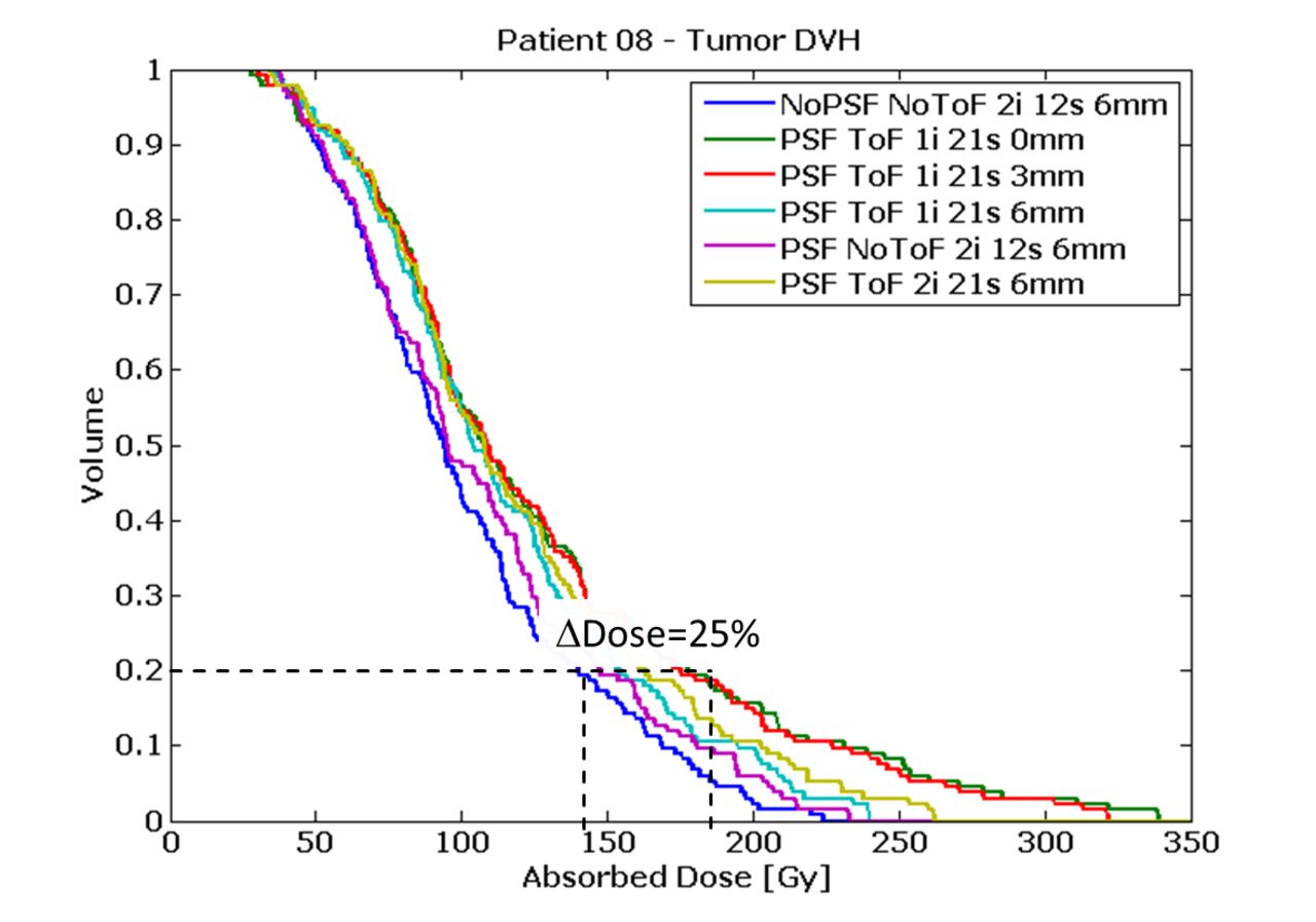
Material and Methods: After SIRT, a PET/CT scan (Siemens Biograph mCT 40, one bed-position, 30 minutes duration) was carried out in 10 patients in order to determine Y-90-resin microsphere distribution. From reconstructed PET data, dose-maps where derived by convolution with pre-calculated dose-voxel-kernels. An MR image was co-registered to PET/CT data and manually segmented for healthy liver (HL) and tumor (TU) regions, for which dose-volume-histograms (DVH) were calculated. DVHs were compared for six different PET reconstruction settings. All reconstructions had in common that corrections for decay, photon attenuation and scatter, and random coincidences were applied. The varied parameters were number of iterations, amount of post-reconstruction Gaussian smoothing, and whether point-spread-function (Siemens TrueX) was modelled or time-of-flight (ToF) information was applied. Evaluated reconstruction settings were: A) No PSF, No ToF, 2 iterations (i), 12 subsets (s), 6mm Gaussian B) PSF, No ToF, 2i, 12s, 6mm C) PSF, ToF, 1i, 21s, 0mm D) PSF, ToF, 1i, 21s, 3mm E) PSF, ToF, 1i, 21s, 6mm F) PSF, ToF, 2i, 21s, 6mm

Results: Reconstruction F) led to smallest deviations between injected and PET-derived activity (7.2±5.5%, ranging 0.5 - 18.8). For F), mean absorbed dose to HL was 26.8±9.2 Gy (15.3-40.1) and 81.8±48.2 Gy (29.2-157.5) for TU. Reconstructions A) and B) led to systematically lower (p<0.05) mean doses than C-F), for HL, as well as for TU. Mean dose differences between different reconstructions were on average 3.9±4.2 % (0.0-15.4) for HL and 6.1±5.5 % (0.0 - 22.4) for TU. Corresponding points on DVHs for different reconstructions showed higher deviations than in average case, especially in higher dose regions.

Conclusions: On general, the accuracy of PET-derived activity could be regarded as sufficient, although some outliers were found. The ToF-technique led to most accurate results. Differences in mean absorbed dose appeared to be minor. However, a detailed comparison of DVHs indicated that choice of reconstruction parameters has a strong influence on exact DVHs shape. References: D'Arienzo, M., et al. (2017). "Phantom validation of quantitative Y-90 PET/CT-based dosimetry in liver radioembolization." EJNMMI Res 7(1): 94.

Figure
  • Download figure
  • Open in new tab
  • Download powerpoint
Previous
Back to top

In this issue

Journal of Nuclear Medicine
Vol. 61, Issue supplement 1
May 1, 2020
  • Table of Contents
  • Index by author
Article Alerts
Sign In to Email Alerts with your Email Address
Email Article

Thank you for your interest in spreading the word on Journal of Nuclear Medicine.

NOTE: We only request your email address so that the person you are recommending the page to knows that you wanted them to see it, and that it is not junk mail. We do not capture any email address.

Enter multiple addresses on separate lines or separate them with commas.
Effect of Reconstruction Settings on PET-Derived Dose-Volume-Histograms after SIRT of the Liver
(Your Name) has sent you a message from Journal of Nuclear Medicine
(Your Name) thought you would like to see the Journal of Nuclear Medicine web site.
Citation Tools
Effect of Reconstruction Settings on PET-Derived Dose-Volume-Histograms after SIRT of the Liver
Marc Roschlein, Theresa Gotz, Christian Schmidkonz, Michael Beck, Torsten Kuwert, Philipp Ritt
Journal of Nuclear Medicine May 2020, 61 (supplement 1) 448;

Citation Manager Formats

  • BibTeX
  • Bookends
  • EasyBib
  • EndNote (tagged)
  • EndNote 8 (xml)
  • Medlars
  • Mendeley
  • Papers
  • RefWorks Tagged
  • Ref Manager
  • RIS
  • Zotero
Share
Effect of Reconstruction Settings on PET-Derived Dose-Volume-Histograms after SIRT of the Liver
Marc Roschlein, Theresa Gotz, Christian Schmidkonz, Michael Beck, Torsten Kuwert, Philipp Ritt
Journal of Nuclear Medicine May 2020, 61 (supplement 1) 448;
del.icio.us logo Digg logo Reddit logo Twitter logo CiteULike logo Facebook logo Google logo Mendeley logo
  • Tweet Widget
  • Facebook Like
  • Google Plus One
Bookmark this article

Jump to section

  • Article
  • Figures & Data
  • Info & Metrics

Related Articles

  • No related articles found.
  • Google Scholar

Cited By...

  • No citing articles found.
  • Google Scholar

More in this TOC Section

Physics, Instrumentation & Data Sciences

  • 3D Structural Convolutional Sparse Coding for PET Image Reconstruction
  • Exploration of Multi-objective Optimization with Genetic Algorithms for PET Image Reconstruction
  • AI-based methods for nuclear-medicine imaging: Need for objective task-specific evaluation
Show more Physics, Instrumentation & Data Sciences

Assessment of Image Quality and Quantitative Accuracy

  • Quantifying rod visibility in standardized SPECT quality assurance images
  • Image texture analysis quantification of concentric ring artifacts in quality assurance scans
  • Characterization of Partial Volume Errors in Coronary Plaque PET Imaging
Show more Assessment of Image Quality and Quantitative Accuracy

Similar Articles

SNMMI

© 2023 Journal of Nuclear Medicine

Powered by HighWire