
Main menu

  • Home
  • Content
    • Current
    • Ahead of print
    • Past Issues
    • JNM Supplement
    • SNMMI Annual Meeting Abstracts
  • Subscriptions
    • Subscribers
    • Institutional and Non-member
    • Rates
    • Corporate & Special Sales
    • Journal Claims
  • Authors
    • Submit to JNM
    • Information for Authors
    • Assignment of Copyright
    • AQARA requirements
  • Info
    • Continuing Education
    • Reviewers
    • Permissions
    • Advertisers
  • About
    • About Us
    • Editorial Board
    • Contact Information
  • More
    • Alerts
    • Feedback
    • Help
    • SNMMI Journals
  • SNMMI
    • JNM
    • JNMT
    • SNMMI Journals
    • SNMMI

User menu

  • Subscribe
  • My alerts
  • Log in
  • My Cart

Search

  • Advanced search
Journal of Nuclear Medicine
  • SNMMI
    • JNM
    • JNMT
    • SNMMI Journals
    • SNMMI
  • Subscribe
  • My alerts
  • Log in
  • My Cart
Journal of Nuclear Medicine

Advanced Search

  • Home
  • Content
    • Current
    • Ahead of print
    • Past Issues
    • JNM Supplement
    • SNMMI Annual Meeting Abstracts
  • Subscriptions
    • Subscribers
    • Institutional and Non-member
    • Rates
    • Corporate & Special Sales
    • Journal Claims
  • Authors
    • Submit to JNM
    • Information for Authors
    • Assignment of Copyright
    • AQARA requirements
  • Info
    • Continuing Education
    • Reviewers
    • Permissions
    • Advertisers
  • About
    • About Us
    • Editorial Board
    • Contact Information
  • More
    • Alerts
    • Feedback
    • Help
    • SNMMI Journals
  • Follow JNM on Twitter
  • Visit JNM on Facebook
  • Join JNM on LinkedIn
  • Subscribe to our RSS feeds
Meeting ReportPhysics, Instrumentation & Data Sciences

PET motion correction using real-time subspace-based MR imaging

Thibault Marin, Yanis Djebra, Paul Han, Isabelle Bloch, Georges El Fakhri, Jinsong Ouyang, Yoann Petibon and Chao Ma
Journal of Nuclear Medicine May 2020, 61 (supplement 1) 373;
Thibault Marin
2Massachusetts General Hospital Boston MA United States
5Massachusetts General Hospital Boston MA United States
1Harvard Medical School Boston MA United States
  • Find this author on Google Scholar
  • Find this author on PubMed
  • Search for this author on this site
Yanis Djebra
2Massachusetts General Hospital Boston MA United States
5Massachusetts General Hospital Boston MA United States
3LTCI, Télécom Paris, Institut Polytechnique de Paris Palaiseau France
  • Find this author on Google Scholar
  • Find this author on PubMed
  • Search for this author on this site
Paul Han
1Harvard Medical School Boston MA United States
2Massachusetts General Hospital Boston MA United States
5Massachusetts General Hospital Boston MA United States
  • Find this author on Google Scholar
  • Find this author on PubMed
  • Search for this author on this site
Isabelle Bloch
4LTCI, Telecom Paris Palaiseau France
  • Find this author on Google Scholar
  • Find this author on PubMed
  • Search for this author on this site
Georges El Fakhri
1Harvard Medical School Boston MA United States
2Massachusetts General Hospital Boston MA United States
5Massachusetts General Hospital Boston MA United States
  • Find this author on Google Scholar
  • Find this author on PubMed
  • Search for this author on this site
Jinsong Ouyang
1Harvard Medical School Boston MA United States
2Massachusetts General Hospital Boston MA United States
5Massachusetts General Hospital Boston MA United States
  • Find this author on Google Scholar
  • Find this author on PubMed
  • Search for this author on this site
Yoann Petibon
2Massachusetts General Hospital Boston MA United States
5Massachusetts General Hospital Boston MA United States
1Harvard Medical School Boston MA United States
  • Find this author on Google Scholar
  • Find this author on PubMed
  • Search for this author on this site
Chao Ma
2Massachusetts General Hospital Boston MA United States
5Massachusetts General Hospital Boston MA United States
1Harvard Medical School Boston MA United States
  • Find this author on Google Scholar
  • Find this author on PubMed
  • Search for this author on this site
  • Article
  • Figures & Data
  • Info & Metrics
Loading

Abstract

373

Introduction: Image quality of PET reconstructions is degraded when motion occurs during the acquisition. Sources of motion include cardiac, respiratory and bulk motion. PET/MR offers a unique opportunity to estimate the motion using MR and correct for it during PET reconstruction. The conventional MR-based motion correction methods first group the k-space MR data into different motion phases (or bins) using navigators or physio signals and then reconstruct images at each motion phase for motion field estimation. However, the binning process assumes periodic motions and could fail when bulk or irregular respiratory/cardiac motion occurs. In this work, we propose to reconstruct dynamic MR images at high frame rate (100 ms/frame) from sparsely sampled (k,t)-space data for motion field measurement in real time. Our method is enabled by a subspace-based MR data acquisition and image reconstruction method.

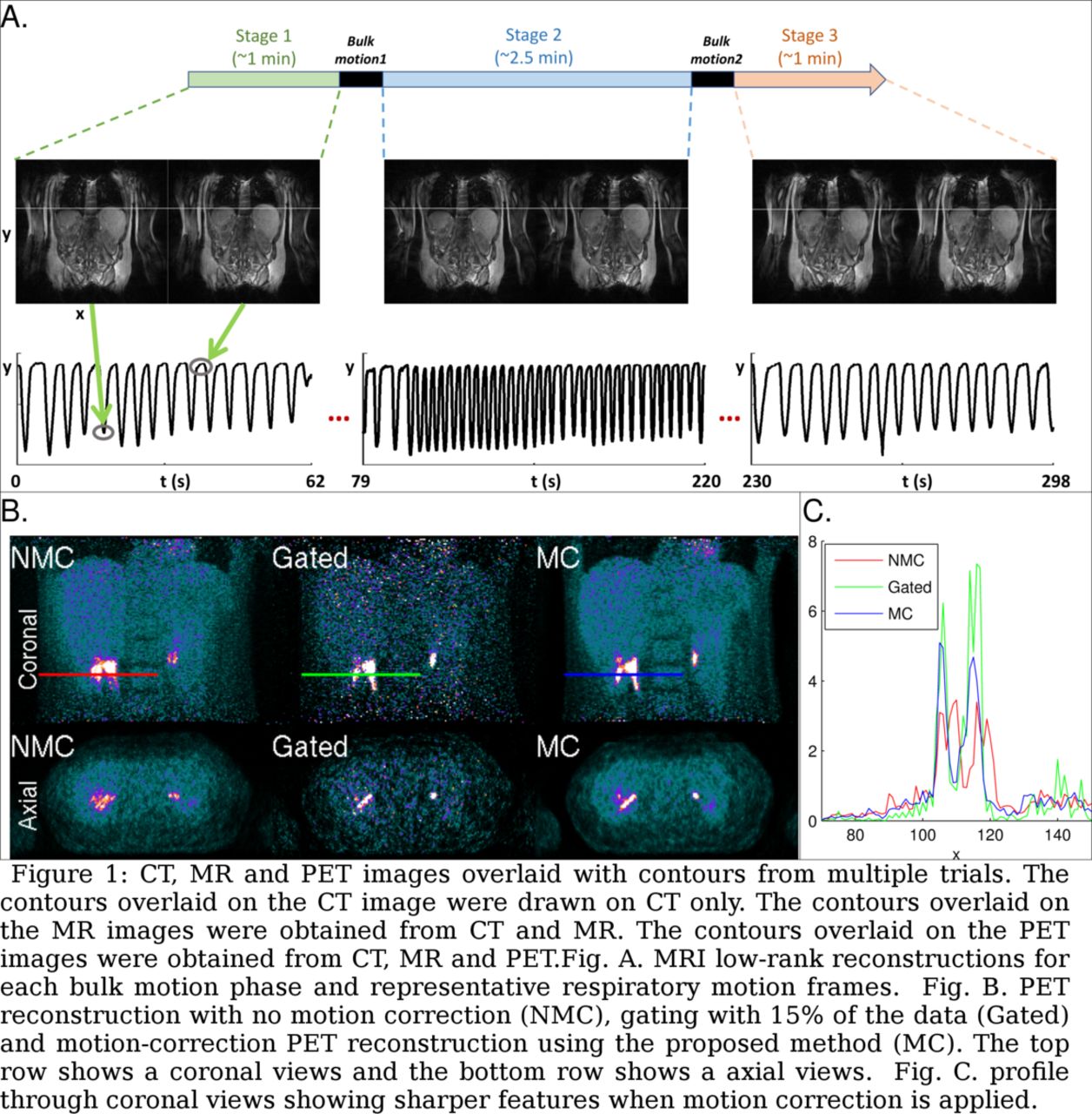
Methods: To evaluate the effect of the proposed MR-based motion correction on PET image quality, an 18F-FDG-PET/MR scan was performed on a healthy subject using a PET/MR scanner (Siemens Biograph mMR). The acquisition time was 5 minutes starting 30 minutes after injection. To assess the effect of both respiratory and bulk motion, the subject was instructed to move twice during the scan. MR acquisition was performed using a spoiled gradient-recalled echo (GRE) sequence (TR/TE = 3/1.6 ms and 7 degree flip angle) with stack-of-stars radial sampling scheme. MR data was acquired with frame rate of 100 ms per frame, consisting of 3 lines for training and the rest for imaging. The spatial resolution was 1.9 mm in-plane and 5 mm through-plane (32 slices). Acquisition with the vendor-provided standard two-point Dixon sequence was performed for attenuation correction. MRI reconstruction from highly undersampled k-space data was performed using a subspace-based image model that leverages a unique property of dynamic MR images, known as partial separability [1]. Iterative reconstruction used ADMM [2] to minimize a cost function composed of a data fidelity term along with a low-rank and spatio-temporal TV penalty. Binning of the MR images was then performed retrospectively from the reconstructed dynamic MR images by first delimiting the bulk motion frames and tracking the position of the tip of the liver for each phase of the bulk motion. A total of 18 bins (6 bins for each of the 3 patient positions) was used. Motion estimation was performed on the binned MR images using B-spline deformation. Finally, PET reconstruction was performed on binned sinograms using the OSEM algorithm [3]. The imaging model for the PET acquisition included motion via a warping operator from the reference bin to each motion bin. 5 iterations and 12 subsets were used for the OSEM algorithm. No scatter correction was performed.

Results: Reconstructed MR images for different phases of the bulk and respiratory motion are shown on Fig.A along with the estimated liver position. Coronal and axial slices through the reconstructed PET volume are shown on Fig.B. The figure shows reconstructions without motion correction (NMC), using gating with 15% of the PET data (Gated) and with motion correction estimated from MRI (MC). The proposed method reduces motion blur artifacts as seen visually from Figs.B and C. Finally, the contrast to noise ratio was computed between the kidney and the surrounding background. The proposed MC method improves the CNR by a factor 2 over NMC and 4 over the Gated reconstruction.

Conclusions: The proposed MR acquisition and reconstruction method recovers high quality MR images in real-time (100 ms per frame) able to capture all types of patient motion. When used for motion correction in PET reconstruction, results demonstrate suppression of motion artifacts. Research support: T32EB013180, R01CA165221, R01HL118261, R21MH121812, R01HL137230 and P41EB022544

Figure
  • Download figure
  • Open in new tab
  • Download powerpoint
Previous
Back to top

In this issue

Journal of Nuclear Medicine
Vol. 61, Issue supplement 1
May 1, 2020
  • Table of Contents
  • Index by author
Article Alerts
Sign In to Email Alerts with your Email Address
Email Article

Thank you for your interest in spreading the word on Journal of Nuclear Medicine.

NOTE: We only request your email address so that the person you are recommending the page to knows that you wanted them to see it, and that it is not junk mail. We do not capture any email address.

Enter multiple addresses on separate lines or separate them with commas.
PET motion correction using real-time subspace-based MR imaging
(Your Name) has sent you a message from Journal of Nuclear Medicine
(Your Name) thought you would like to see the Journal of Nuclear Medicine web site.
Citation Tools
PET motion correction using real-time subspace-based MR imaging
Thibault Marin, Yanis Djebra, Paul Han, Isabelle Bloch, Georges El Fakhri, Jinsong Ouyang, Yoann Petibon, Chao Ma
Journal of Nuclear Medicine May 2020, 61 (supplement 1) 373;

Citation Manager Formats

  • BibTeX
  • Bookends
  • EasyBib
  • EndNote (tagged)
  • EndNote 8 (xml)
  • Medlars
  • Mendeley
  • Papers
  • RefWorks Tagged
  • Ref Manager
  • RIS
  • Zotero
Share
PET motion correction using real-time subspace-based MR imaging
Thibault Marin, Yanis Djebra, Paul Han, Isabelle Bloch, Georges El Fakhri, Jinsong Ouyang, Yoann Petibon, Chao Ma
Journal of Nuclear Medicine May 2020, 61 (supplement 1) 373;
Twitter logo Facebook logo LinkedIn logo Mendeley logo
  • Tweet Widget
  • Facebook Like
  • Google Plus One
Bookmark this article

Jump to section

  • Article
  • Figures & Data
  • Info & Metrics

Related Articles

  • No related articles found.
  • Google Scholar

Cited By...

  • No citing articles found.
  • Google Scholar

More in this TOC Section

Physics, Instrumentation & Data Sciences

  • 3D Structural Convolutional Sparse Coding for PET Image Reconstruction
  • Exploration of Multi-objective Optimization with Genetic Algorithms for PET Image Reconstruction
  • AI-based methods for nuclear-medicine imaging: Need for objective task-specific evaluation
Show more Physics, Instrumentation & Data Sciences

Motion Correction PET

  • Optical facial feature-based motion tracking and correction method for PET brain studies
  • Impact of low coincidence count density on Quantification when using Elastic Motion Compensation in PET/CT imaging
  • Data-driven respiratory gating for the uEXPLORER with fast dynamics
Show more Motion Correction PET

Similar Articles

SNMMI

© 2023 Journal of Nuclear Medicine

Powered by HighWire