
Main menu

  • Home
  • Content
    • Current
    • Ahead of print
    • Past Issues
    • JNM Supplement
    • SNMMI Annual Meeting Abstracts
  • Subscriptions
    • Subscribers
    • Institutional and Non-member
    • Rates
    • Corporate & Special Sales
    • Journal Claims
  • Authors
    • Submit to JNM
    • Information for Authors
    • Assignment of Copyright
    • AQARA requirements
  • Info
    • Permissions
    • Advertisers
    • Continuing Education
  • About
    • About Us
    • Editorial Board
    • Contact Information
  • More
    • Alerts
    • Feedback
    • Help
    • SNMMI Journals
  • SNMMI
    • JNM
    • JNMT
    • SNMMI Journals
    • SNMMI

User menu

  • Subscribe
  • My alerts
  • Log in
  • My Cart

Search

  • Advanced search
Journal of Nuclear Medicine
  • SNMMI
    • JNM
    • JNMT
    • SNMMI Journals
    • SNMMI
  • Subscribe
  • My alerts
  • Log in
  • My Cart
Journal of Nuclear Medicine

Advanced Search

  • Home
  • Content
    • Current
    • Ahead of print
    • Past Issues
    • JNM Supplement
    • SNMMI Annual Meeting Abstracts
  • Subscriptions
    • Subscribers
    • Institutional and Non-member
    • Rates
    • Corporate & Special Sales
    • Journal Claims
  • Authors
    • Submit to JNM
    • Information for Authors
    • Assignment of Copyright
    • AQARA requirements
  • Info
    • Permissions
    • Advertisers
    • Continuing Education
  • About
    • About Us
    • Editorial Board
    • Contact Information
  • More
    • Alerts
    • Feedback
    • Help
    • SNMMI Journals
  • Follow JNM on Twitter
  • Visit JNM on Facebook
  • Join JNM on LinkedIn
  • Subscribe to our RSS feeds
Meeting ReportPhysics, Instrumentation & Data Sciences

10-fold reduction of scan time with deep learning reconstruction of Coronary PET images

Martin Lyngby Lassen, Frederic Commandeur, Jacek Kwiecinski, Damini Dey, Sebastien Cadet, Guido Germano, Daniel Berman, Marc Dweck, David Newby and Piotr Slomka
Journal of Nuclear Medicine May 2019, 60 (supplement 1) 244;
Martin Lyngby Lassen
1Cedars-Sinai Medical Center Los Angeles CA United States
2Cedars-Sinai Medical Center Los Angeles CA United States
  • Find this author on Google Scholar
  • Find this author on PubMed
  • Search for this author on this site
Frederic Commandeur
1Cedars-Sinai Medical Center Los Angeles CA United States
2Cedars-Sinai Medical Center Los Angeles CA United States
  • Find this author on Google Scholar
  • Find this author on PubMed
  • Search for this author on this site
Jacek Kwiecinski
1Cedars-Sinai Medical Center Los Angeles CA United States
2Cedars-Sinai Medical Center Los Angeles CA United States
  • Find this author on Google Scholar
  • Find this author on PubMed
  • Search for this author on this site
Damini Dey
1Cedars-Sinai Medical Center Los Angeles CA United States
2Cedars-Sinai Medical Center Los Angeles CA United States
  • Find this author on Google Scholar
  • Find this author on PubMed
  • Search for this author on this site
Sebastien Cadet
1Cedars-Sinai Medical Center Los Angeles CA United States
2Cedars-Sinai Medical Center Los Angeles CA United States
  • Find this author on Google Scholar
  • Find this author on PubMed
  • Search for this author on this site
Guido Germano
1Cedars-Sinai Medical Center Los Angeles CA United States
2Cedars-Sinai Medical Center Los Angeles CA United States
  • Find this author on Google Scholar
  • Find this author on PubMed
  • Search for this author on this site
Daniel Berman
1Cedars-Sinai Medical Center Los Angeles CA United States
2Cedars-Sinai Medical Center Los Angeles CA United States
  • Find this author on Google Scholar
  • Find this author on PubMed
  • Search for this author on this site
Marc Dweck
3University of Edinburgh Edinburgh United Kingdom
  • Find this author on Google Scholar
  • Find this author on PubMed
  • Search for this author on this site
David Newby
3University of Edinburgh Edinburgh United Kingdom
  • Find this author on Google Scholar
  • Find this author on PubMed
  • Search for this author on this site
Piotr Slomka
1Cedars-Sinai Medical Center Los Angeles CA United States
2Cedars-Sinai Medical Center Los Angeles CA United States
  • Find this author on Google Scholar
  • Find this author on PubMed
  • Search for this author on this site
  • Article
  • Figures & Data
  • Info & Metrics
Loading

Abstract

244

Introduction: Coronary plaque 18F-Sodium Fluoride (18F-NaF) images need to be acquired over long time (up to 30 minutes) to obtain diagnostic image quality due to low signal and noise in the images but this is inconvenient to patient, decreases patient throughput and can result in patient motion. In this study we investigate the feasibility of reconstructing images equivalent to 30 min 18F-NaF-PET acquisitions using 1/10th of the acquired counts using deep learning (DL) computer techniques.

Methods: To train the DL model, we included 132 patients recruited for 30 min long 18F-NaF coronary plaque 250 MBq PET imaging in a hybrid PET/CT Angiography (CTA) protocol. Two image sets were reconstructed for each scan, a static image using all the data (Full-dose) and an image set using 10% of the acquired PET counts (Low-dose) using list mode data. The DL model (U-net) was trained to predict the Full-dose scan from the Low-dose scans slice by slice (creating third image dataset - Predicted High Dose), using 10-fold cross validation for parameter optimization. Lesion uptake was quantified from spherical Volume of Interest (VOI) (radius=5mm), while background activities were obtained in the right atrium using a cylindrical VOI (length=15mm, radius=10mm). Lesions with target-to-background ratios (TBR) ≥1.25 were considered 18F-NaF-active, while TBR<1.25 were considered 18F-NaF-negative. All lesions were classified from the Full-dose data sets. A separate test group consisting of 20 patients with known coronary artery disease were analyzed in this study with the DL reconstruction. We report the number of coronary lesions as well as the signal-to-noise ratios (SNR) obtained for the three datasets (Full-dose, Low-dose and Predicted-High-Dose).

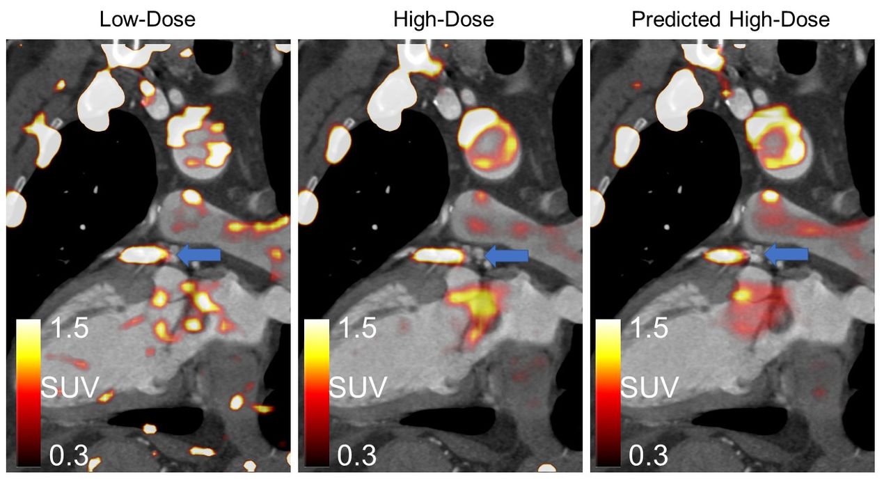
Results: A total of 55 coronary lesions (25 18F-NaF-positive) were identified on the 20 Full dose scans. The Low-dose datasets had significantly lower SNR (8.9±4.8), in the lesions compared to Full-dose data (19.5±12.8), p<0.001, while no difference were observed for the Predicted-High-Dose SNR (16.1±12.1, p=0.14). Figure shows examples of High-Dose, Low-Dose and Predicted-High-Dose reconstructions.

Conclusions: Using DL, it is possible to reconstruct images from 3-minute scan data with SNR equivalent to 30 min long acquisitions. This technique may help reducing the long acquisition times in current coronary PET clinical studies.

Figure
  • Download figure
  • Open in new tab
  • Download powerpoint
Previous
Back to top

In this issue

Journal of Nuclear Medicine
Vol. 60, Issue supplement 1
May 1, 2019
  • Table of Contents
  • Index by author
Article Alerts
Sign In to Email Alerts with your Email Address
Email Article

Thank you for your interest in spreading the word on Journal of Nuclear Medicine.

NOTE: We only request your email address so that the person you are recommending the page to knows that you wanted them to see it, and that it is not junk mail. We do not capture any email address.

Enter multiple addresses on separate lines or separate them with commas.
10-fold reduction of scan time with deep learning reconstruction of Coronary PET images
(Your Name) has sent you a message from Journal of Nuclear Medicine
(Your Name) thought you would like to see the Journal of Nuclear Medicine web site.
Citation Tools
10-fold reduction of scan time with deep learning reconstruction of Coronary PET images
Martin Lyngby Lassen, Frederic Commandeur, Jacek Kwiecinski, Damini Dey, Sebastien Cadet, Guido Germano, Daniel Berman, Marc Dweck, David Newby, Piotr Slomka
Journal of Nuclear Medicine May 2019, 60 (supplement 1) 244;

Citation Manager Formats

  • BibTeX
  • Bookends
  • EasyBib
  • EndNote (tagged)
  • EndNote 8 (xml)
  • Medlars
  • Mendeley
  • Papers
  • RefWorks Tagged
  • Ref Manager
  • RIS
  • Zotero
Share
10-fold reduction of scan time with deep learning reconstruction of Coronary PET images
Martin Lyngby Lassen, Frederic Commandeur, Jacek Kwiecinski, Damini Dey, Sebastien Cadet, Guido Germano, Daniel Berman, Marc Dweck, David Newby, Piotr Slomka
Journal of Nuclear Medicine May 2019, 60 (supplement 1) 244;
del.icio.us logo Digg logo Reddit logo Twitter logo CiteULike logo Facebook logo Google logo Mendeley logo
  • Tweet Widget
  • Facebook Like
  • Google Plus One
Bookmark this article

Jump to section

  • Article
  • Figures & Data
  • Info & Metrics

Related Articles

  • No related articles found.
  • Google Scholar

Cited By...

  • No citing articles found.
  • Google Scholar

More in this TOC Section

Physics, Instrumentation & Data Sciences

  • Exploration of Multi-objective Optimization with Genetic Algorithms for PET Image Reconstruction
  • AI-based methods for nuclear-medicine imaging: Need for objective task-specific evaluation
  • Ultra-Fast Reconstruction of Short List-Mode PET Data Frames for Real-Time Visualization and Processing
Show more Physics, Instrumentation & Data Sciences

Image Generation: PET Image Quality

  • Image Quality evaluation of analytic reconstruction algorithms for long axial field-of-view PET scanner
  • Evaluation of Joint Reconstruction of Activity and Attenuation in Time-of-Flight PET
Show more Image Generation: PET Image Quality

Similar Articles

SNMMI

© 2023 Journal of Nuclear Medicine

Powered by HighWire