
Main menu

  • Home
  • Content
    • Current
    • Ahead of print
    • Past Issues
    • JNM Supplement
    • SNMMI Annual Meeting Abstracts
  • Subscriptions
    • Subscribers
    • Institutional and Non-member
    • Rates
    • Corporate & Special Sales
    • Journal Claims
  • Authors
    • Submit to JNM
    • Information for Authors
    • Assignment of Copyright
    • AQARA requirements
  • Info
    • Permissions
    • Advertisers
    • Continuing Education
  • About
    • About Us
    • Editorial Board
    • Contact Information
  • More
    • Alerts
    • Feedback
    • Help
    • SNMMI Journals
  • SNMMI
    • JNM
    • JNMT
    • SNMMI Journals
    • SNMMI

User menu

  • Subscribe
  • My alerts
  • Log in
  • My Cart

Search

  • Advanced search
Journal of Nuclear Medicine
  • SNMMI
    • JNM
    • JNMT
    • SNMMI Journals
    • SNMMI
  • Subscribe
  • My alerts
  • Log in
  • My Cart
Journal of Nuclear Medicine

Advanced Search

  • Home
  • Content
    • Current
    • Ahead of print
    • Past Issues
    • JNM Supplement
    • SNMMI Annual Meeting Abstracts
  • Subscriptions
    • Subscribers
    • Institutional and Non-member
    • Rates
    • Corporate & Special Sales
    • Journal Claims
  • Authors
    • Submit to JNM
    • Information for Authors
    • Assignment of Copyright
    • AQARA requirements
  • Info
    • Permissions
    • Advertisers
    • Continuing Education
  • About
    • About Us
    • Editorial Board
    • Contact Information
  • More
    • Alerts
    • Feedback
    • Help
    • SNMMI Journals
  • Follow JNM on Twitter
  • Visit JNM on Facebook
  • Join JNM on LinkedIn
  • Subscribe to our RSS feeds
Meeting ReportOncology: Clinical Therapy and Diagnosis

Hepatic arterial tree segmentation: Towards patient-specific dosimetry for liver cancer radioembolization

Amirtaha Taebi, Bahman Roudsari, Catherine Vu, Simon Cherry and Emilie Roncali
Journal of Nuclear Medicine May 2019, 60 (supplement 1) 122;
Amirtaha Taebi
1Department of Biomedical Engineering University of California, Davis Davis CA United States
  • Find this author on Google Scholar
  • Find this author on PubMed
  • Search for this author on this site
Bahman Roudsari
2Department of Radiology University of California, Davis Davis CA United States
  • Find this author on Google Scholar
  • Find this author on PubMed
  • Search for this author on this site
Catherine Vu
2Department of Radiology University of California, Davis Davis CA United States
  • Find this author on Google Scholar
  • Find this author on PubMed
  • Search for this author on this site
Simon Cherry
2Department of Radiology University of California, Davis Davis CA United States
1Department of Biomedical Engineering University of California, Davis Davis CA United States
  • Find this author on Google Scholar
  • Find this author on PubMed
  • Search for this author on this site
Emilie Roncali
1Department of Biomedical Engineering University of California, Davis Davis CA United States
  • Find this author on Google Scholar
  • Find this author on PubMed
  • Search for this author on this site
  • Article
  • Figures & Data
  • Info & Metrics
Loading

Abstract

122

Objectives: Liver cancer is among the deadliest cancers and its mortality rate is increasing in the United States [1]. Yttrium-90 (Y-90) radioembolization is being increasingly used for the treatment of advanced liver cancer. Accurate pretreatment dosimetry is necessary to determine the Y-90 activity to inject in order to maximize the dose to the tumor while limiting the dose to surrounding healthy parenchyma. Current dosimetry methods are not accurate nor precise, because they do not consider the non-uniform Y-90 microsphere distribution in the hepatic arterial (HA) tree as well as HA anatomy variations among the patients [2]. We are addressing this problem by developing a patient-specific approach combining computational fluid dynamics (CFD) simulation carried out for each patient’s anatomy and Y-90 microsphere decay physics to estimate the amount of dose delivered to different liver tissues more accurately. This study aims at providing a pipeline for extracting HA geometry from cone beam computed tomography (CBCT) images as the first step of our dosimetry approach.

Methods: A total of two exams from liver cancer patients were included in this retrospective study. The first subject had multiple hypervascular tumors mainly in the right lobe of the liver. The second patient had a hypermetabolic tumor involving the majority of right hepatic lobe. Breath-hold CBCT was performed using a Siemens Artis Zeego angiography system (6s, 198° coverage with a 0.5° angular sampling). The images were converted into 3D axial images using syngo DynaCT which included a total of 185 1024×1024 images (pixel size of ~0.25 mm) covering the abdominal region. After selecting the volume of interest and performing simple thresholding of the raw images, a fast-marching method was used to extract the closed isosurfaces at a specific gray level of 250-315. The largest connected region was then found to identify the HA tubular surface and remove other unwanted detected regions. The HA surface was smoothed in 30 iterations using Taubin’s algorithm with a passband of 0.01. The Voronoi diagram of the arteries was then used to calculate the centerlines and largest inscribed spheres. The radius of these spheres was used to create a tubular surface around the centerline trajectories. The analysis was done in Matlab and Python library vmtk.

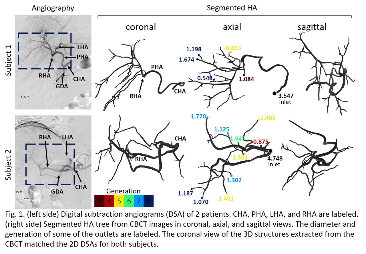
Results: Subjects angiograms are shown on the left side of Fig. 1 where the common, proper, right, and left hepatic arteries (CHA, PHA, RHA, and LHA) as well as gastroduodenal artery (GDA) are labeled. The segmented HAs are shown on the right side of Fig 1. The segmented structures included one inlet at the CHA, and 23 and 46 outlets for subject 1 and 2, respectively. The inlet diameter was 3.5 and 4.7 mm, respectively. These values are in agreement with reported CHA diameter ranges [3]. The smallest detected outlet diameter was ~0.5 mm, indicating the ability of our method to segment small vessels. The outlet generation is also shown with different colors such that CHA was considered as the first generation and the generation number was increased by one after each bifurcation. Results showed that the proposed pipeline can be successfully applied to segment the HA tree in different anatomies allowing for more efficient dosimetry compared to current clinical methods.

Conclusions: We proposed a pipeline for segmentation of HA tree from CBCT images as the first step of developing a new patient-specific dosimetry protocol for liver cancer radioembolization planning. This new approach is based on CFD simulation and Y-90 radioactive decay physics. Our proposed pipeline provides the geometry (i.e. fluid domain of interest) needed for the CFD simulation for each patient. Future works will focus on predicting the distribution of the radioactive microspheres in the HA branches through CFD simulation, which in turn can help developing a more patient-specific dosimetry approach. Research Support: NIH R35CA197608 and CCSG P30 (NCI P30CA093373).

Figure
  • Download figure
  • Open in new tab
  • Download powerpoint
Previous
Back to top

In this issue

Journal of Nuclear Medicine
Vol. 60, Issue supplement 1
May 1, 2019
  • Table of Contents
  • Index by author
Article Alerts
Sign In to Email Alerts with your Email Address
Email Article

Thank you for your interest in spreading the word on Journal of Nuclear Medicine.

NOTE: We only request your email address so that the person you are recommending the page to knows that you wanted them to see it, and that it is not junk mail. We do not capture any email address.

Enter multiple addresses on separate lines or separate them with commas.
Hepatic arterial tree segmentation: Towards patient-specific dosimetry for liver cancer radioembolization
(Your Name) has sent you a message from Journal of Nuclear Medicine
(Your Name) thought you would like to see the Journal of Nuclear Medicine web site.
Citation Tools
Hepatic arterial tree segmentation: Towards patient-specific dosimetry for liver cancer radioembolization
Amirtaha Taebi, Bahman Roudsari, Catherine Vu, Simon Cherry, Emilie Roncali
Journal of Nuclear Medicine May 2019, 60 (supplement 1) 122;

Citation Manager Formats

  • BibTeX
  • Bookends
  • EasyBib
  • EndNote (tagged)
  • EndNote 8 (xml)
  • Medlars
  • Mendeley
  • Papers
  • RefWorks Tagged
  • Ref Manager
  • RIS
  • Zotero
Share
Hepatic arterial tree segmentation: Towards patient-specific dosimetry for liver cancer radioembolization
Amirtaha Taebi, Bahman Roudsari, Catherine Vu, Simon Cherry, Emilie Roncali
Journal of Nuclear Medicine May 2019, 60 (supplement 1) 122;
Twitter logo Facebook logo LinkedIn logo Mendeley logo
  • Tweet Widget
  • Facebook Like
  • Google Plus One
Bookmark this article

Jump to section

  • Article
  • Figures & Data
  • Info & Metrics

Related Articles

  • No related articles found.
  • Google Scholar

Cited By...

  • No citing articles found.
  • Google Scholar

More in this TOC Section

Oncology: Clinical Therapy and Diagnosis

  • Added value of single photon emission computed tomography (SPECT) / Computed Tomography (CT) with radioiodine whole body scan in follow up of patients with differentiated thyroid cancer
  • Prognostic value of pre-and post-induction chemotherapy FDG PET/CT in locally advanced oropharyngeal cancer patients.
  • Assessment of incidental pituitary uptake on FDG PET/CT scan: Digital vs. conventional PET/CT
Show more Oncology: Clinical Therapy and Diagnosis

Image Guided Therapy

  • Real-time pharmacodynamic evaluation of a therapeutic system XC- inhibitor using [18F]FSPG-PET
  • CD54-targeted PET for predicting the abscopal effect of local radiotherapy
  • Immuno-PET imaging of receptor activator of the nuclear factor k B ligand (RANKL) in the tumour microenvironment using [68Ga]Ga-NOTA-denosumab
Show more Image Guided Therapy

Similar Articles

SNMMI

© 2023 Journal of Nuclear Medicine

Powered by HighWire