
Main menu

  • Home
  • Content
    • Current
    • Ahead of print
    • Past Issues
    • JNM Supplement
    • SNMMI Annual Meeting Abstracts
  • Subscriptions
    • Subscribers
    • Rates
    • Journal Claims
    • Institutional and Non-member
  • Authors
    • Submit to JNM
    • Information for Authors
    • Assignment of Copyright
    • AQARA requirements
  • Info
    • Permissions
    • Advertisers
    • Continuing Education
    • Corporate & Special Sales
  • About
    • About Us
    • Editorial Board
    • Editorial Contact
  • More
    • Alerts
    • Feedback
    • Help
    • SNMMI Journals
  • SNMMI
    • JNM
    • JNMT
    • SNMMI Journals
    • SNMMI

User menu

  • Subscribe
  • My alerts
  • Log in
  • My Cart

Search

  • Advanced search
Journal of Nuclear Medicine
  • SNMMI
    • JNM
    • JNMT
    • SNMMI Journals
    • SNMMI
  • Subscribe
  • My alerts
  • Log in
  • My Cart
Journal of Nuclear Medicine

Advanced Search

  • Home
  • Content
    • Current
    • Ahead of print
    • Past Issues
    • JNM Supplement
    • SNMMI Annual Meeting Abstracts
  • Subscriptions
    • Subscribers
    • Rates
    • Journal Claims
    • Institutional and Non-member
  • Authors
    • Submit to JNM
    • Information for Authors
    • Assignment of Copyright
    • AQARA requirements
  • Info
    • Permissions
    • Advertisers
    • Continuing Education
    • Corporate & Special Sales
  • About
    • About Us
    • Editorial Board
    • Editorial Contact
  • More
    • Alerts
    • Feedback
    • Help
    • SNMMI Journals
  • Follow SNMMI on Twitter
  • Visit SNMMI on Facebook
Research ArticleCardiology

Deep Learning Analysis of Upright-Supine High-Efficiency SPECT Myocardial Perfusion Imaging for Prediction of Obstructive Coronary Artery Disease: A Multicenter Study

Julian Betancur, Lien-Hsin Hu, Frederic Commandeur, Tali Sharir, Andrew J. Einstein, Mathews B. Fish, Terrence D. Ruddy, Philipp A. Kaufmann, Albert J. Sinusas, Edward J. Miller, Timothy M. Bateman, Sharmila Dorbala, Marcelo Di Carli, Guido Germano, Yuka Otaki, Joanna X. Liang, Balaji K. Tamarappoo, Damini Dey, Daniel S. Berman and Piotr J. Slomka
Journal of Nuclear Medicine May 2019, 60 (5) 664-670; DOI: https://doi.org/10.2967/jnumed.118.213538
Julian Betancur
1Division of Nuclear Medicine, Department of Imaging, and Departments of Medicine and Biomedical Sciences, Cedars-Sinai Medical Center, Los Angeles, California
  • Find this author on Google Scholar
  • Find this author on PubMed
  • Search for this author on this site
Lien-Hsin Hu
1Division of Nuclear Medicine, Department of Imaging, and Departments of Medicine and Biomedical Sciences, Cedars-Sinai Medical Center, Los Angeles, California
  • Find this author on Google Scholar
  • Find this author on PubMed
  • Search for this author on this site
Frederic Commandeur
1Division of Nuclear Medicine, Department of Imaging, and Departments of Medicine and Biomedical Sciences, Cedars-Sinai Medical Center, Los Angeles, California
  • Find this author on Google Scholar
  • Find this author on PubMed
  • Search for this author on this site
Tali Sharir
2Department of Nuclear Cardiology, Assuta Medical Centers, Tel Aviv, Israel
3Ben Gurion University of the Negev, Beer Sheba, Israel
  • Find this author on Google Scholar
  • Find this author on PubMed
  • Search for this author on this site
Andrew J. Einstein
4Division of Cardiology, Department of Medicine, Columbia University Irving Medical Center and New York–Presbyterian Hospital, New York, New York
5Department of Radiology, Columbia University Irving Medical Center and New York–Presbyterian Hospital, New York, New York
  • Find this author on Google Scholar
  • Find this author on PubMed
  • Search for this author on this site
Mathews B. Fish
6Oregon Heart and Vascular Institute, Sacred Heart Medical Center, Springfield, Oregon
  • Find this author on Google Scholar
  • Find this author on PubMed
  • Search for this author on this site
Terrence D. Ruddy
7Division of Cardiology, University of Ottawa Heart Institute, Ottawa, Ontario, Canada
  • Find this author on Google Scholar
  • Find this author on PubMed
  • Search for this author on this site
Philipp A. Kaufmann
8Department of Nuclear Medicine, Cardiac Imaging, University Hospital Zurich, Zurich, Switzerland
  • Find this author on Google Scholar
  • Find this author on PubMed
  • Search for this author on this site
Albert J. Sinusas
9Section of Cardiovascular Medicine, Department of Internal Medicine, Yale University School of Medicine, New Haven, Connecticut
  • Find this author on Google Scholar
  • Find this author on PubMed
  • Search for this author on this site
Edward J. Miller
9Section of Cardiovascular Medicine, Department of Internal Medicine, Yale University School of Medicine, New Haven, Connecticut
  • Find this author on Google Scholar
  • Find this author on PubMed
  • Search for this author on this site
Timothy M. Bateman
10Cardiovascular Imaging Technologies LLC, Kansas City, Missouri; and
  • Find this author on Google Scholar
  • Find this author on PubMed
  • Search for this author on this site
Sharmila Dorbala
11Division of Nuclear Medicine and Molecular Imaging, Department of Radiology, Brigham and Women’s Hospital, Boston, Massachusetts
  • Find this author on Google Scholar
  • Find this author on PubMed
  • Search for this author on this site
Marcelo Di Carli
11Division of Nuclear Medicine and Molecular Imaging, Department of Radiology, Brigham and Women’s Hospital, Boston, Massachusetts
  • Find this author on Google Scholar
  • Find this author on PubMed
  • Search for this author on this site
Guido Germano
1Division of Nuclear Medicine, Department of Imaging, and Departments of Medicine and Biomedical Sciences, Cedars-Sinai Medical Center, Los Angeles, California
  • Find this author on Google Scholar
  • Find this author on PubMed
  • Search for this author on this site
Yuka Otaki
1Division of Nuclear Medicine, Department of Imaging, and Departments of Medicine and Biomedical Sciences, Cedars-Sinai Medical Center, Los Angeles, California
  • Find this author on Google Scholar
  • Find this author on PubMed
  • Search for this author on this site
Joanna X. Liang
1Division of Nuclear Medicine, Department of Imaging, and Departments of Medicine and Biomedical Sciences, Cedars-Sinai Medical Center, Los Angeles, California
  • Find this author on Google Scholar
  • Find this author on PubMed
  • Search for this author on this site
Balaji K. Tamarappoo
1Division of Nuclear Medicine, Department of Imaging, and Departments of Medicine and Biomedical Sciences, Cedars-Sinai Medical Center, Los Angeles, California
  • Find this author on Google Scholar
  • Find this author on PubMed
  • Search for this author on this site
Damini Dey
1Division of Nuclear Medicine, Department of Imaging, and Departments of Medicine and Biomedical Sciences, Cedars-Sinai Medical Center, Los Angeles, California
  • Find this author on Google Scholar
  • Find this author on PubMed
  • Search for this author on this site
Daniel S. Berman
1Division of Nuclear Medicine, Department of Imaging, and Departments of Medicine and Biomedical Sciences, Cedars-Sinai Medical Center, Los Angeles, California
  • Find this author on Google Scholar
  • Find this author on PubMed
  • Search for this author on this site
Piotr J. Slomka
1Division of Nuclear Medicine, Department of Imaging, and Departments of Medicine and Biomedical Sciences, Cedars-Sinai Medical Center, Los Angeles, California
  • Find this author on Google Scholar
  • Find this author on PubMed
  • Search for this author on this site
  • Article
  • Figures & Data
  • Supplemental
  • Info & Metrics
  • PDF
Loading

Article Figures & Data

Figures

  • Tables
  • Additional Files
  • FIGURE 1.
    • Download figure
    • Open in new tab
    • Download powerpoint
    FIGURE 1.

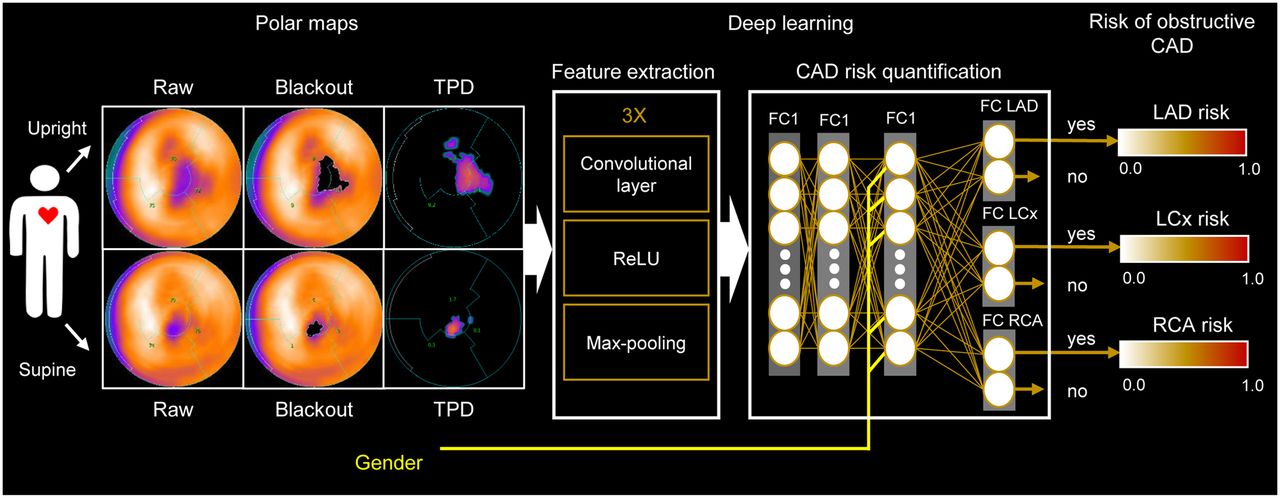
    DL prediction of obstructive CAD from upright and supine MPI. A deep convolutional neural network trained from obstructive stenosis correlations by ICA was used to simultaneously estimate probability of obstructive CAD for LAD, LCx, and RCA territories from upright and supine polar MPI maps. Maximum probability was retained as probability of patient disease. FC = fully connected layer; Max-pooling = function that returns maximum value for image patch; ReLU = rectified linear unit.

  • FIGURE 2.
    • Download figure
    • Open in new tab
    • Download powerpoint
    FIGURE 2.

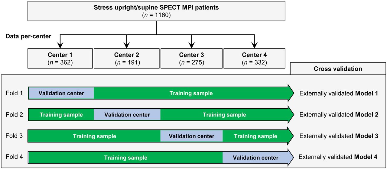
    Leave-one-center-out cross-validation. Input stress MPI datasets are divided by center (4 in total). Four folds are built, each containing training sample made up of images from 3 centers and validation sample with images from remaining center. This procedure allows external validation of 4 DL models trained separately in each fold.

  • FIGURE 3.
    • Download figure
    • Open in new tab
    • Download powerpoint
    FIGURE 3.

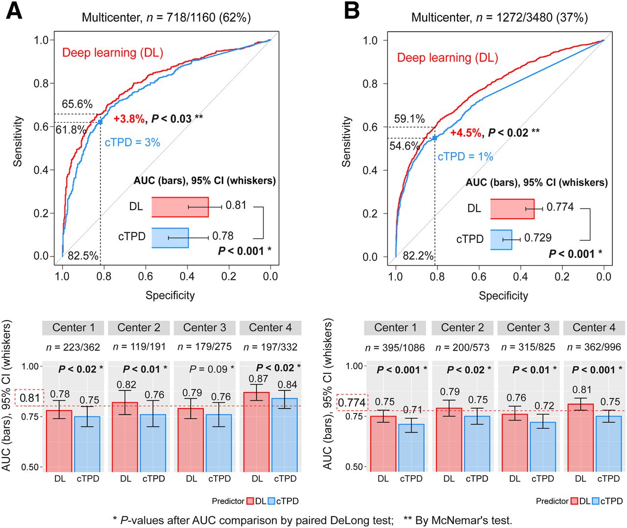
    DL prediction of obstructive CAD from 4 externally validated models with merged data for 4 centers. Per-patient (A) and per-vessel (B) DL predictions of obstructive CAD from upright and supine images (DL, red) are compared with prediction of obstructive CAD by combined upright-supine TPD (cTPD, blue). AUC per center was externally validated using CAD scores from 4 different DL models (1 per center) with each model trained with data from other 3 centers. Red dotted line (bottom) shows overall multicenter AUC. CI = confidence interval; ROC = receiver operating characteristic.
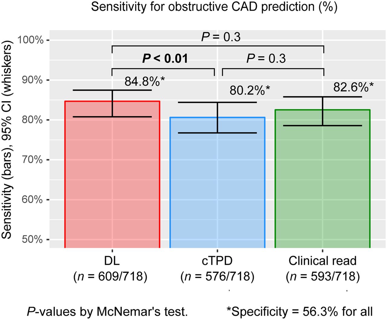
  • FIGURE 4.
    • Download figure
    • Open in new tab
    • Download powerpoint
    FIGURE 4.

    Sensitivity for prediction of obstructive CAD. Per-patient DL prediction of obstructive CAD by DL computed from upright and supine MPI (DL, red) had higher sensitivity than prediction by cTPD (blue) and same sensitivity as on-site clinical readers (green). DL cutoff was set to 0.29, and cTPD cutoff was set to 0.62% to exhibit same specificity as normal or probably-normal clinical read. CI = confidence interval.

  • FIGURE 5.
    • Download figure
    • Open in new tab
    • Download powerpoint
    FIGURE 5.

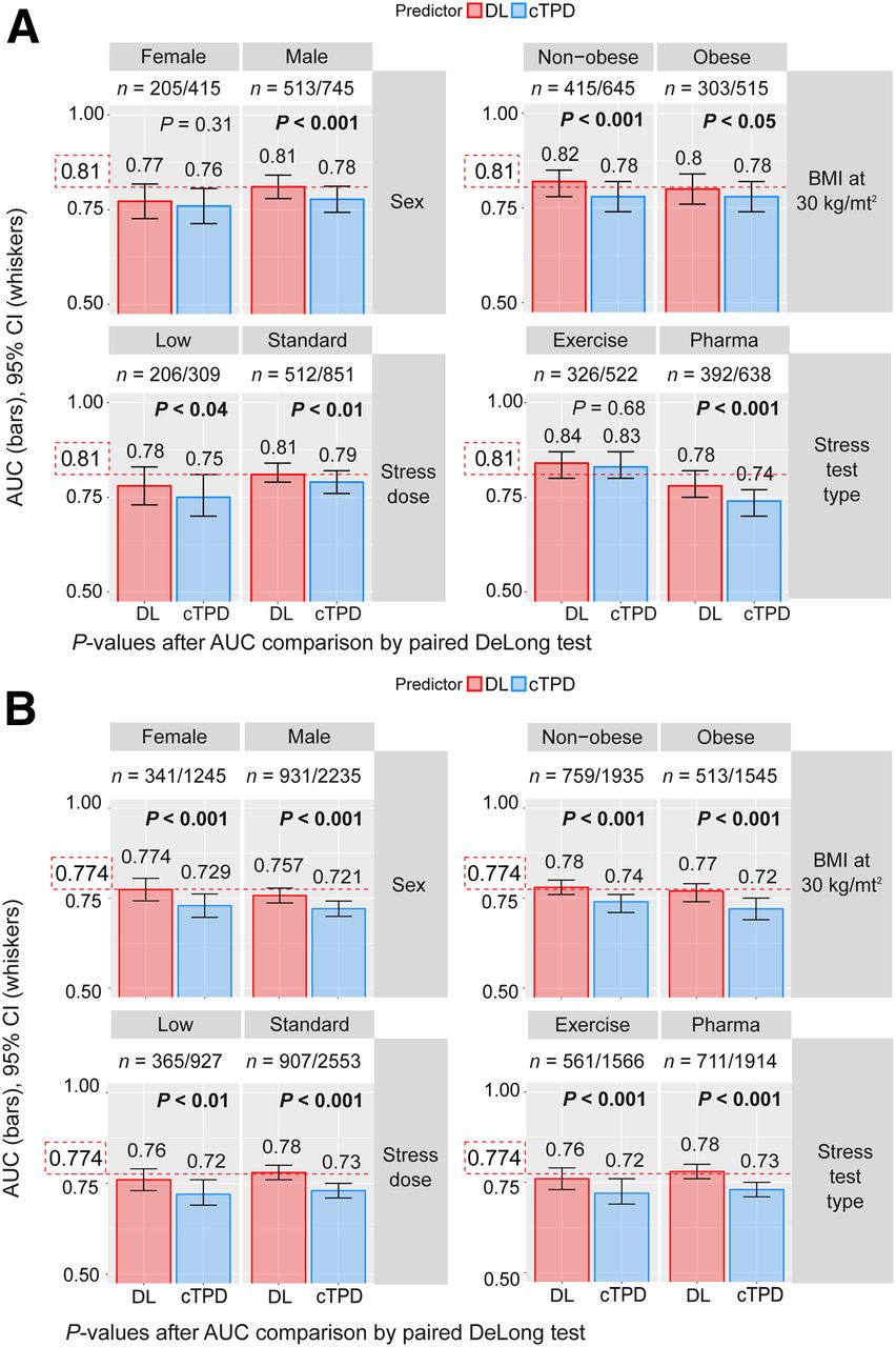
    Prediction of obstructive CAD per subpopulation. REFINE-SPECT subpopulations were defined by sex (F, M), obesity (nonobese: body mass index < 30 kg/m2, obese: body mass index ≥ 30 kg/m2), stress imaging activity (low: patients undergoing stress-first/stress-only MPI; standard: patients undergoing rest-first/2-d MPI), and stress test type (exercise, pharmacologic). Red dotted line shows overall multicenter AUC as reported in Figure 3. BMI = body mass index; CI = confidence interval.

  • FIGURE 6.
    • Download figure
    • Open in new tab
    • Download powerpoint
    FIGURE 6.

    Prediction of obstructive CAD from upright and supine stress MPI. Short/long axis views, polar maps depicting normalized radiotracer count distribution and perfusion defects (top), and predictions by cTPD and DL (bottom) are shown for 2 patients with obstructive CAD. (A) In 79-y-old man (85% proximal LAD stenosis) quantified with normal cTPD (per-patient cTPD < 3% and per-vessel cTPD < 1%), DL correctly identified LAD disease. Patient had body mass index of 30 kg/m2 and diabetes and underwent exercise stress MPI. (B) In 62-y-old woman (70% mid LAD stenosis, 95% proximal LCX stenosis, and 80% proximal RCA stenosis) with cTPD abnormal for 1 vessel only, DL correctly identified triple-vessel disease. Patient had body mass index of 25 kg/m2, dyslipidemia, and family history of cardiac disease and underwent exercise stress MPI. BMI = body mass index.

Tables

  • Figures
  • Additional Files
    • View popup
    TABLE 1

    Baseline Characteristics of Studied Population

    CharacteristicOverall, n = 1,160Nonobstructive CAD, n = 442 (38.1%)Obstructive CAD, n = 718 (61.9%)P
    Age (y)64.3 ± 11.562.2 ± 1265.6 ± 11.1<0.0001
    Sex
     Male745 (64.2)232 (52.5)513 (71.5)<0.0001
     Female415 (35.8)210 (47.5)205 (28.5)<0.0001
    Weight (kg)88.5 ± 21.989.4 ± 23.687.9 ± 20.80.278
    Body mass index (kg/m2)30 ± 6.530.8 ± 7.529.5 ± 5.7<0.01
    Diabetes mellitus351 (30.3)121 (27.4)230 (32)0.09
    Hypertension841 (72.5)304 (68.8)537 (74.8)<0.05
    Dyslipidemia780 (67.2)281 (63.6)499 (69.5)<0.05
    Smoking221 (19.1)83 (18.8)138 (19.2)0.85
    Stress test type
     Exercise522 (45)196 (44.3)326 (45.4)0.73
     Exercise + pharmacologic164 (14.1)48 (10.9)116 (16.2)<0.05
     Pharmacologic474 (40.9)198 (44.8)276 (38.4)<0.05
    Imaging protocol
    Stress only48 (4.1)19 (4.3)29 (4)0.83
    Same day stress and rest1,073 (92.5)403 (91.2)670 (93.3)0.18
     Stress-first261 (22.5)84 (19.0)177 (24.7)<0.05
     Rest-first812 (70.0)319 (72.2)493 (68.7)<0.05
    Two-day stress and rest39 (3.4)20 (4.5)19 (2.7)0.09
    • Qualitative data are expressed as numbers followed by percentages in parentheses; continuous data are expressed as mean ± SD.

    • View popup
    TABLE 2

    Prevalence of Obstructive CAD

    Prevalence
    Obstructive diseaseOverall multicenter, n = 1,160Center 1, n = 362Center 2, n = 191Center 3, n = 275Center 4, n = 332P
    No disease442 (38.1)139 (38.4)72 (37.8)96 (34.9)135 (40.7)0.54
    One-vessel disease321 (27.7)100 (27.6)62 (32.5)79 (28.7)80 (24.1)0.22
    Double-vessel disease240 (20.7)74 (20.4)33 (17.3)64 (23.3)69 (20.8)0.48
    Triple-vessel disease157 (13.5)49 (13.6)24 (11.0)36 (13.1)48 (14.5)0.93
    Per-patient718 (61.9)223 (61.6)119 (62.3)179 (65.1)197 (59.3)0.54
    LAD509 (43.9)163 (45.0)84 (44.0)124 (45.1)138 (41.6)0.78
    LCx384 (33.1)121 (33.4)60 (31.4)92 (33.5)111 (33.4)0.96
    RCA379 (32.7)111 (30.7)56 (29.3)99 (36)113 (34.0)0.35
    Per-vessel (LAD + LCx + RCA)1,272/3,480 (36.6)395/1,086 (36.4)200/573 (34.9)315/825 (38.2)362/996 (36.4)0.65
    • Data are numbers followed by percentages in parentheses.

    • View popup
    TABLE 3

    Radiotracer Activity for Stress Image Acquisition

    Image protocolInjected activity (MBq)
    Same-day protocols, n = 1,073 (92.5%)
    Stress-first protocol, n = 261 (24.3%)213.1 ± 87.3
    Rest-first protocol, n = 812 (75.7%)103 ± 384
    Two-day protocol, n = 39 (3.4%)682.3 ± 481.44
    Stress-only protocol, n = 48 (4.1%)260.2 ± 486.5
    Overall, n = 1,160804 ± 494
    • Data are mean ± SD.

Additional Files

  • Figures
  • Tables
  • Supplemental Data

    Files in this Data Supplement:

    • Supplemental Data
PreviousNext
Back to top

In this issue

Journal of Nuclear Medicine: 60 (5)
Journal of Nuclear Medicine
Vol. 60, Issue 5
May 1, 2019
  • Table of Contents
  • Table of Contents (PDF)
  • About the Cover
  • Index by author
Print
Download PDF
Article Alerts
Sign In to Email Alerts with your Email Address
Email Article

Thank you for your interest in spreading the word on Journal of Nuclear Medicine.

NOTE: We only request your email address so that the person you are recommending the page to knows that you wanted them to see it, and that it is not junk mail. We do not capture any email address.

Enter multiple addresses on separate lines or separate them with commas.
Deep Learning Analysis of Upright-Supine High-Efficiency SPECT Myocardial Perfusion Imaging for Prediction of Obstructive Coronary Artery Disease: A Multicenter Study
(Your Name) has sent you a message from Journal of Nuclear Medicine
(Your Name) thought you would like to see the Journal of Nuclear Medicine web site.
Citation Tools
Deep Learning Analysis of Upright-Supine High-Efficiency SPECT Myocardial Perfusion Imaging for Prediction of Obstructive Coronary Artery Disease: A Multicenter Study
Julian Betancur, Lien-Hsin Hu, Frederic Commandeur, Tali Sharir, Andrew J. Einstein, Mathews B. Fish, Terrence D. Ruddy, Philipp A. Kaufmann, Albert J. Sinusas, Edward J. Miller, Timothy M. Bateman, Sharmila Dorbala, Marcelo Di Carli, Guido Germano, Yuka Otaki, Joanna X. Liang, Balaji K. Tamarappoo, Damini Dey, Daniel S. Berman, Piotr J. Slomka
Journal of Nuclear Medicine May 2019, 60 (5) 664-670; DOI: 10.2967/jnumed.118.213538

Citation Manager Formats

  • BibTeX
  • Bookends
  • EasyBib
  • EndNote (tagged)
  • EndNote 8 (xml)
  • Medlars
  • Mendeley
  • Papers
  • RefWorks Tagged
  • Ref Manager
  • RIS
  • Zotero
Share
Deep Learning Analysis of Upright-Supine High-Efficiency SPECT Myocardial Perfusion Imaging for Prediction of Obstructive Coronary Artery Disease: A Multicenter Study
Julian Betancur, Lien-Hsin Hu, Frederic Commandeur, Tali Sharir, Andrew J. Einstein, Mathews B. Fish, Terrence D. Ruddy, Philipp A. Kaufmann, Albert J. Sinusas, Edward J. Miller, Timothy M. Bateman, Sharmila Dorbala, Marcelo Di Carli, Guido Germano, Yuka Otaki, Joanna X. Liang, Balaji K. Tamarappoo, Damini Dey, Daniel S. Berman, Piotr J. Slomka
Journal of Nuclear Medicine May 2019, 60 (5) 664-670; DOI: 10.2967/jnumed.118.213538
del.icio.us logo Digg logo Reddit logo Twitter logo CiteULike logo Facebook logo Google logo Mendeley logo
  • Tweet Widget
  • Facebook Like
  • Google Plus One

Jump to section

  • Article
    • Abstract
    • MATERIALS AND METHODS
    • RESULTS
    • DISCUSSION
    • CONCLUSION
    • DISCLOSURE
    • Acknowledgments
    • Footnotes
    • REFERENCES
  • Figures & Data
  • Supplemental
  • Info & Metrics
  • PDF

Related Articles

  • This Month in JNM
  • PubMed
  • Google Scholar

Cited By...

  • Automated interpretation of the coronary angioscopy with deep convolutional neural networks
  • Proposed Requirements for Cardiovascular Imaging-Related Machine Learning Evaluation (PRIME): A Checklist: Reviewed by the American College of Cardiology Healthcare Innovation Council
  • The Future of Nuclear Medicine Depends on the Quality of Its Research: Researchers at the University of Heidelberg Receive the Award for Best Article of the Year
  • Automated interpretation of the coronary angioscopy with deep convolutional neural networks
  • Functionalized mesoporous silica nanoparticles for innovative boron-neutron capture therapy of resistant cancers
  • Artificial Intelligence in Nuclear Medicine
  • Intelligent Imaging: Artificial Intelligence Augmented Nuclear Medicine
  • Solid-State Detector SPECT Myocardial Perfusion Imaging
  • Google Scholar

More in this TOC Section

Cardiology

  • Molecular Imaging of Fibroblast Activity After Myocardial Infarction Using a 68Ga-Labeled Fibroblast Activation Protein Inhibitor, FAPI-04
  • Imaging of Chemotherapy-Induced Acute Cardiotoxicity with 18F-Labeled Lipophilic Cations
  • Assessment of Myocardial CZT SPECT Recording in a Forward-Leaning Bikerlike Position
Show more Cardiology

Clinical

  • Nondisplaceable Binding Is a Potential Confounding Factor in 11C-PBR28 Translocator Protein PET Studies
  • Comparison of Interim PET Response to Second-Line Versus First-Line Treatment in Classic Hodgkin Lymphoma: Contribution to the Development of Response Criteria for Relapsed or Progressive Disease
  • Dissimilar DNA Damage to Blood Lymphocytes After 177Lu-Labeled DOTATOC or Prostate-Specific Membrane Antigen Therapy
Show more Clinical

Similar Articles

Keywords

  • obstructive coronary artery disease
  • SPECT myocardial perfusion imaging
  • deep learning
  • convolutional neural network
  • total perfusion deficit
SNMMI

© 2021 Journal of Nuclear Medicine

Powered by HighWire