
Main menu

  • Home
  • Content
    • Current
    • Ahead of print
    • Past Issues
    • JNM Supplement
    • SNMMI Annual Meeting Abstracts
  • Subscriptions
    • Subscribers
    • Institutional and Non-member
    • Rates
    • Corporate & Special Sales
    • Journal Claims
  • Authors
    • Submit to JNM
    • Information for Authors
    • Assignment of Copyright
    • AQARA requirements
  • Info
    • Continuing Education
    • Reviewers
    • Permissions
    • Advertisers
  • About
    • About Us
    • Editorial Board
    • Contact Information
  • More
    • Alerts
    • Feedback
    • Help
    • SNMMI Journals
  • SNMMI
    • JNM
    • JNMT
    • SNMMI Journals
    • SNMMI

User menu

  • Subscribe
  • My alerts
  • Log in
  • My Cart

Search

  • Advanced search
Journal of Nuclear Medicine
  • SNMMI
    • JNM
    • JNMT
    • SNMMI Journals
    • SNMMI
  • Subscribe
  • My alerts
  • Log in
  • My Cart
Journal of Nuclear Medicine

Advanced Search

  • Home
  • Content
    • Current
    • Ahead of print
    • Past Issues
    • JNM Supplement
    • SNMMI Annual Meeting Abstracts
  • Subscriptions
    • Subscribers
    • Institutional and Non-member
    • Rates
    • Corporate & Special Sales
    • Journal Claims
  • Authors
    • Submit to JNM
    • Information for Authors
    • Assignment of Copyright
    • AQARA requirements
  • Info
    • Continuing Education
    • Reviewers
    • Permissions
    • Advertisers
  • About
    • About Us
    • Editorial Board
    • Contact Information
  • More
    • Alerts
    • Feedback
    • Help
    • SNMMI Journals
  • Visit JNM on Facebook
  • Join JNM on LinkedIn
  • Follow JNM on Twitter
  • Subscribe to our RSS feeds
Research ArticlePulmonary

Glycoprotein IIb/IIIa Receptor Imaging with 18F-GP1 PET for Acute Venous Thromboembolism: An Open-Label, Nonrandomized, Phase 1 Study

Chanwoo Kim, Jae Seung Lee, Youngjin Han, Sun Young Chae, Soyoung Jin, Changhwan Sung, Hye Joo Son, Seung Jun Oh, Sang Ju Lee, Jungsu S. Oh, Yong-Pil Cho, Tae-Won Kwon, Deok Hee Lee, Seongsoo Jang, Bohyun Kim, Norman Koglin, Mathias Berndt, Andrew W. Stephens and Dae Hyuk Moon
Journal of Nuclear Medicine February 2019, 60 (2) 244-249; DOI: https://doi.org/10.2967/jnumed.118.212084
Chanwoo Kim
1Department of Nuclear Medicine, Asan Medical Center, University of Ulsan College of Medicine, Seoul, Republic of Korea
  • Find this author on Google Scholar
  • Find this author on PubMed
  • Search for this author on this site
Jae Seung Lee
2Department of Pulmonology, Asan Medical Center, University of Ulsan College of Medicine, Seoul, Republic of Korea
  • Find this author on Google Scholar
  • Find this author on PubMed
  • Search for this author on this site
Youngjin Han
3Department of Vascular Surgery, Asan Medical Center, University of Ulsan College of Medicine, Seoul, Republic of Korea
  • Find this author on Google Scholar
  • Find this author on PubMed
  • Search for this author on this site
Sun Young Chae
1Department of Nuclear Medicine, Asan Medical Center, University of Ulsan College of Medicine, Seoul, Republic of Korea
  • Find this author on Google Scholar
  • Find this author on PubMed
  • Search for this author on this site
Soyoung Jin
4Department of Nuclear Medicine, Nowon Eulji Medical Center, Eulji University, Seoul, Republic of Korea
  • Find this author on Google Scholar
  • Find this author on PubMed
  • Search for this author on this site
Changhwan Sung
1Department of Nuclear Medicine, Asan Medical Center, University of Ulsan College of Medicine, Seoul, Republic of Korea
  • Find this author on Google Scholar
  • Find this author on PubMed
  • Search for this author on this site
Hye Joo Son
1Department of Nuclear Medicine, Asan Medical Center, University of Ulsan College of Medicine, Seoul, Republic of Korea
  • Find this author on Google Scholar
  • Find this author on PubMed
  • Search for this author on this site
Seung Jun Oh
1Department of Nuclear Medicine, Asan Medical Center, University of Ulsan College of Medicine, Seoul, Republic of Korea
  • Find this author on Google Scholar
  • Find this author on PubMed
  • Search for this author on this site
Sang Ju Lee
1Department of Nuclear Medicine, Asan Medical Center, University of Ulsan College of Medicine, Seoul, Republic of Korea
  • Find this author on Google Scholar
  • Find this author on PubMed
  • Search for this author on this site
Jungsu S. Oh
1Department of Nuclear Medicine, Asan Medical Center, University of Ulsan College of Medicine, Seoul, Republic of Korea
  • Find this author on Google Scholar
  • Find this author on PubMed
  • Search for this author on this site
Yong-Pil Cho
3Department of Vascular Surgery, Asan Medical Center, University of Ulsan College of Medicine, Seoul, Republic of Korea
  • Find this author on Google Scholar
  • Find this author on PubMed
  • Search for this author on this site
Tae-Won Kwon
3Department of Vascular Surgery, Asan Medical Center, University of Ulsan College of Medicine, Seoul, Republic of Korea
  • Find this author on Google Scholar
  • Find this author on PubMed
  • Search for this author on this site
Deok Hee Lee
5Department of Radiology, Asan Medical Center, University of Ulsan College of Medicine, Seoul, Republic of Korea
  • Find this author on Google Scholar
  • Find this author on PubMed
  • Search for this author on this site
Seongsoo Jang
6Department of Laboratory Medicine, Asan Medical Center, University of Ulsan College of Medicine, Seoul, Republic of Korea
  • Find this author on Google Scholar
  • Find this author on PubMed
  • Search for this author on this site
Bohyun Kim
7Department of Laboratory Medicine, Soonchunhyang University Hospital Cheonan, Soonchunhyang University College of Medicine, Cheonan, Chungcheongnam-do, Republic of Korea; and
  • Find this author on Google Scholar
  • Find this author on PubMed
  • Search for this author on this site
Norman Koglin
8Piramal Imaging GmbH (now Life Molecular Imaging GmbH), Berlin, Germany
  • Find this author on Google Scholar
  • Find this author on PubMed
  • Search for this author on this site
Mathias Berndt
8Piramal Imaging GmbH (now Life Molecular Imaging GmbH), Berlin, Germany
  • Find this author on Google Scholar
  • Find this author on PubMed
  • Search for this author on this site
Andrew W. Stephens
8Piramal Imaging GmbH (now Life Molecular Imaging GmbH), Berlin, Germany
  • Find this author on Google Scholar
  • Find this author on PubMed
  • Search for this author on this site
Dae Hyuk Moon
1Department of Nuclear Medicine, Asan Medical Center, University of Ulsan College of Medicine, Seoul, Republic of Korea
  • Find this author on Google Scholar
  • Find this author on PubMed
  • Search for this author on this site
  • Article
  • Figures & Data
  • Supplemental
  • Info & Metrics
  • PDF
Loading

Article Figures & Data

Figures

  • Tables
  • Additional Files
  • FIGURE 1.
    • Download figure
    • Open in new tab
    • Download powerpoint
    FIGURE 1.

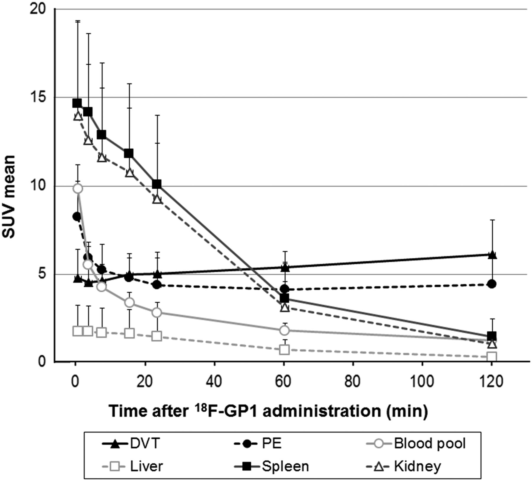
    18F-GP1 biodistribution as function of time. Kidney, spleen, and blood-pool activities show high initial uptake followed by gradual washout, whereas DVT and PE demonstrate rapid initial accumulation followed by plateau phase, with minimal decrease of activity until 120 min after 18F-GP1 injection.

  • FIGURE 2.
    • Download figure
    • Open in new tab
    • Download powerpoint
    FIGURE 2.

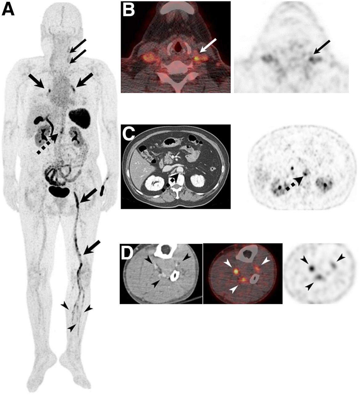
    18F-GP1 PET/CT and CT images of 55-y-old man with DVT and PE. (A) Anterior maximum-intensity projections of 18F-GP1 PET/CT over 120 min show positive 18F-GP1 accumulation in pulmonary arteries (arrows) and in proximal (dotted arrows) and distal (arrowheads) veins of leg, which are gradually distinct on late images as 18F-GP1 activity from other organs is excreted via both urinary and hepatobiliary tracts. (B–D) Transaxial CT images (left) clearly show pulmonary emboli in right main and left lower lobar pulmonary arteries (B, arrows) and thrombus in right popliteal vein (C, dotted arrow). Positive 18F-GP1 uptake (right) at 120 min after injection is seen at corresponding vessels (B and C, solid and dotted arrows, respectively). Additional positive 18F-GP1 uptake (right) is observed in left peroneal and gastrocnemius veins (D, arrowheads), but no corresponding filling defects are seen on CT venography (D, left).

  • FIGURE 3.
    • Download figure
    • Open in new tab
    • Download powerpoint
    FIGURE 3.

    18F-GP1 PET/CT and CT images of 69-y-old woman with DVT and PE. (A) Anterior maximum-intensity projection of 18F-GP1 PET/CT at 120 min after injection shows multiple areas of increased uptake in both pulmonary arteries and veins of left lower extremity at DVT and PE sites (thick arrows), as well as foci of abnormal 18F-GP1 uptake in left common carotid artery (thin arrows); left anterior tibial, posterior tibial and peroneal veins (arrowheads); and abdominal aorta (dotted arrow). (B–D) Foci of abnormal 18F-GP1 uptake are seen in left common carotid artery (B, right, arrow); abdominal aorta (C, right, dotted arrow); and left anterior tibial, posterior tibial, and peroneal veins (D, middle and right, arrowheads). Contrast-enhanced CT images reveal low-density, noncalcified plaque in abdominal aorta (C, left, dotted arrow) but no filling defects in distal veins of left lower extremity (D, left, arrowheads). No further imaging studies were performed to characterize left carotid uptake.

  • FIGURE 4.
    • Download figure
    • Open in new tab
    • Download powerpoint
    FIGURE 4.

    Relationship between P-selectin expression on circulating platelets and 18F-GP1 uptake. Scattergram shows positive correlation between highest SUVmax of 18F-GP1 among all reference lesions at 120 min after injection and percentage of P-selectin–positive platelets measured by flow cytometry using CD62P monoclonal antibody.

Tables

  • Figures
  • Additional Files
    • View popup
    TABLE 1

    Baseline Characteristics of Patients with Acute DVT or PE

    CharacteristicDVT (n = 10)PE (n = 10)Total
    Demographic
     Age (y)59.9 ± 13.858.4 ± 18.159.2 ± 15.7
     Male sex5 (50%)4 (40%)9 (45%)
     Non-Hispanic/Latino and Asian (Korean)10 (100%)10 (100%)20 (100%)
    Clinical feature
     Duration of signs and symptoms (d)*18.8 ± 11.49.5 ± 8.714.2 ± 11.0
     Active cancer1 (10%)1 (10%)2 (10%)
     Paralysis, paresis, or recent immobilization1 (10%)None1 (5%)
     Recently bedridden > 3 d or major surgery3 (30%)1 (10%)4 (20%)
     Estrogen use by womenNone1 (10%)1 (5%)
     Body mass index ≥ 30 kg/m21 (10%)2 (20%)3 (15%)
    Laboratory test (d-dimer [μg/mL FEU])9.7 ± 5.47.0 ± 4.78.4 ± 5.1
    Treatment before 18F-GP1 PET/CT
     Duration of anticoagulant therapy (d)†2.8 ± 1.13.2 ± 1.13.0 ± 1.1
     Prior antiplatelet therapy4 (40%)1 (10%)5 (25%)
    • ↵* Duration before 18F-GP1 PET/CT.

    • ↵† Duration of anticoagulation therapy before 18F-GP1 PET/CT or interval between standard imaging studies and 18F-GP1 PET/CT.

    • FEU = fibrinogen-equivalent units.

    • Qualitative data are expressed as numbers, followed by percentages in parentheses; continuous data are expressed as mean ± SD.

Additional Files

  • Figures
  • Tables
  • Supplemental Data

    Files in this Data Supplement:

    • Supplemental Data
PreviousNext
Back to top

In this issue

Journal of Nuclear Medicine: 60 (2)
Journal of Nuclear Medicine
Vol. 60, Issue 2
February 1, 2019
  • Table of Contents
  • Table of Contents (PDF)
  • About the Cover
  • Index by author
Print
Download PDF
Article Alerts
Sign In to Email Alerts with your Email Address
Email Article

Thank you for your interest in spreading the word on Journal of Nuclear Medicine.

NOTE: We only request your email address so that the person you are recommending the page to knows that you wanted them to see it, and that it is not junk mail. We do not capture any email address.

Enter multiple addresses on separate lines or separate them with commas.
Glycoprotein IIb/IIIa Receptor Imaging with 18F-GP1 PET for Acute Venous Thromboembolism: An Open-Label, Nonrandomized, Phase 1 Study
(Your Name) has sent you a message from Journal of Nuclear Medicine
(Your Name) thought you would like to see the Journal of Nuclear Medicine web site.
Citation Tools
Glycoprotein IIb/IIIa Receptor Imaging with 18F-GP1 PET for Acute Venous Thromboembolism: An Open-Label, Nonrandomized, Phase 1 Study
Chanwoo Kim, Jae Seung Lee, Youngjin Han, Sun Young Chae, Soyoung Jin, Changhwan Sung, Hye Joo Son, Seung Jun Oh, Sang Ju Lee, Jungsu S. Oh, Yong-Pil Cho, Tae-Won Kwon, Deok Hee Lee, Seongsoo Jang, Bohyun Kim, Norman Koglin, Mathias Berndt, Andrew W. Stephens, Dae Hyuk Moon
Journal of Nuclear Medicine Feb 2019, 60 (2) 244-249; DOI: 10.2967/jnumed.118.212084

Citation Manager Formats

  • BibTeX
  • Bookends
  • EasyBib
  • EndNote (tagged)
  • EndNote 8 (xml)
  • Medlars
  • Mendeley
  • Papers
  • RefWorks Tagged
  • Ref Manager
  • RIS
  • Zotero
Share
Glycoprotein IIb/IIIa Receptor Imaging with 18F-GP1 PET for Acute Venous Thromboembolism: An Open-Label, Nonrandomized, Phase 1 Study
Chanwoo Kim, Jae Seung Lee, Youngjin Han, Sun Young Chae, Soyoung Jin, Changhwan Sung, Hye Joo Son, Seung Jun Oh, Sang Ju Lee, Jungsu S. Oh, Yong-Pil Cho, Tae-Won Kwon, Deok Hee Lee, Seongsoo Jang, Bohyun Kim, Norman Koglin, Mathias Berndt, Andrew W. Stephens, Dae Hyuk Moon
Journal of Nuclear Medicine Feb 2019, 60 (2) 244-249; DOI: 10.2967/jnumed.118.212084
Twitter logo Facebook logo LinkedIn logo Mendeley logo
  • Tweet Widget
  • Facebook Like
  • Google Plus One
Bookmark this article

Jump to section

  • Article
    • Abstract
    • MATERIALS AND METHODS
    • RESULTS
    • DISCUSSION
    • CONCLUSION
    • DISCLOSURE
    • Footnotes
    • REFERENCES
  • Figures & Data
  • Supplemental
  • Info & Metrics
  • PDF

Related Articles

  • This Month in JNM
  • PubMed
  • Google Scholar

Cited By...

  • The Future of Nuclear Medicine Depends on the Quality of Its Research: Researchers at the University of Heidelberg Receive the Award for Best Article of the Year
  • Google Scholar

More in this TOC Section

Pulmonary

  • Lung perfusion quantification using a SPECT/CT-based semi-automated segmentation workflow rather than planar perfusion scintigraphy
  • Time-reduction for simultaneous dual isotope lung scintigraphy using 3D-ring CZT SPECT/CT.
  • SPECT VQ and CTPA agreement for diagnosis of pulmonary embolism during the COVID-19 pandemic : a single-institution experience
Show more Pulmonary

Clinical

  • Lung perfusion quantification using a SPECT/CT-based semi-automated segmentation workflow rather than planar perfusion scintigraphy
  • Time-reduction for simultaneous dual isotope lung scintigraphy using 3D-ring CZT SPECT/CT.
  • SPECT VQ and CTPA agreement for diagnosis of pulmonary embolism during the COVID-19 pandemic : a single-institution experience
Show more Clinical

Similar Articles

Keywords

  • deep vein thrombosis
  • pulmonary embolism
  • positron emission tomography
  • 18F-GP1
  • platelet activation
SNMMI

© 2023 Journal of Nuclear Medicine

Powered by HighWire