


Abstract
56
Objectives: Tumor-associated activated macrophages (TAMS) play a critical role in tumorigenesis by perpetuating inflammation via cytokine release and contributing to neovascularization and inhibition of immune surveillance. Specific detection of such activated macrophage infiltration can provide valuable immunodiagnostic insight toward disease status and therapeutic response. Unlike 18FDG uptake, which is based on the Warburg effect, 99mTc-tilmanocept (TCT) is a radiopharmaceutical that binds to the mannose receptor (CD206) highly expressed by TAMS with high affinity (KD = 2.76 x 10⁻¹¹ M). Thus, FDG and TCT interrogate different elements of the malignancy and the tumor progression process. Here we present initial safety data and imaging findings from an exploratory study of Navidea’s Phase I dose escalation trial of intravenous (IV) TCT in ML-CRC subjects. Methods: Subjects with primary colorectal cancer with synchronous liver metastasis (ML-CRC) are eligible for this study. This is a single-center, open-label, within-subject exploratory study of TCT in the localization of liver metastasis in ML-CRC subjects. Up to 6 participants receive a single IV dose of 99mTc-labeled TCT (2 mCi) with a mass dose of 50 μg, and a second cohort of up to 6 subjects receive a single IV dose of 99mTc-labeled TCT (2 mCi) with a mass dose of 200 μg. Both groups are imaged at 4-6 hours post-injection with SPECT/CT of the abdomen including the liver. This study is designed to evaluate the safety and tolerability of a single dose of TCT administered IV. Additionally, the tumor uptake of TCT-SPECT/CT is compared to FDG-PET/CT in each subject.
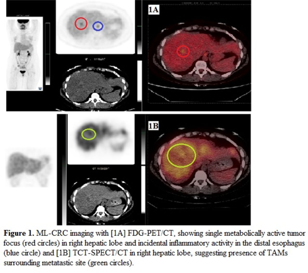
Results: IV injection of TCT was well-tolerated. No adverse drug reactions were observed. The FDG-PET/CT image (1A, red circles) shows a single metabolically active tumor focus in the right hepatic lobe. Incidental inflammatory activity is identified in the distal esophagus (1A, blue circle). The TCT-SPECT/CT image (1B, green circles), in addition to prior histological assessments in other tumors, suggests the presence of TAMs around the metastasis (CD206+ macrophages).
Conclusion: TCT and FDG are both molecular markers relevant to tumor biology and progression: TAMS vs. aerobic glycolysis. Initial results suggest that TCT binding may detect TAMS surrounding liver metastases. The diagnostic, prognostic, and therapeutic implications of TAM imaging in ML-CRC remains to be determined, but TCT-based agents may serve to aid in targeting TAMs with a therapeutic payload where response may be directly monitored through TCT imaging. This study is currently enrolling additional patients. Research Support: This study was supported by a grant from the National Cancer Institute/NIH, Grant R44CA162783 ClinicalTrials.gov Identifier: NCT03029988.

In this issue


Jump to section
Related Articles
Cited By...
- No citing articles found.