
Main menu

  • Home
  • Content
    • Current
    • Ahead of print
    • Past Issues
    • JNM Supplement
    • SNMMI Annual Meeting Abstracts
  • Subscriptions
    • Subscribers
    • Institutional and Non-member
    • Rates
    • Corporate & Special Sales
    • Journal Claims
  • Authors
    • Submit to JNM
    • Information for Authors
    • Assignment of Copyright
    • AQARA requirements
  • Info
    • Permissions
    • Advertisers
    • Continuing Education
  • About
    • About Us
    • Editorial Board
    • Contact Information
  • More
    • Alerts
    • Feedback
    • Help
    • SNMMI Journals
  • SNMMI
    • JNM
    • JNMT
    • SNMMI Journals
    • SNMMI

User menu

  • Subscribe
  • My alerts
  • Log in
  • My Cart

Search

  • Advanced search
Journal of Nuclear Medicine
  • SNMMI
    • JNM
    • JNMT
    • SNMMI Journals
    • SNMMI
  • Subscribe
  • My alerts
  • Log in
  • My Cart
Journal of Nuclear Medicine

Advanced Search

  • Home
  • Content
    • Current
    • Ahead of print
    • Past Issues
    • JNM Supplement
    • SNMMI Annual Meeting Abstracts
  • Subscriptions
    • Subscribers
    • Institutional and Non-member
    • Rates
    • Corporate & Special Sales
    • Journal Claims
  • Authors
    • Submit to JNM
    • Information for Authors
    • Assignment of Copyright
    • AQARA requirements
  • Info
    • Permissions
    • Advertisers
    • Continuing Education
  • About
    • About Us
    • Editorial Board
    • Contact Information
  • More
    • Alerts
    • Feedback
    • Help
    • SNMMI Journals
  • Follow JNM on Twitter
  • Visit JNM on Facebook
  • Join JNM on LinkedIn
  • Subscribe to our RSS feeds
Meeting ReportInstrumentation & Data Analysis Track

Dynamic FDG brain PET imaging using Helmet_PET: Model corrected blood input function to compute FDG uptake rate during an ambulatory upright task

Qiao Huang, Chris Bauer, Alexander Stolin, Thorsten Wuest, Samantha Melroy, Stan Majewski, Julie Brefczynski-Lewis and Bijoy Kundu
Journal of Nuclear Medicine May 2018, 59 (supplement 1) 427;
Qiao Huang
2Radiology and medical imaging University of Virginia Charlottesville VA United States
  • Find this author on Google Scholar
  • Find this author on PubMed
  • Search for this author on this site
Chris Bauer
3West Virginia University Mogantown WV United States
  • Find this author on Google Scholar
  • Find this author on PubMed
  • Search for this author on this site
Alexander Stolin
4West Virginia University Morgantown WV United States
  • Find this author on Google Scholar
  • Find this author on PubMed
  • Search for this author on this site
Thorsten Wuest
3West Virginia University Mogantown WV United States
  • Find this author on Google Scholar
  • Find this author on PubMed
  • Search for this author on this site
Samantha Melroy
3West Virginia University Mogantown WV United States
  • Find this author on Google Scholar
  • Find this author on PubMed
  • Search for this author on this site
Stan Majewski
1University of Virginia Charlottesville VA United States
  • Find this author on Google Scholar
  • Find this author on PubMed
  • Search for this author on this site
Julie Brefczynski-Lewis
3West Virginia University Mogantown WV United States
  • Find this author on Google Scholar
  • Find this author on PubMed
  • Search for this author on this site
Bijoy Kundu
1University of Virginia Charlottesville VA United States
  • Find this author on Google Scholar
  • Find this author on PubMed
  • Search for this author on this site
  • Article
  • Figures & Data
  • Info & Metrics
Loading

Abstract

427

Objectives: The objective of this work was to compute the rate of cerebral FDG uptake from dynamic PET images obtained using the prototype helmet PET brain imager (Helmet_PET).

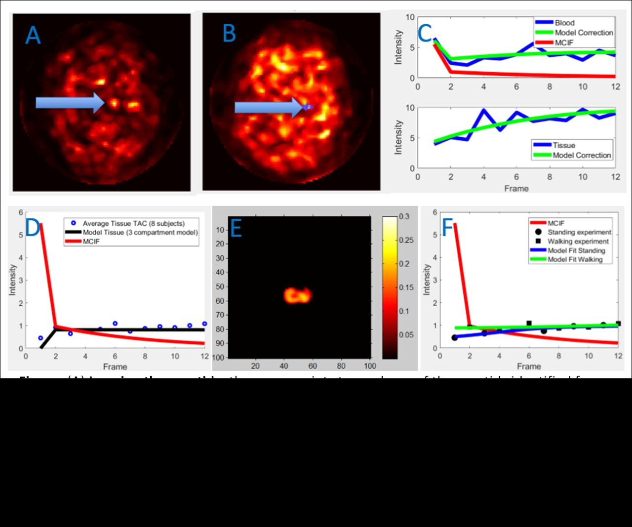
Methods: In the pilot FDG brain PET imaging experiment, using the small field-of-view (5 cm) wearable Helmet_PET imager [1], dynamic data in 8 subjects were obtained in an upright position in list mode (LM) from the point of injection of ~1-2 mCi of FDG. Each of the subjects followed the repeated two-cycle protocol: walking in place for 30 seconds, stopping for 30 seconds, again walking for 30 seconds, etc, over a period of 6 minutes total. The LM data was reconstructed using MLEM algorithm into twelve 30 sec time bins (24 slices per time frame). The reconstructed spatial resolution was 2x2 mm2 (100 by 100 pixels) with a slice thickness also of 2 mm. Two additional subjects aside from the 8 original subjects were imaged with the imager low enough to capture the carotid arteries in the FOV in LM format from the point of injection to obtain image derived blood input function. These subjects also underwent whole brain PET/CT imaging. The carotid arteries were first identified in representative MR images and then located in corresponding PET slices at the early time points (first 30 second frame) when FDG is in the blood and a region of interest (ROI) drawn to obtain arterial input function (blood time activity curve (TAC)). In addition, an ROI was drawn in the surrounding tissue in the 6 minute time frame to obtain tissue TAC. A model corrected blood input function (MCIF) was generated using a dual output model, wherein, the blood input function corrected for spill-over and partial volume averaging was obtained. The MCIF was then used to compute rate of FDG uptake (Ki) for average TAC obtained from 8 subjects for a region of interest (leg motor cortex) using a FDG transport model correcting for spill-out from the blood and also radioactivity recovery of the tissue of interest [2] . The walking and standing time frames (alternate frames) were also isolated for each of the 8-subjects imaged and average TACs generated. The average rate of FDG uptake for walking in place (Ki-walking) and standing in place (Ki-standing) with standard error was computed.

Results: The Ki for the tissue of interest (leg-related motor cortex) averaged over 8-subjects was computed to be 0.13 ml/min/g. The average Ki-standing and Ki-walking was computed to be 0.12±0.014 ml/min/g and 0.14±0.021 ml/min/g respectively (p=0.57).

Conclusions: Summarizing this pilot Helmet_PET experiment for a walking-in-place task, the data trend of the two-mode motion protocol suggests that even with a bolus injection, task-related motor cortex activity can be differentiated using the changing rate of FDG uptake. In the future, superior results may be obtained using a continuous infusion ligand delivery, as has been utilized in other FDG studies. Grant support: NIH R24 MH106057 (JBL); Radiology start-up at UVA (BK).

Figure
  • Download figure
  • Open in new tab
  • Download powerpoint
Previous
Back to top

In this issue

Journal of Nuclear Medicine
Vol. 59, Issue supplement 1
May 1, 2018
  • Table of Contents
  • Index by author
Article Alerts
Sign In to Email Alerts with your Email Address
Email Article

Thank you for your interest in spreading the word on Journal of Nuclear Medicine.

NOTE: We only request your email address so that the person you are recommending the page to knows that you wanted them to see it, and that it is not junk mail. We do not capture any email address.

Enter multiple addresses on separate lines or separate them with commas.
Dynamic FDG brain PET imaging using Helmet_PET: Model corrected blood input function to compute FDG uptake rate during an ambulatory upright task
(Your Name) has sent you a message from Journal of Nuclear Medicine
(Your Name) thought you would like to see the Journal of Nuclear Medicine web site.
Citation Tools
Dynamic FDG brain PET imaging using Helmet_PET: Model corrected blood input function to compute FDG uptake rate during an ambulatory upright task
Qiao Huang, Chris Bauer, Alexander Stolin, Thorsten Wuest, Samantha Melroy, Stan Majewski, Julie Brefczynski-Lewis, Bijoy Kundu
Journal of Nuclear Medicine May 2018, 59 (supplement 1) 427;

Citation Manager Formats

  • BibTeX
  • Bookends
  • EasyBib
  • EndNote (tagged)
  • EndNote 8 (xml)
  • Medlars
  • Mendeley
  • Papers
  • RefWorks Tagged
  • Ref Manager
  • RIS
  • Zotero
Share
Dynamic FDG brain PET imaging using Helmet_PET: Model corrected blood input function to compute FDG uptake rate during an ambulatory upright task
Qiao Huang, Chris Bauer, Alexander Stolin, Thorsten Wuest, Samantha Melroy, Stan Majewski, Julie Brefczynski-Lewis, Bijoy Kundu
Journal of Nuclear Medicine May 2018, 59 (supplement 1) 427;
Twitter logo Facebook logo LinkedIn logo Mendeley logo
  • Tweet Widget
  • Facebook Like
  • Google Plus One
Bookmark this article

Jump to section

  • Article
  • Figures & Data
  • Info & Metrics

Related Articles

  • No related articles found.
  • Google Scholar

Cited By...

  • No citing articles found.
  • Google Scholar

More in this TOC Section

Instrumentation & Data Analysis Track

  • Deep Learning Based Kidney Segmentation for Glomerular Filtration Rate Measurement Using Quantitative SPECT/CT
  • Comparison of 22 partial volume correction methods for amyloid PET imaging with 11C-PiB
  • The Benefit of Time-of-Flight in Digital Photon Counting PET Imaging: Physics and Clinical Evaluation
Show more Instrumentation & Data Analysis Track

Quantitative Imaging

  • Performance evaluation and quantitative accuracy of a multi-pinhole small animal SPECT/CT scanner for theranostic Lu-177 imaging
  • A Preclinical SPECT System with Optimized Data Acquisition for Imaging Larger Animals
Show more Quantitative Imaging

Similar Articles

SNMMI

© 2023 Journal of Nuclear Medicine

Powered by HighWire