


Abstract
1667
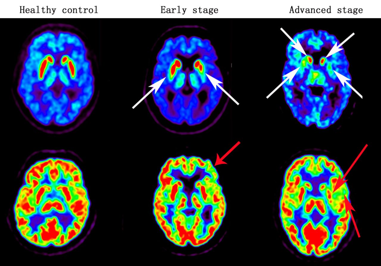
Objectives: To evaluate the clinical application value of 18F-FDG cerebral metabolism and vesicular monoamine transporter type 2 (VMAT II) imaging positron emission tomography (PET) imaging in diagnosing and assessing the severity of Parkinson’s disease (PD).Methods:[18F]9-fluoropropyl-(+)-dihydrotetrabenazine([18F]FP-(+)-DTBZ) PET was used to explore the characteristics of VMAT II imaging. 30 patients with PD and 10 healthy controls were recruited for the 18F-FDG and 18F-FP-DTBZ PET scans in 2 days. According to Hoehn-Yahr (H&Y) scale and Unified Parkinson’s Disease Rating Scale motor score (UPDRS III), the patients were further divided into 2 subgroups of early stage (H&Y: 1-2, n=19) and advanced stage (H&Y: 2.5-5, n=11).The 18F-FP-DTBZ uptake of caudate, anterior putamen and posterior putamen were obtained by volumes of interest (VOIs)-based on image analyses. The specific uptake ratio (SUR) of each VOI was calculated as target uptake / reference uptake using the occipital reference region normalized 18F-FP-DTBZ images for each participant. The 18F-FDG uptake of caudate, putamen and cerebral cortex were also obtained by cerebellum as reference.Results: 86.7% and 20% patients were cortices and unilateral putamen hypometabolism in 18F-FDG PET images (p<0.01, p<0.05). The mean reductions of VMAT II density for the caudate, anterior putamen and posterior putamen were 24.60%, 48.00%, and 57.50% for the advanced stage (p<0.01). However, there was no significant difference in the caudate for the early stage (p=0.252). Anterior putamen and posterior putamen were reduced by 27.30% and 36.40%, respectively (p<0.01). The SURs of caudate, anterior putamen and posterior putamen exhibited significantly negative correlations (r: -0.574 ~ -0.807; p<0.01) to H&Y scores and UPDRS III scores. Conclusions: Combined application of 18F-FDG cerebral metabolism and VMAT II PET imaging is a potential imaging biomarker in the diagnosis and monitoring the severity of PD, or some other diseases with further exploration.

In this issue


Jump to section
Related Articles
Cited By...
- No citing articles found.