
Main menu

  • Home
  • Content
    • Current
    • Ahead of print
    • Past Issues
    • JNM Supplement
    • SNMMI Annual Meeting Abstracts
  • Subscriptions
    • Subscribers
    • Institutional and Non-member
    • Rates
    • Corporate & Special Sales
    • Journal Claims
  • Authors
    • Submit to JNM
    • Information for Authors
    • Assignment of Copyright
    • AQARA requirements
  • Info
    • Permissions
    • Advertisers
    • Continuing Education
  • About
    • About Us
    • Editorial Board
    • Contact Information
  • More
    • Alerts
    • Feedback
    • Help
    • SNMMI Journals
  • SNMMI
    • JNM
    • JNMT
    • SNMMI Journals
    • SNMMI

User menu

  • Subscribe
  • My alerts
  • Log in
  • My Cart

Search

  • Advanced search
Journal of Nuclear Medicine
  • SNMMI
    • JNM
    • JNMT
    • SNMMI Journals
    • SNMMI
  • Subscribe
  • My alerts
  • Log in
  • My Cart
Journal of Nuclear Medicine

Advanced Search

  • Home
  • Content
    • Current
    • Ahead of print
    • Past Issues
    • JNM Supplement
    • SNMMI Annual Meeting Abstracts
  • Subscriptions
    • Subscribers
    • Institutional and Non-member
    • Rates
    • Corporate & Special Sales
    • Journal Claims
  • Authors
    • Submit to JNM
    • Information for Authors
    • Assignment of Copyright
    • AQARA requirements
  • Info
    • Permissions
    • Advertisers
    • Continuing Education
  • About
    • About Us
    • Editorial Board
    • Contact Information
  • More
    • Alerts
    • Feedback
    • Help
    • SNMMI Journals
  • Follow JNM on Twitter
  • Visit JNM on Facebook
  • Join JNM on LinkedIn
  • Subscribe to our RSS feeds
Meeting ReportOncology, Basic Science Track

Targeting VPAC1 receptors for imaging glioblastoma

Sushil Tripathi, Rhonda Kean, D. Craig Hooper, Yuan Yuan Jin, Eric Wickstrom and Mathew Thakur
Journal of Nuclear Medicine May 2018, 59 (supplement 1) 1262;
Sushil Tripathi
4Radiology Thomas Jefferson University Philadelphia PA United States
  • Find this author on Google Scholar
  • Find this author on PubMed
  • Search for this author on this site
Rhonda Kean
3Cancer Biology Thomas Jefferson University Philadelphia PA United States
  • Find this author on Google Scholar
  • Find this author on PubMed
  • Search for this author on this site
D. Craig Hooper
1Sidney Kimmel Cancer Center Philadelphia PA United States
3Cancer Biology Thomas Jefferson University Philadelphia PA United States
  • Find this author on Google Scholar
  • Find this author on PubMed
  • Search for this author on this site
Yuan Yuan Jin
2Biochemistry and Molecular Biology Thomas Jefferson University Philadelphia PA United States
  • Find this author on Google Scholar
  • Find this author on PubMed
  • Search for this author on this site
Eric Wickstrom
1Sidney Kimmel Cancer Center Philadelphia PA United States
2Biochemistry and Molecular Biology Thomas Jefferson University Philadelphia PA United States
  • Find this author on Google Scholar
  • Find this author on PubMed
  • Search for this author on this site
Mathew Thakur
1Sidney Kimmel Cancer Center Philadelphia PA United States
4Radiology Thomas Jefferson University Philadelphia PA United States
  • Find this author on Google Scholar
  • Find this author on PubMed
  • Search for this author on this site
  • Article
  • Figures & Data
  • Info & Metrics
Loading

Abstract

1262

Objectives: Scintigraphic imaging of human glioblastoma continues to be challenging. Objective was to develop a genomic tracer to efficiently image glioblastoma at an early stage.

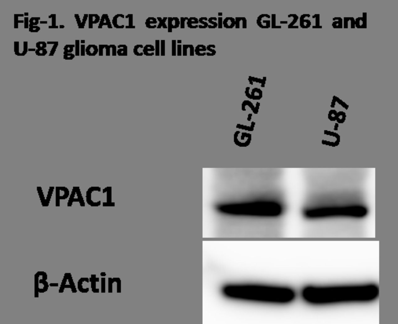
Methods: Expression of VPAC1 receptors on the mouse GL-261 and human U-87 glioma cell lines was determined by western blot. Cell binding assay was performed by incubating 64Cu-TP3805 with 1.2 x 10⁶ GL-261 and U-87 cells. GL-261 cell line which mimics human glioblastoma was chosen and was grown in tissue culture. 105 GL-261 cells were then implanted in the right brain lobe of T-bet knockout C57BL/6 mice (N=5). Tumors were allowed to grow for 2-3 weeks. PET/CT imaging and tissue distribution studies were performed using F-18-FDG and 24 hrs later using Cu-64-TP3805, which has been shown in our laboratory to efficiently image breast and prostate cancers in humans. On day 10 to 14 after GL-261 implantation mice were injected i.v., first with 150 µCi of F-18-FDG and 24 hrs later with 150 µCi of Cu-64-TP3805. Mice were imaged using Micro-PET/CT 2 hr later, sacrificed and % ID/g of tumor and normal brain were calculated. To prepare Cu-64-TP3805, 30 µL of 64CuCl2 in 0.1M HC1, was added to peptide-conjugate (20 µg) in 200 µL of 0.2M glycine buffer (pH=9.27), and incubated at 70º C for 90 min. The radiochemical purity was determined by radio-HPLC.

Results: The labeling efficiency of 64-TP3805 as determined by radio-HPLC was 96.3±0.5% with retention time for 64Cu-peptide at 5.3 min and for free 64CuCl2 at 3.4 min. We found strong signal for VPAC1 receptor on western blot for GL-261 and U-87 cells (Fig-1). The cell binding for 64Cu-peptide was 86±1.5% for both the cell lines. Micro-PET/CT image analyses and tissue distribution showed that the Cu-64-TP3805 tumor uptake at 2 hrs post injection was 7±2% ID/g as compared to 0.96±0.3% ID/g for normal brain. The normal tissue distribution showed a similar pattern as observed in PET imaging. F-18-FDG brain images were unclear for tumor imaging. Conclusion: Western blot studies confirmed high VPAC1 expression on the glioblastoma cell lines examined. 64Cu-TP3805 showed excellent labeling efficiency, and high uptake in glioblastoma. Targeting VPAC1 receptors using 64Cu-TP3805 for PET imaging of glioblastoma is a promising approach and calls for further investigation. Acknowledgements: The research, in part, was supported by NIH/NCI R01CA157372 (MLT), NIH/NCI 1S10OD012406 (MLT) and NIH/NCI S10RR23709 (MLT).

Figure
  • Download figure
  • Open in new tab
  • Download powerpoint
Previous
Back to top

In this issue

Journal of Nuclear Medicine
Vol. 59, Issue supplement 1
May 1, 2018
  • Table of Contents
  • Index by author
Article Alerts
Sign In to Email Alerts with your Email Address
Email Article

Thank you for your interest in spreading the word on Journal of Nuclear Medicine.

NOTE: We only request your email address so that the person you are recommending the page to knows that you wanted them to see it, and that it is not junk mail. We do not capture any email address.

Enter multiple addresses on separate lines or separate them with commas.
Targeting VPAC1 receptors for imaging glioblastoma
(Your Name) has sent you a message from Journal of Nuclear Medicine
(Your Name) thought you would like to see the Journal of Nuclear Medicine web site.
Citation Tools
Targeting VPAC1 receptors for imaging glioblastoma
Sushil Tripathi, Rhonda Kean, D. Craig Hooper, Yuan Yuan Jin, Eric Wickstrom, Mathew Thakur
Journal of Nuclear Medicine May 2018, 59 (supplement 1) 1262;

Citation Manager Formats

  • BibTeX
  • Bookends
  • EasyBib
  • EndNote (tagged)
  • EndNote 8 (xml)
  • Medlars
  • Mendeley
  • Papers
  • RefWorks Tagged
  • Ref Manager
  • RIS
  • Zotero
Share
Targeting VPAC1 receptors for imaging glioblastoma
Sushil Tripathi, Rhonda Kean, D. Craig Hooper, Yuan Yuan Jin, Eric Wickstrom, Mathew Thakur
Journal of Nuclear Medicine May 2018, 59 (supplement 1) 1262;
del.icio.us logo Digg logo Reddit logo Twitter logo Facebook logo Google logo Mendeley logo
  • Tweet Widget
  • Facebook Like
  • Google Plus One
Bookmark this article

Jump to section

  • Article
  • Figures & Data
  • Info & Metrics

Related Articles

  • No related articles found.
  • Google Scholar

Cited By...

  • No citing articles found.
  • Google Scholar

More in this TOC Section

Oncology, Basic Science Track

  • Imaging adult glioma with 68Ga-citrate PET/MR
  • Evaluation of L-1-[18F]Fluoroethyl-Tryptophan for PET Imaging of Cancer
  • Pretargeted radioimmunotherapy with 225Ac-proteus-DOTA hapten.
Show more Oncology, Basic Science Track

Basic Science Posters (Oncology)

  • Imaging adult glioma with 68Ga-citrate PET/MR
  • Evaluation of L-1-[18F]Fluoroethyl-Tryptophan for PET Imaging of Cancer
  • HER2-targeted SPECT imaging of nude mice with colon cancer xenografts using 125I-Herceptin
Show more Basic Science Posters (Oncology)

Similar Articles

SNMMI

© 2022 Journal of Nuclear Medicine

Powered by HighWire