
Main menu

  • Home
  • Content
    • Current
    • Ahead of print
    • Past Issues
    • JNM Supplement
    • SNMMI Annual Meeting Abstracts
  • Subscriptions
    • Subscribers
    • Institutional and Non-member
    • Rates
    • Corporate & Special Sales
    • Journal Claims
  • Authors
    • Submit to JNM
    • Information for Authors
    • Assignment of Copyright
    • AQARA requirements
  • Info
    • Permissions
    • Advertisers
    • Continuing Education
  • About
    • About Us
    • Editorial Board
    • Contact Information
  • More
    • Alerts
    • Feedback
    • Help
    • SNMMI Journals
  • SNMMI
    • JNM
    • JNMT
    • SNMMI Journals
    • SNMMI

User menu

  • Subscribe
  • My alerts
  • Log in
  • My Cart

Search

  • Advanced search
Journal of Nuclear Medicine
  • SNMMI
    • JNM
    • JNMT
    • SNMMI Journals
    • SNMMI
  • Subscribe
  • My alerts
  • Log in
  • My Cart
Journal of Nuclear Medicine

Advanced Search

  • Home
  • Content
    • Current
    • Ahead of print
    • Past Issues
    • JNM Supplement
    • SNMMI Annual Meeting Abstracts
  • Subscriptions
    • Subscribers
    • Institutional and Non-member
    • Rates
    • Corporate & Special Sales
    • Journal Claims
  • Authors
    • Submit to JNM
    • Information for Authors
    • Assignment of Copyright
    • AQARA requirements
  • Info
    • Permissions
    • Advertisers
    • Continuing Education
  • About
    • About Us
    • Editorial Board
    • Contact Information
  • More
    • Alerts
    • Feedback
    • Help
    • SNMMI Journals
  • Follow JNM on Twitter
  • Visit JNM on Facebook
  • Join JNM on LinkedIn
  • Subscribe to our RSS feeds
Meeting ReportNeurosciences Track

Evaluation of Intranasal Insulin Administration on Regional Metabolic Changes in Rat Brain with Cognitive testing during Uptake using [18F] FDG PET following moderate CCI

Shalini Jaiswal, Nicole Hockenbury, Catherine Meyer, Andrew Hoy, Bernard Dardzinski and Kimberly Byrnes
Journal of Nuclear Medicine May 2017, 58 (supplement 1) 655;
Shalini Jaiswal
5HJF, Uniformed Services University of Health Sciences Bethesda MD United States
1Center for Neuroscience and Regenerative Medicine Bethesda MD United States
  • Find this author on Google Scholar
  • Find this author on PubMed
  • Search for this author on this site
Nicole Hockenbury
2Anatomy, Physiology and Genetics Center for Neuroscience and Regenerative Medicine Bethesda MD United States
6Anatomy Physiology and Genetics HJF, Uniformed Services University of Health Sciences Bethesda MD United States
  • Find this author on Google Scholar
  • Find this author on PubMed
  • Search for this author on this site
Catherine Meyer
5HJF, Uniformed Services University of Health Sciences Bethesda MD United States
1Center for Neuroscience and Regenerative Medicine Bethesda MD United States
  • Find this author on Google Scholar
  • Find this author on PubMed
  • Search for this author on this site
Andrew Hoy
8Radiology and Radiological Sciences Uniformed Services University of Health Sciences Bethesda MD United States
4Radiology Center for Neuroscience and Regenerative Medicine, USU Bethesda MD United States
  • Find this author on Google Scholar
  • Find this author on PubMed
  • Search for this author on this site
Bernard Dardzinski
8Radiology and Radiological Sciences Uniformed Services University of Health Sciences Bethesda MD United States
4Radiology Center for Neuroscience and Regenerative Medicine, USU Bethesda MD United States
  • Find this author on Google Scholar
  • Find this author on PubMed
  • Search for this author on this site
Kimberly Byrnes
7Anatomy Physiology and Genetics Uniformed Services University of Health Sciences Bethesda MD United States
3Anatomy, Physiology and Genetics Center for Neuroscience and Regenerative Medicine, USU Bethesda MD United States
  • Find this author on Google Scholar
  • Find this author on PubMed
  • Search for this author on this site
  • Article
  • Figures & Data
  • Info & Metrics
Loading

Abstract

655

Objectives: Traumatic Brain Injury (TBI) can lead to long term cognitive deficits. These cognitive impairments are often associated with complex metabolic alterations (1). Currently there is no treatment for the cellular and metabolic dysfunction that occurs after injury. The objective of this pilot study was to trace regional metabolic changes due to intranasal insulin / saline treatment in the injured rat brain with novel object recognition test during uptake (2,3).

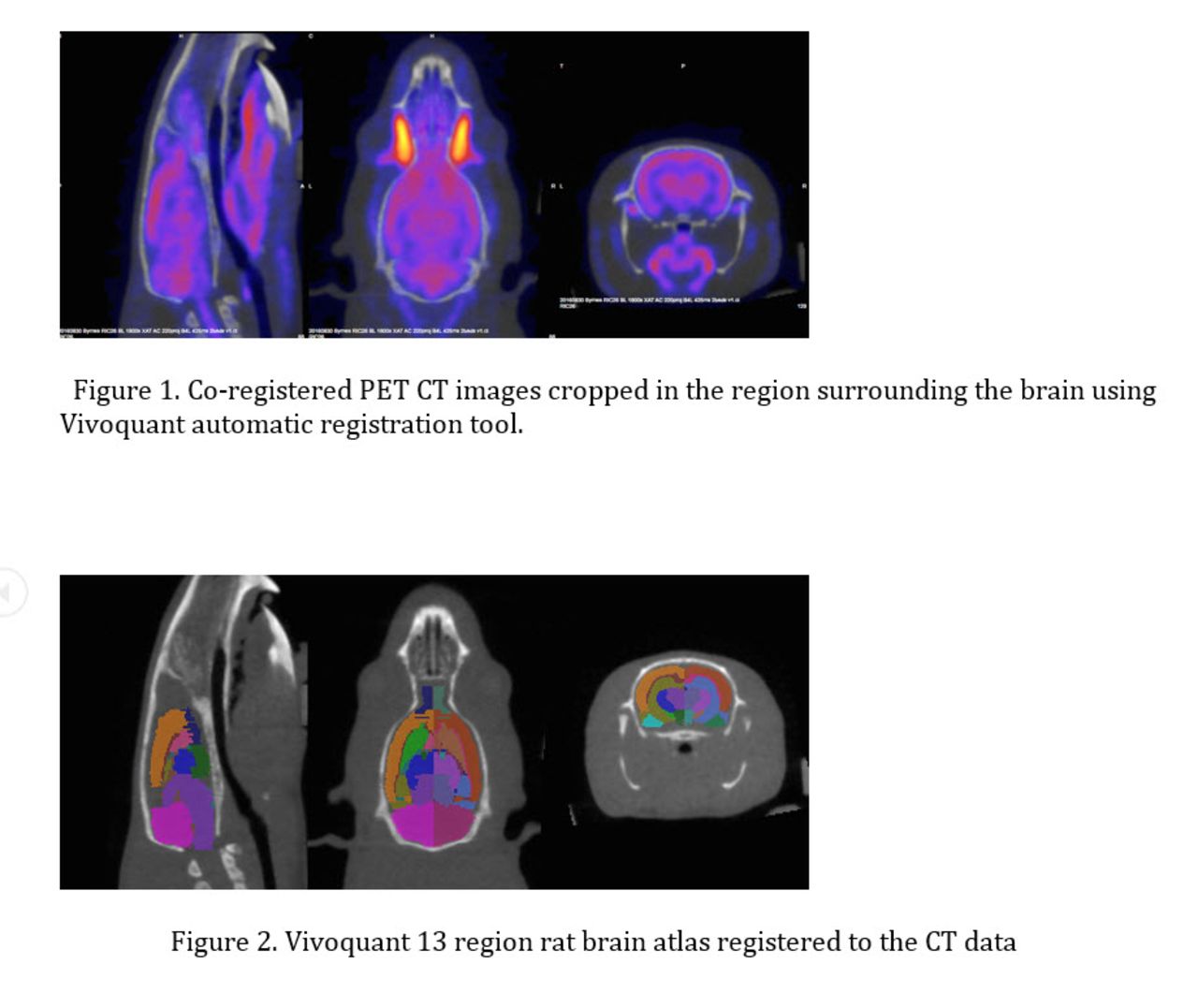
Methods: Adult male Sprague Dawley rats were imaged at 3 time points [(baseline, prior to injury), day 2 and day 10 (post injury)]. The animals were given intranasal insulin (n=7) or saline (n=5) treatment beginning at 24 hours after injury and continuing daily for 14 days. For PET/CT imaging, [18F] FDG (1.8 ± 0.06 mCi) was injected in the anesthetized animals via lateral tail vein. For the conscious uptake, the animals were kept in a separate cage (without food and water) on a heating pad (37°C). Cognitive testing (novel object recognition) was performed during the uptake period. For PET/CT imaging (Siemens Inveon pre-clinical system, Erlangen, Germany), the animals were anesthetized after 30 minutes of conscious uptake. Total uptake time before PET acquisition was 45 minutes. Static PET scans were acquired for 30 minutes followed by CT scans for anatomical localization and scatter and attenuation correction. The PET data were reconstructed as a single static frame with OSEM3D/MAP algorithm and corrected for scatter and attenuation. The CT images were reconstructed using Feldkamp algorithm, downsample2 with beam hardening and HU corrections applied. The reconstructed PET/CT data were processed and analyzed using Vivoquant Software version 2.5 (inviCRO, Boston MA). The PET data were registered to the 13 regions rat brain atlas by way of CT using an automatic algorithm (Figure 1, 2). The data analysis was performed using a semi quantitative method. Standardized Uptake Value (SUV - activity concentration in a specific region normalized to the total injected activity and body weight) normalized to the whole brain (SUVw) was analyzed in 13 different brain regions. All statistical analyses were conducted using Graph Pad Prism software version 7.01

Results: 1. There were no significant changes in the baseline scans of insulin versus saline treated animals (multiple t-tests using Holm-Sidak method with alpha = 0.05). 2. Significant changes in regional brain glucose metabolism were found in insulin and saline treated groups after injury (from baseline to day 2 and /or day 10, repeated measures two-way ANOVA, Sidak’s multiple comparisons tests). Insulin Group i) Hypermetabolism was seen in hypothalamus, midbrain and olfactory regions on day 2 when compared to baseline scans, returning to baseline scans by day 10. ii) Significant decrease in glucose metabolism was detected in cortex and corpus callosum on day 2 when compared to baseline scans. Saline Group i) A significant decrease in cortical uptake was observed at day 2 and day 10 compared to baseline. ii) Increased uptake was evident in hypothalamus at day 2 and day 10 when compared to baseline scans. iii) Significant increase in uptake was observed in olfactory and hippocampal regions on day 2 compared to baseline and remained elevated at day 10. iv) Increase in glucose uptake in corpus callosum is apparent only at day 10. v) Increased uptake in cerebellum and midbrain were also noticed at day 2 compared to baseline but returns to baseline by day 10.

Conclusion: Our findings demonstrate that metabolic changes due to TBI and treatment can be evaluated using [18F] FDG imaging. Regional brain glucose uptake changes after TBI revert back to normal (baseline) in the insulin treated group by day 10 compared to saline treated animals. This suggests that insulin treatment post-TBI may mediate metabolic changes associated with injury. Research Support: This project is supported by CNRM and HJF.

Figure
  • Download figure
  • Open in new tab
  • Download powerpoint
Previous
Back to top

In this issue

Journal of Nuclear Medicine
Vol. 58, Issue supplement 1
May 1, 2017
  • Table of Contents
  • Index by author
Article Alerts
Sign In to Email Alerts with your Email Address
Email Article

Thank you for your interest in spreading the word on Journal of Nuclear Medicine.

NOTE: We only request your email address so that the person you are recommending the page to knows that you wanted them to see it, and that it is not junk mail. We do not capture any email address.

Enter multiple addresses on separate lines or separate them with commas.
Evaluation of Intranasal Insulin Administration on Regional Metabolic Changes in Rat Brain with Cognitive testing during Uptake using [18F] FDG PET following moderate CCI
(Your Name) has sent you a message from Journal of Nuclear Medicine
(Your Name) thought you would like to see the Journal of Nuclear Medicine web site.
Citation Tools
Evaluation of Intranasal Insulin Administration on Regional Metabolic Changes in Rat Brain with Cognitive testing during Uptake using [18F] FDG PET following moderate CCI
Shalini Jaiswal, Nicole Hockenbury, Catherine Meyer, Andrew Hoy, Bernard Dardzinski, Kimberly Byrnes
Journal of Nuclear Medicine May 2017, 58 (supplement 1) 655;

Citation Manager Formats

  • BibTeX
  • Bookends
  • EasyBib
  • EndNote (tagged)
  • EndNote 8 (xml)
  • Medlars
  • Mendeley
  • Papers
  • RefWorks Tagged
  • Ref Manager
  • RIS
  • Zotero
Share
Evaluation of Intranasal Insulin Administration on Regional Metabolic Changes in Rat Brain with Cognitive testing during Uptake using [18F] FDG PET following moderate CCI
Shalini Jaiswal, Nicole Hockenbury, Catherine Meyer, Andrew Hoy, Bernard Dardzinski, Kimberly Byrnes
Journal of Nuclear Medicine May 2017, 58 (supplement 1) 655;
del.icio.us logo Digg logo Reddit logo Twitter logo Facebook logo Google logo Mendeley logo
  • Tweet Widget
  • Facebook Like
  • Google Plus One
Bookmark this article

Jump to section

  • Article
  • Figures & Data
  • Info & Metrics

Related Articles

  • No related articles found.
  • Google Scholar

Cited By...

  • No citing articles found.
  • Google Scholar

More in this TOC Section

Neurosciences Track

  • Impact of cognitive reserve in frontotemporal dementia illustrated by FDG-PET.
  • Quantification of brain cholinergic denervation in dementia with Lewy bodies using PET imaging with 18F-FEOBV
  • Kinetic evaluation of [18F]MOZAT PET imaging in humans.
Show more Neurosciences Track

Basic Science III: Preclinical Neuroimaging: Novel Methods & Applications

  • Validation of an image derived input function estimation method on PET/MR
  • Mu opioid receptor SNP alters neuroadaptation of the 5-HT1A serotonergic system following fixed-dose ethanol exposure in non-human primates
  • PET/fMRI reveals no functional response to 5-HT4 drugs even at high receptor occupancy
Show more Basic Science III: Preclinical Neuroimaging: Novel Methods & Applications

Similar Articles

SNMMI

© 2022 Journal of Nuclear Medicine

Powered by HighWire