


Abstract
647
Objectives Alkaline protease (AP) reduces the progress of TNF-α-induced mouse inflammatory bowel disease. FDG-PET provides accurate, non-invasive visualization of inflammation. We investigated whether FDG-PET imaging is useful to detect therapeutic response of alkaline protease in acute inflammatory bowel disease (IBD).
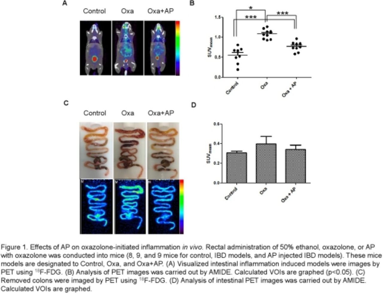
Methods Rectal administration of the hapten reagent oxazolone dissolved in ethanol induces a severe IBD in C56BL/6 mice. The mice are divided into three groups: Control, Oxa, and Oxa+AP administered mice. After 4 hours of oxazolone administration, FDG-PET images were acquired using small animal PET/X-ray scanner. PET data were acquired from whole body approximately 60 min after intravenous administration of 50 μCi of 18F-FDG. The mice were sacrificed and the colon was removed. Ex vivo PET images were acquired. To determine whether AP blocks TNF-α activity in this acute inflammation induced mice, samples of their intestines were collected. And then TNF-α immunostaining of intestines was carried out.
Results Oxazolone treated mouse model showed swelled intestine and darker color than the other group. Immunohistochemistry assay showed less TNF-α staining in villous epithelium of group Oxa+AP than of group Oxa. FDG uptake increased 2.0 times of control when oxazolone had treated (p=0.016). However, FDG uptake increased 1.4 times of control when oxazolone and AP administered together (p=0.001).
Conclusions Pretreatment of alkaline protease reduced not only TNF-α expression but also FDG uptake. FDG-PET shows therapeutic effect of AP in mouse IBD.
In this issue


Jump to section
Related Articles
Cited By...
- No citing articles found.