
Main menu

  • Home
  • Content
    • Current
    • Ahead of print
    • Past Issues
    • JNM Supplement
    • SNMMI Annual Meeting Abstracts
  • Subscriptions
    • Subscribers
    • Rates
    • Journal Claims
    • Institutional and Non-member
  • Authors
    • Submit to JNM
    • Information for Authors
    • Assignment of Copyright
    • AQARA requirements
  • Info
    • Permissions
    • Advertisers
    • Continuing Education
    • Corporate & Special Sales
  • About
    • About Us
    • Editorial Board
    • Editorial Contact
  • More
    • Alerts
    • Feedback
    • Help
    • SNMMI Journals
  • SNMMI
    • JNM
    • JNMT
    • SNMMI Journals
    • SNMMI

User menu

  • Subscribe
  • My alerts
  • Log in
  • My Cart

Search

  • Advanced search
Journal of Nuclear Medicine
  • SNMMI
    • JNM
    • JNMT
    • SNMMI Journals
    • SNMMI
  • Subscribe
  • My alerts
  • Log in
  • My Cart
Journal of Nuclear Medicine

Advanced Search

  • Home
  • Content
    • Current
    • Ahead of print
    • Past Issues
    • JNM Supplement
    • SNMMI Annual Meeting Abstracts
  • Subscriptions
    • Subscribers
    • Rates
    • Journal Claims
    • Institutional and Non-member
  • Authors
    • Submit to JNM
    • Information for Authors
    • Assignment of Copyright
    • AQARA requirements
  • Info
    • Permissions
    • Advertisers
    • Continuing Education
    • Corporate & Special Sales
  • About
    • About Us
    • Editorial Board
    • Editorial Contact
  • More
    • Alerts
    • Feedback
    • Help
    • SNMMI Journals
  • Follow SNMMI on Twitter
  • Visit SNMMI on Facebook
Meeting ReportNeurosciences Track

The impact of partial volume effect correction on brain network analysis using AV-45 PET data.

Jiarui Yang, Chenhui Hu, Ning Guo, Joyita Dutta, Georges El Fakhri and Quanzheng Li
Journal of Nuclear Medicine May 2016, 57 (supplement 2) 459;
Jiarui Yang
4Division of Nuclear Medicine & Molecular Imaging Massachusetts General Hospital Boston MA United States
  • Find this author on Google Scholar
  • Find this author on PubMed
  • Search for this author on this site
Chenhui Hu
1SEAS Harvard University Boston MA United States
  • Find this author on Google Scholar
  • Find this author on PubMed
  • Search for this author on this site
Ning Guo
2Massachusetts General Hospital Boston MA United States
3Massachusetts General Hospital Boston MA United States
  • Find this author on Google Scholar
  • Find this author on PubMed
  • Search for this author on this site
Joyita Dutta
6MGH / Harvard / UMass Lowell Boston MA United States
  • Find this author on Google Scholar
  • Find this author on PubMed
  • Search for this author on this site
Georges El Fakhri
5Massachusetts General Hospital, Harvard Medical Sc Boston MA United States
  • Find this author on Google Scholar
  • Find this author on PubMed
  • Search for this author on this site
Quanzheng Li
2Massachusetts General Hospital Boston MA United States
3Massachusetts General Hospital Boston MA United States
  • Find this author on Google Scholar
  • Find this author on PubMed
  • Search for this author on this site
  • Article
  • Figures & Data
  • Info & Metrics
Loading

Abstract

459

Objectives The quantitative accuracy of PET is degraded by partial volume effects caused by limited spatial resolution of PET scanners. The goal of this study was to evaluate the effectiveness of partial volume correction (PVC) [1] on AV-45 PET image and consequently its impact on brain network analysis. Brain network at different stages of Alzheimer’s disease (AD) was constructed to demonstrate of the impact of PVC.

Methods The AV-45 PET scans of 416 subjects in four different stages of AD, including NC (Normal Control), EMCI (Early Mild Cognitive Impairment), LMCI (Late Mild Cognitive Impairment) and AD, in ADNI (Alzheimer’s Disease Neuroimaging Initiative) database were analyzed. After image registration to MNI-152 brain template, PVC was performed on all images. Then brain networks of four stages of AD were generated from both pre- and post-partial volume corrected images. For each stage and each group (pre and post PVC) we then randomly selected 90% subjects to generate brain network and repeated 200 times to obtain 200 brain networks of each group. We performed permutation test on these 200 networks and did t-test on all different edges within stages.

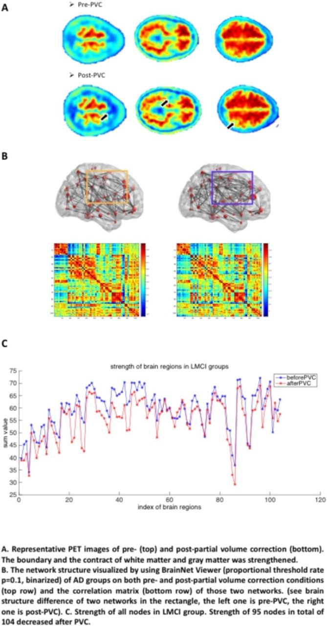
Results Spatial resolution of PET images was improved after PVC: the boundary of gray matter and white matter was strengthened and the details became more visible. Mean network degrees of LMCI group decreased significantly (from 11.0769±7.9717 to 8.4423±5.7502, network absolute threshold rate r=0.8) while other groups remained almost the same. The strength of nodes of brain networks also changed after PVC, especially in LMCI group, where 95 of total 104 regions decreased and 30.53% of them decreased more than 10%. Mean node strength of LMCI group decreased from 59.5318±8.2607 to 55.6213±8.4887 after PVC. 59 more edges (22.61% of insignificant edges before PVC) of whole brain network in LMCI and AD groups showed significant difference (p=0.05) after PVC, 118 more edges (47.98%) in EMCI and LMCI groups and 96 more edges (76.19%) in EMCI and AD groups, which suggests the effectiveness of PVC in distinguishing EMCI, LMCI and AD patients.

Conclusions Partial volume correction can improve the quantitative accuracy of AV-45 PET and consequently the accuracy of brain network analysis, especially in distinguishing EMCI, LMCI and AD patients.$$graphic_{E39B9FCF-A106-410A-BB22-CD3F11EB848F}$$

Figure1
  • Download figure
  • Open in new tab
  • Download powerpoint
Previous
Back to top

In this issue

Journal of Nuclear Medicine
Vol. 57, Issue supplement 2
May 1, 2016
  • Table of Contents
  • Index by author
Article Alerts
Sign In to Email Alerts with your Email Address
Email Article

Thank you for your interest in spreading the word on Journal of Nuclear Medicine.

NOTE: We only request your email address so that the person you are recommending the page to knows that you wanted them to see it, and that it is not junk mail. We do not capture any email address.

Enter multiple addresses on separate lines or separate them with commas.
The impact of partial volume effect correction on brain network analysis using AV-45 PET data.
(Your Name) has sent you a message from Journal of Nuclear Medicine
(Your Name) thought you would like to see the Journal of Nuclear Medicine web site.
Citation Tools
The impact of partial volume effect correction on brain network analysis using AV-45 PET data.
Jiarui Yang, Chenhui Hu, Ning Guo, Joyita Dutta, Georges El Fakhri, Quanzheng Li
Journal of Nuclear Medicine May 2016, 57 (supplement 2) 459;

Citation Manager Formats

  • BibTeX
  • Bookends
  • EasyBib
  • EndNote (tagged)
  • EndNote 8 (xml)
  • Medlars
  • Mendeley
  • Papers
  • RefWorks Tagged
  • Ref Manager
  • RIS
  • Zotero
Share
The impact of partial volume effect correction on brain network analysis using AV-45 PET data.
Jiarui Yang, Chenhui Hu, Ning Guo, Joyita Dutta, Georges El Fakhri, Quanzheng Li
Journal of Nuclear Medicine May 2016, 57 (supplement 2) 459;
del.icio.us logo Digg logo Reddit logo Twitter logo CiteULike logo Facebook logo Google logo Mendeley logo
  • Tweet Widget
  • Facebook Like
  • Google Plus One

Jump to section

  • Article
  • Figures & Data
  • Info & Metrics

Related Articles

  • No related articles found.
  • Google Scholar

Cited By...

  • No citing articles found.
  • Google Scholar

More in this TOC Section

Neurosciences Track

  • Impact of cognitive reserve in frontotemporal dementia illustrated by FDG-PET.
  • Quantification of brain cholinergic denervation in dementia with Lewy bodies using PET imaging with 18F-FEOBV
  • Kinetic evaluation of [18F]MOZAT PET imaging in humans.
Show more Neurosciences Track

Tau imaging and Amyloid imaging 1

  • Robustness of 18F-Florbetaben SUVR cutoff quantification across reference regions and standards of truth
  • New method of quantification of tau pathology with 11C-PBB3 PET using reference tissue extracted from cerebral cortical gray matter
  • PET assessment of human cerebral tau deposition using [18F]AV-1451 - comparison of quantification methods
Show more Tau imaging and Amyloid imaging 1

Similar Articles

SNMMI

© 2021 Journal of Nuclear Medicine

Powered by HighWire