


Article Figures & Data
Figures
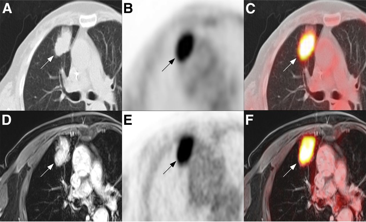
- FIGURE 1.
In 65-y-old woman with lung cancer, 38-mm tumor (arrows) is seen in right upper lobe of lung on CT component (A) of PET/CT (C) and on MRI component (D) of PET/MRI (F). SUVmax of strong 18F-FDG uptake on PET component is 17.3 for PET/CT (B) and 19.4 for PET/MRI (E).
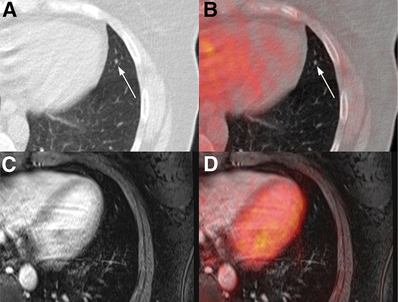
- FIGURE 2.
In 46-y-old woman with breast cancer, 4-mm nodule (arrows) is seen in lingula segment of left upper lobe of lung on CT component (A) of PET/CT (B) but not on MRI component (C) of PET/MRI (D). No 18F-FDG uptake is seen on PET component of either PET/CT (C) or PET/MRI (D).
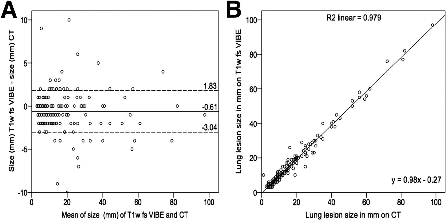
- FIGURE 3.
(A) For each of 161 lung lesions, difference between size on MRI component of PET/MRI and size on CT component of PET/CT is plotted against mean size, which was −0.61 mm (95% confidence interval, 1.83 and −3.04 mm). (B) Linear regression plot demonstrates correlation between size on MRI and size on CT (r = 0.98; P < 0.001). T1w fs = T1-weighted fat saturation.
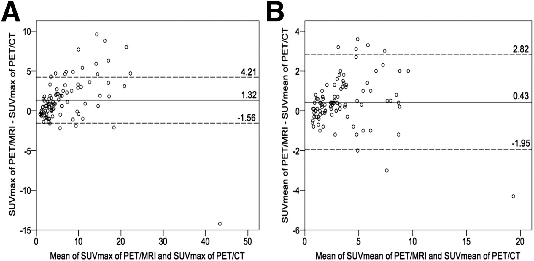
- FIGURE 4.
(A) For each of 161 lung lesions, difference in SUVmax (A) and SUVmean (B) between PET component of PET/MRI and PET component of PET/CT is plotted against mean difference, which was 1.32 for SUVmax (95% confidence interval, 4.21 and −1.56) and 0.43 for SUVmean (95% confidence interval, 2.82 and −1.95).


Tables
Type n Lung cancer 28 Lymphoma 21 Breast cancer 18 Uterine cancer 12 Ovarian cancer 10 Cancer of unknown primary 8 Malignant melanoma 4 Head and neck cancer 4 Gastrointestinal cancer 3 Malignant mesothelioma 3 Other (<3 cases/type) 10 Lesions detected (n) Modality Total Most likely benign Indeterminate Suggestive of malignancy PET/CT 241 110 (46%) 31 (13%) 100 (41%) PET/MRI 161 41 (25%) 14 (9%) 106 (66%)


