
Main menu

  • Home
  • Content
    • Current
    • Ahead of print
    • Past Issues
    • JNM Supplement
    • SNMMI Annual Meeting Abstracts
  • Subscriptions
    • Subscribers
    • Institutional and Non-member
    • Rates
    • Corporate & Special Sales
    • Journal Claims
  • Authors
    • Submit to JNM
    • Information for Authors
    • Assignment of Copyright
    • AQARA requirements
  • Info
    • Continuing Education
    • Reviewers
    • Permissions
    • Advertisers
  • About
    • About Us
    • Editorial Board
    • Contact Information
  • More
    • Alerts
    • Feedback
    • Help
    • SNMMI Journals
  • SNMMI
    • JNM
    • JNMT
    • SNMMI Journals
    • SNMMI

User menu

  • Subscribe
  • My alerts
  • Log in
  • My Cart

Search

  • Advanced search
Journal of Nuclear Medicine
  • SNMMI
    • JNM
    • JNMT
    • SNMMI Journals
    • SNMMI
  • Subscribe
  • My alerts
  • Log in
  • My Cart
Journal of Nuclear Medicine

Advanced Search

  • Home
  • Content
    • Current
    • Ahead of print
    • Past Issues
    • JNM Supplement
    • SNMMI Annual Meeting Abstracts
  • Subscriptions
    • Subscribers
    • Institutional and Non-member
    • Rates
    • Corporate & Special Sales
    • Journal Claims
  • Authors
    • Submit to JNM
    • Information for Authors
    • Assignment of Copyright
    • AQARA requirements
  • Info
    • Continuing Education
    • Reviewers
    • Permissions
    • Advertisers
  • About
    • About Us
    • Editorial Board
    • Contact Information
  • More
    • Alerts
    • Feedback
    • Help
    • SNMMI Journals
  • Visit JNM on Facebook
  • Join JNM on LinkedIn
  • Follow JNM on Twitter
  • Subscribe to our RSS feeds
Research ArticleClinical Investigations

Mapping α2 Adrenoceptors of the Human Brain with 11C-Yohimbine

Adjmal Nahimi, Steen Jakobsen, Ole L. Munk, Kim Vang, Jenny A. Phan, Anders Rodell and Albert Gjedde
Journal of Nuclear Medicine March 2015, 56 (3) 392-398; DOI: https://doi.org/10.2967/jnumed.114.145565
Adjmal Nahimi
1Department of Nuclear Medicine and PET Centre, Aarhus University Hospitals, Aarhus, Denmark
  • Find this author on Google Scholar
  • Find this author on PubMed
  • Search for this author on this site
Steen Jakobsen
1Department of Nuclear Medicine and PET Centre, Aarhus University Hospitals, Aarhus, Denmark
  • Find this author on Google Scholar
  • Find this author on PubMed
  • Search for this author on this site
Ole L. Munk
1Department of Nuclear Medicine and PET Centre, Aarhus University Hospitals, Aarhus, Denmark
  • Find this author on Google Scholar
  • Find this author on PubMed
  • Search for this author on this site
Kim Vang
1Department of Nuclear Medicine and PET Centre, Aarhus University Hospitals, Aarhus, Denmark
  • Find this author on Google Scholar
  • Find this author on PubMed
  • Search for this author on this site
Jenny A. Phan
1Department of Nuclear Medicine and PET Centre, Aarhus University Hospitals, Aarhus, Denmark
  • Find this author on Google Scholar
  • Find this author on PubMed
  • Search for this author on this site
Anders Rodell
1Department of Nuclear Medicine and PET Centre, Aarhus University Hospitals, Aarhus, Denmark
  • Find this author on Google Scholar
  • Find this author on PubMed
  • Search for this author on this site
Albert Gjedde
1Department of Nuclear Medicine and PET Centre, Aarhus University Hospitals, Aarhus, Denmark
2Department of Neuroscience and Pharmacology, University of Copenhagen, Copenhagen, Denmark
3Department of Radiology and Radiological Science, Johns Hopkins University, Baltimore, Maryland; and
4Department of Neurology, McGill University, Montreal, Quebec, Canada
  • Find this author on Google Scholar
  • Find this author on PubMed
  • Search for this author on this site
  • Article
  • Figures & Data
  • Info & Metrics
  • PDF
Loading

Article Figures & Data

Figures

  • Tables
  • FIGURE 1.
    • Download figure
    • Open in new tab
    • Download powerpoint
    FIGURE 1.

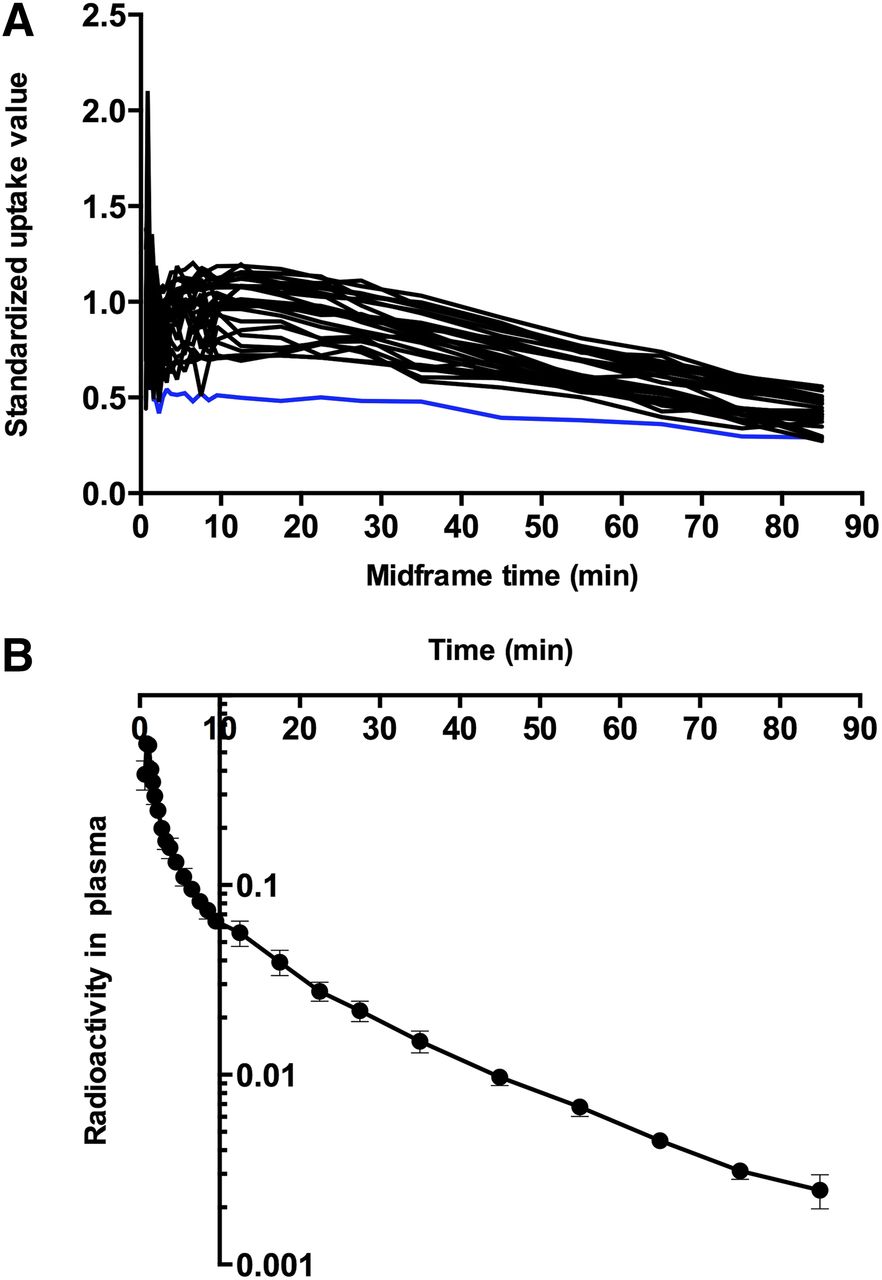
    (A) Examples of standardized uptake value curves for all 27 VOIs of 11C-yohimbine presented for subject 5 (abscissa is mid-frame time [minutes] and ordinate is standardized uptake value of radioactivity in VOIs). Blue line shows corpus callosum, which was used as reference region for calculation of binding potentials (BPND). (B) Radioactivity in plasma, expressed as fraction of radioactivity in VOIs, is shown on ordinate, and abscissa represents time (minutes).

  • FIGURE 2.
    • Download figure
    • Open in new tab
    • Download powerpoint
    FIGURE 2.

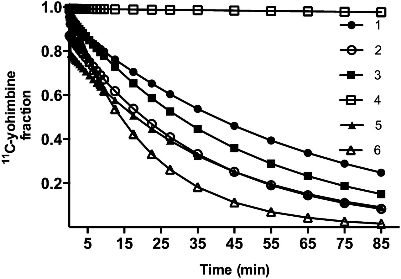
    Fractions of unchanged 11C-yohimbine were interpolated to match time points of tissue time–activity curves, presented here for all 6 subjects. Ordinate represents intact 11C-yohimbine, and abscissa shows time (minutes).

  • FIGURE 3.
    • Download figure
    • Open in new tab
    • Download powerpoint
    FIGURE 3.

    Six linearized solutions were used to derive linear regression estimates of kinetic parameters, using metabolite-corrected plasma curves as input function: plot N1 (A), plot N2 (B), plot P1 (C), plot P2 (D), plot P3 (E), plot P4 (F). Blue lines represent corpus callosum.

  • FIGURE 4.
    • Download figure
    • Open in new tab
    • Download powerpoint
    FIGURE 4.

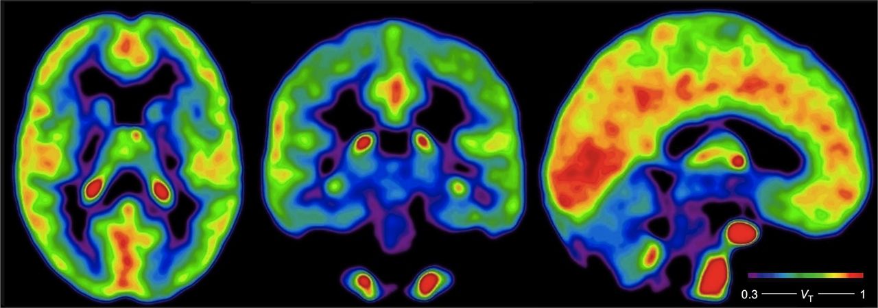
    Average parametric images of voxelwise Embedded Image estimates in 5 of 6 subjects, estimated with P2 plot (Logan plot). Color bar gives estimates of Embedded Image in units of mL cm−3.

Tables

  • Figures
    • View popup
    TABLE 1

    Injected Radioactivity Dose, Yohimbine Mass, and CYP2D6 Status

    SubjectMBqYohimbine (nmol)CYP2D6
    164130.31 × 2 (F)
    24414.64 × 1 (M)
    363571 × 4 (M)
    471521.44 × 4 (N)
    550711.22 × 5 (M)
    6169112 × 2 (F)
    • F, M, and N = fast, moderate, and nonmetabolizers, respectively, of 11C-yohimbine.

    • View popup
    TABLE 2

    Kinetic Parameters of 11C-Yohimbine

    VOIVT (mL/cm3)K1 (mL/cm3/min)Embedded Image (min−1)Binding potential
    Amygdala, L0.55 ± 0.110.014 ± 0.0020.026 ± 0.0020.22 ± 0.12
    Amygdala, R0.55 ± 0.100.014 ± 0.0020.025 ± 0.0010.23 ± 0.07
    Caudate nucleus, L0.55 ± 0.100.016 ± 0.0030.029 ± 0.0020.24 ± 0.13
    Caudate nucleus, R0.56 ± 0.120.016 ± 0.0030.028 ± 0.0020.23 ± 0.11
    Cerebellum, L0.57 ± 0.100.019 ± 0.0030.032 ± 0.0020.28 ± 0.11
    Cerebellum, R0.59 ± 0.110.020 ± 0.0030.032 ± 0.0020.32 ± 0.11
    Corpus callosum0.46 ± 0.090.009 ± 0.0010.019 ± 0.003—
    Frontal lobe, L0.80 ± 0.160.020 ± 0.0030.023 ± 0.0010.77 ± 0.14
    Frontal lobe, R0.82 ± 0.160.020 ± 0.0030.023 ± 0.0010.81 ± 0.13
    Gyrus cinguli, L0.80 ± 0.160.020 ± 0.0030.025 ± 0.0010.77 ± 0.14
    Gyrus cinguli, R0.80 ± 0.150.020 ± 0.0030.024 ± 0.0010.77 ± 0.14
    Hippocampus, L0.77 ± 0.130.016 ± 0.0030.021 ± 0.0030.73 ± 0.14
    Hippocampus, R0.82 ± 0.160.014 ± 0.0020.018 ± 0.0030.82 ± 0.21
    Insula, L0.72 ± 0.140.017 ± 0.0030.023 ± 0.0010.61 ± 0.15
    Insula, R0.73 ± 0.130.017 ± 0.0030.023 ± 0.0010.62 ± 0.12
    Occipital lobe, L0.78 ± 0.150.020 ± 0.0030.024 ± 0.0010.73 ± 0.14
    Occipital lobe, R0.82 ± 0.170.020 ± 0.0040.024 ± 0.0010.80 ± 0.12
    Parietal lobe, L0.78 ± 0.150.019 ± 0.0030.023 ± 0.0010.72 ± 0.13
    Parietal lobe, R0.79 ± 0.160.019 ± 0.0030.023 ± 0.0010.75 ± 0.11
    Putamen, L0.62 ± 0.120.021 ± 0.0040.032 ± 0.0020.40 ± 0.15
    Putamen, R0.60 ± 0.120.020 ± 0.0040.033 ± 0.0020.33 ± 0.12
    Temporal lobe, L0.75 ± 0.130.017 ± 0.0030.022 ± 0.0010.67 ± 0.13
    Temporal lobe, R0.75 ± 0.140.017 ± 0.0030.022 ± 0.0010.66 ± 0.12
    Thalamus, L0.62 ± 0.120.018 ± 0.0030.029 ± 0.0020.37 ± 0.09
    Thalamus, R0.65 ± 0.100.019 ± 0.0040.027 ± 0.0020.46 ± 0.11
    White matter, L0.59 ± 0.110.012 ± 0.0020.020 ± 0.0010.30 ± 0.10
    White matter, R0.59 ± 0.120.012 ± 0.0020.019 ± 0.0010.30 ± 0.09
    • Data are mean ± SEM.

PreviousNext
Back to top

In this issue

Journal of Nuclear Medicine: 56 (3)
Journal of Nuclear Medicine
Vol. 56, Issue 3
March 1, 2015
  • Table of Contents
  • Table of Contents (PDF)
  • About the Cover
  • Index by author
Print
Download PDF
Article Alerts
Sign In to Email Alerts with your Email Address
Email Article

Thank you for your interest in spreading the word on Journal of Nuclear Medicine.

NOTE: We only request your email address so that the person you are recommending the page to knows that you wanted them to see it, and that it is not junk mail. We do not capture any email address.

Enter multiple addresses on separate lines or separate them with commas.
Mapping α2 Adrenoceptors of the Human Brain with 11C-Yohimbine
(Your Name) has sent you a message from Journal of Nuclear Medicine
(Your Name) thought you would like to see the Journal of Nuclear Medicine web site.
Citation Tools
Mapping α2 Adrenoceptors of the Human Brain with 11C-Yohimbine
Adjmal Nahimi, Steen Jakobsen, Ole L. Munk, Kim Vang, Jenny A. Phan, Anders Rodell, Albert Gjedde
Journal of Nuclear Medicine Mar 2015, 56 (3) 392-398; DOI: 10.2967/jnumed.114.145565

Citation Manager Formats

  • BibTeX
  • Bookends
  • EasyBib
  • EndNote (tagged)
  • EndNote 8 (xml)
  • Medlars
  • Mendeley
  • Papers
  • RefWorks Tagged
  • Ref Manager
  • RIS
  • Zotero
Share
Mapping α2 Adrenoceptors of the Human Brain with 11C-Yohimbine
Adjmal Nahimi, Steen Jakobsen, Ole L. Munk, Kim Vang, Jenny A. Phan, Anders Rodell, Albert Gjedde
Journal of Nuclear Medicine Mar 2015, 56 (3) 392-398; DOI: 10.2967/jnumed.114.145565
Twitter logo Facebook logo LinkedIn logo Mendeley logo
  • Tweet Widget
  • Facebook Like
  • Google Plus One
Bookmark this article

Jump to section

  • Article
    • Abstract
    • MATERIALS AND METHODS
    • RESULTS
    • DISCUSSION
    • CONCLUSION
    • DISCLOSURE
    • Footnotes
    • REFERENCES
  • Figures & Data
  • Info & Metrics
  • PDF

Related Articles

  • This Month in JNM
  • PubMed
  • Google Scholar

Cited By...

  • Amplification and Suppression of Distinct Brain-wide Activity Patterns by Catecholamines
  • Amplification and Suppression of Distinct Brainwide Activity Patterns by Catecholamines
  • Noradrenergic Deficits in Parkinson Disease Imaged with 11C-MeNER
  • Catecholaminergic Neuromodulation Shapes Intrinsic MRI Functional Connectivity in the Human Brain
  • Google Scholar

More in this TOC Section

  • Prognostic Role of 68Ga-PSMA11 PET–Based Response in Patients with Prostate Cancer Undergoing Taxane-Based Chemotherapy
  • Radioimmunoscintigraphy and Pretreatment Dosimetry of 131I-Omburtamab for Planning Treatment of Leptomeningeal Disease
  • Efficacy and Safety of 124I-MIBG Dosimetry-Guided High-Activity 131I-MIBG Therapy of Advanced Pheochromocytoma or Neuroblastoma
Show more Clinical Investigations

Similar Articles

Keywords

  • 11C-yohimbine
  • α2 adrenoceptors
  • positron emission tomography
  • brain
  • noradrenaline
SNMMI

© 2023 Journal of Nuclear Medicine

Powered by HighWire