
Main menu

  • Home
  • Content
    • Current
    • Ahead of print
    • Past Issues
    • JNM Supplement
    • SNMMI Annual Meeting Abstracts
    • Continuing Education
    • JNM Podcasts
  • Subscriptions
    • Subscribers
    • Institutional and Non-member
    • Rates
    • Journal Claims
    • Corporate & Special Sales
  • Authors
    • Submit to JNM
    • Information for Authors
    • Assignment of Copyright
    • AQARA requirements
  • Info
    • Reviewers
    • Permissions
    • Advertisers
  • About
    • About Us
    • Editorial Board
    • Contact Information
  • More
    • Alerts
    • Feedback
    • Help
    • SNMMI Journals
  • SNMMI
    • JNM
    • JNMT
    • SNMMI Journals
    • SNMMI

User menu

  • Subscribe
  • My alerts
  • Log in
  • My Cart

Search

  • Advanced search
Journal of Nuclear Medicine
  • SNMMI
    • JNM
    • JNMT
    • SNMMI Journals
    • SNMMI
  • Subscribe
  • My alerts
  • Log in
  • My Cart
Journal of Nuclear Medicine

Advanced Search

  • Home
  • Content
    • Current
    • Ahead of print
    • Past Issues
    • JNM Supplement
    • SNMMI Annual Meeting Abstracts
    • Continuing Education
    • JNM Podcasts
  • Subscriptions
    • Subscribers
    • Institutional and Non-member
    • Rates
    • Journal Claims
    • Corporate & Special Sales
  • Authors
    • Submit to JNM
    • Information for Authors
    • Assignment of Copyright
    • AQARA requirements
  • Info
    • Reviewers
    • Permissions
    • Advertisers
  • About
    • About Us
    • Editorial Board
    • Contact Information
  • More
    • Alerts
    • Feedback
    • Help
    • SNMMI Journals
  • View or Listen to JNM Podcast
  • Visit JNM on Facebook
  • Join JNM on LinkedIn
  • Follow JNM on Twitter
  • Subscribe to our RSS feeds
Research ArticleBasic Science Investigations

Glial Activation in the Early Stages of Brain Metastasis: TSPO as a Diagnostic Biomarker

Emma R. O’Brien, Veerle Kersemans, Matthew Tredwell, Begona Checa, Sébastien Serres, Manuel Sarmiento Soto, Veronique Gouverneur, David Leppert, Daniel C. Anthony and Nicola R. Sibson
Journal of Nuclear Medicine February 2014, 55 (2) 275-280; DOI: https://doi.org/10.2967/jnumed.113.127449
Emma R. O’Brien
1CR-United Kingdom/MRC Gray Institute for Radiation Oncology and Biology, Department of Oncology, University of Oxford, Churchill Hospital, Oxford, United Kingdom
  • Find this author on Google Scholar
  • Find this author on PubMed
  • Search for this author on this site
Veerle Kersemans
1CR-United Kingdom/MRC Gray Institute for Radiation Oncology and Biology, Department of Oncology, University of Oxford, Churchill Hospital, Oxford, United Kingdom
  • Find this author on Google Scholar
  • Find this author on PubMed
  • Search for this author on this site
Matthew Tredwell
2Chemistry Research Laboratory, University of Oxford, Oxford, United Kingdom
  • Find this author on Google Scholar
  • Find this author on PubMed
  • Search for this author on this site
Begona Checa
2Chemistry Research Laboratory, University of Oxford, Oxford, United Kingdom
  • Find this author on Google Scholar
  • Find this author on PubMed
  • Search for this author on this site
Sébastien Serres
1CR-United Kingdom/MRC Gray Institute for Radiation Oncology and Biology, Department of Oncology, University of Oxford, Churchill Hospital, Oxford, United Kingdom
  • Find this author on Google Scholar
  • Find this author on PubMed
  • Search for this author on this site
Manuel Sarmiento Soto
1CR-United Kingdom/MRC Gray Institute for Radiation Oncology and Biology, Department of Oncology, University of Oxford, Churchill Hospital, Oxford, United Kingdom
  • Find this author on Google Scholar
  • Find this author on PubMed
  • Search for this author on this site
Veronique Gouverneur
2Chemistry Research Laboratory, University of Oxford, Oxford, United Kingdom
  • Find this author on Google Scholar
  • Find this author on PubMed
  • Search for this author on this site
David Leppert
3Department of Neurology, University Hospitals Basel, Basle, Switzerland; and
  • Find this author on Google Scholar
  • Find this author on PubMed
  • Search for this author on this site
Daniel C. Anthony
4Department of Pharmacology, University of Oxford, Oxford, United Kingdom
  • Find this author on Google Scholar
  • Find this author on PubMed
  • Search for this author on this site
Nicola R. Sibson
1CR-United Kingdom/MRC Gray Institute for Radiation Oncology and Biology, Department of Oncology, University of Oxford, Churchill Hospital, Oxford, United Kingdom
  • Find this author on Google Scholar
  • Find this author on PubMed
  • Search for this author on this site
  • Article
  • Figures & Data
  • Supplemental
  • Info & Metrics
  • PDF
Loading

Article Figures & Data

Figures

  • Additional Files
  • FIGURE 1.
    • Download figure
    • Open in new tab
    • Download powerpoint
    FIGURE 1.

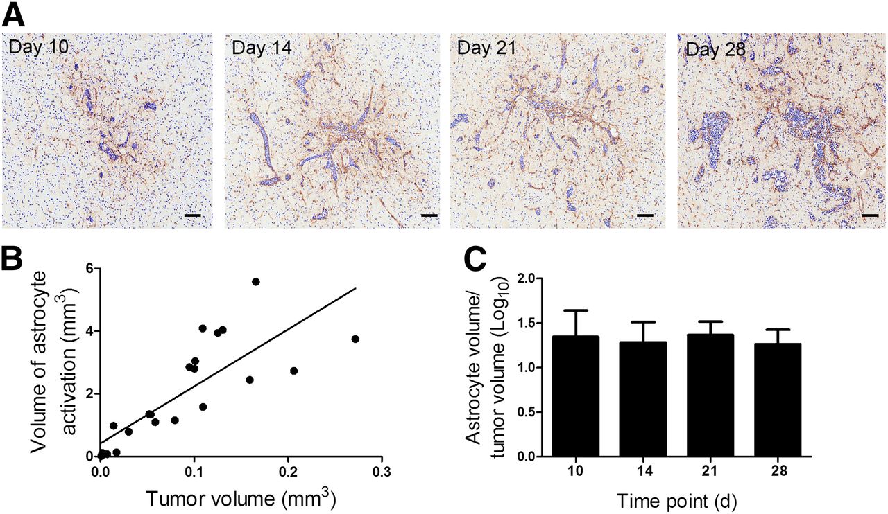
    (A) Photomicrographs of brain sections from animals at days 10, 14, 21, and 28 after intracerebral injection of 4T1-GFP cells. Reactive astrocytes were identified by GFAP immunoreactivity (brown stain), and sections were counterstained with cresyl violet. Scale bar = 100 μm. (B) Significant positive correlation was found between volume of astrocyte activation and tumor volume (y = 18.2x + 0.415; r2 = 0.657; P < 0.0001). (C) No significant differences were found in ratio of astrocyte to tumor volume over time (n = 4–5 per group).

  • FIGURE 2.
    • Download figure
    • Open in new tab
    • Download powerpoint
    FIGURE 2.

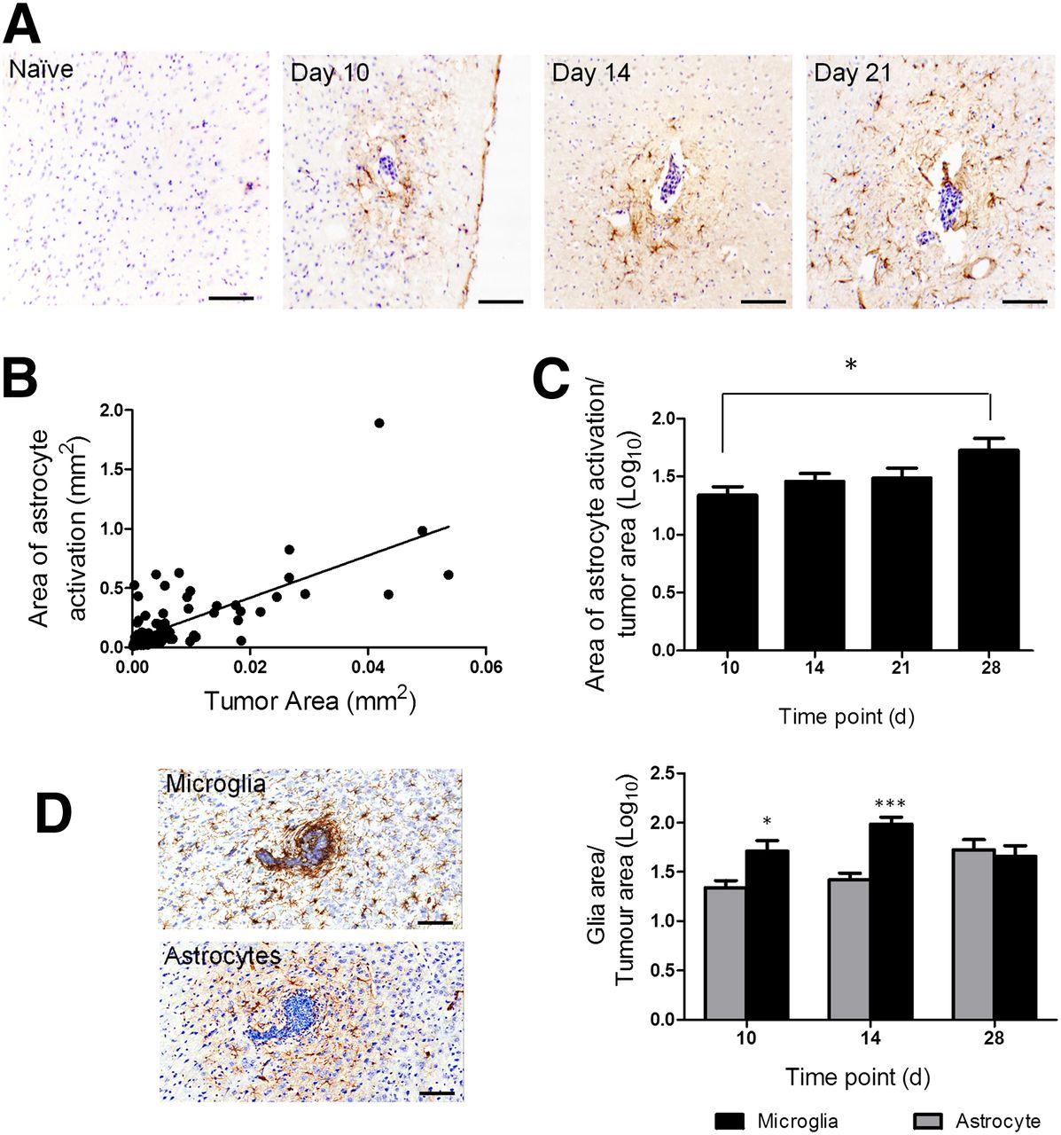
    (A) Photomicrographs of brain sections from a naive BALB/c mouse and at days 10, 14, and 28 after intracardiac injection of 4T1-GFP cells. Reactive astrocytes were identified by GFAP immunoreactivity (brown stain), and sections were counterstained with cresyl violet. Scale bar = 100 μm. (B) Significant positive correlation was found between extent of astrocyte activation and tumor area (y = 11x + 0.006; r2 = 0.6; P < 0.0001). (C) Significant increase in ratio of astrocyte to tumor area was found over time (*P < 0.05, n = 5–6 per group). (D) Photomicrographs comparing microglial infiltration of metastases, detected by Iba-1 immunoreactivity, with astrocyte activation (brown stain in each case). Graph shows quantitation of microglial (black bars) and astrocyte (gray bars) activation at days 10 (tumor number = 16), 14 (n = 39), and 28 (n = 7). *P < 0.05. ***P < 0.0001.

  • FIGURE 3.
    • Download figure
    • Open in new tab
    • Download powerpoint
    FIGURE 3.

    Confocal microscopy images obtained from mouse brain 14 d after intrastriatal injection of 4T1-GFP cells (A) or 21 d after intracardiac injection (B) demonstrating TSPO colocalization with either astrocyte reactivity (GFAP) surrounding metastases or reactive microglia in tumor periphery (IBA-1). Scale bar = 50 μm.

  • FIGURE 4.
    • Download figure
    • Open in new tab
    • Download powerpoint
    FIGURE 4.

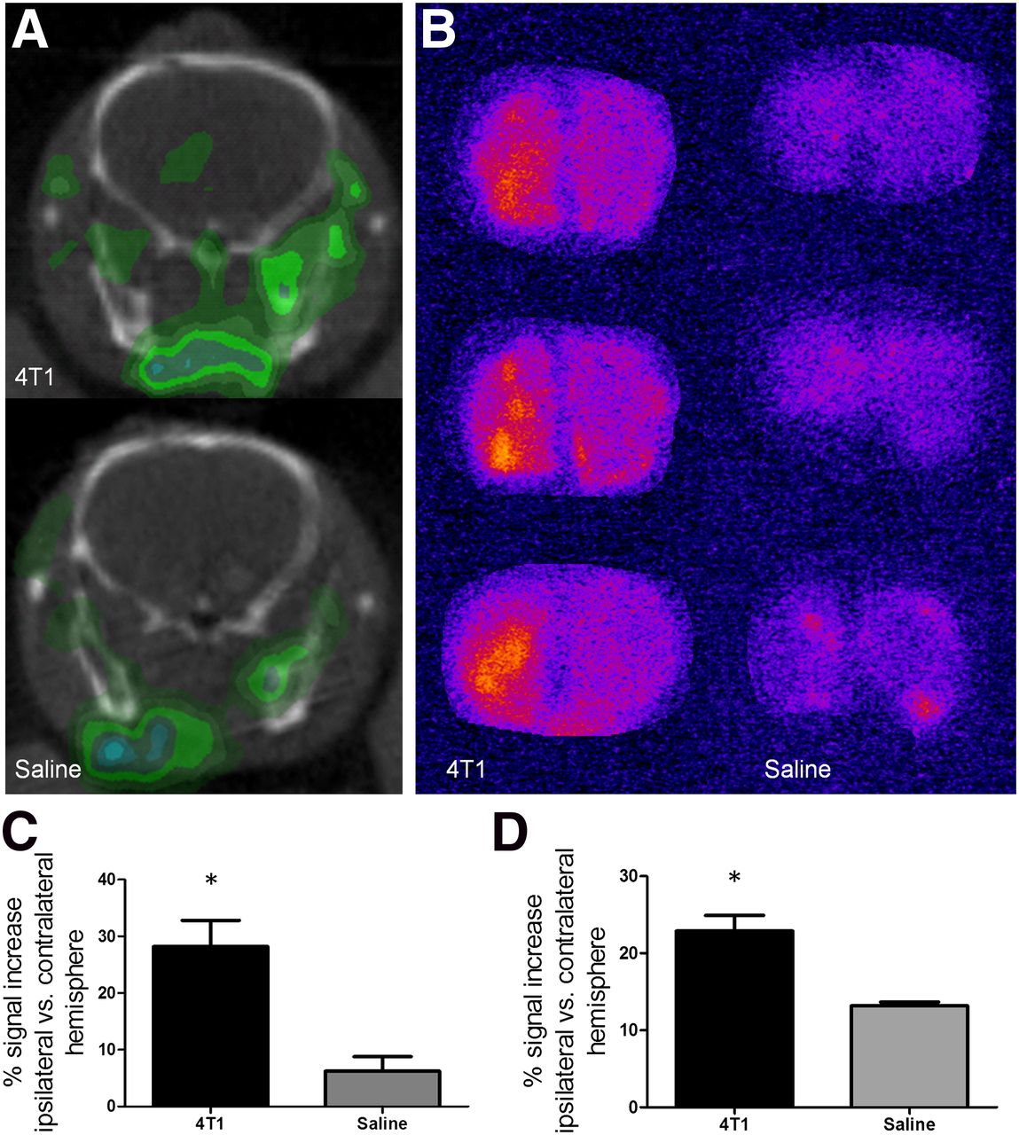
    (A) Representative SPECT images at injection site (left hemisphere) from 4T1-GFP–injected mouse and saline-injected control. (B) 123I-DPA713 binding was confirmed by autoradiography in 4T1-GFP–injected animals as compared with saline-injected animals. (C–D) Graphs show percentage signal increase in injected hemisphere, compared with control contralateral hemisphere for 4T1-GFP– (n = 6) and saline- (n = 3) injected animals for either SPECT (C) or autoradiography (D) data. *P < 0.05.

  • FIGURE 5.
    • Download figure
    • Open in new tab
    • Download powerpoint
    FIGURE 5.

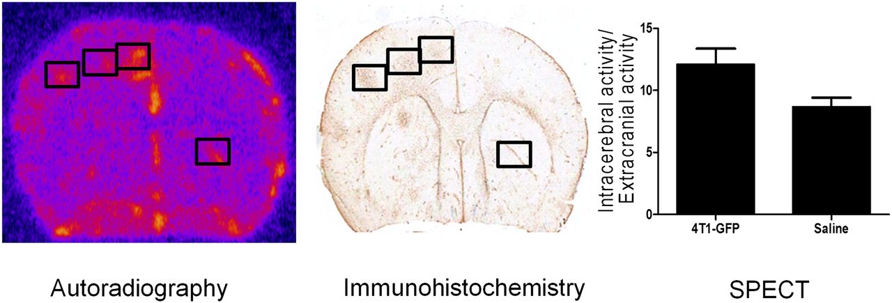
    Representative autoradiography image from mouse brain obtained 21 d after intracardiac injection of 4T1-GFP cells; areas of increased signal correlated spatially with immunohistochemical detection of tumor burden, as seen in immunohistochemistry image. Graph shows quantification of 123I-DPA713 binding (normalized activity), as determined by SPECT, in animals injected intracardially with either 4T1-GFP cells or saline.

Additional Files

  • Figures
  • Supplemental Data

    Files in this Data Supplement:

    • Supplemental Data
PreviousNext
Back to top

In this issue

Journal of Nuclear Medicine: 55 (2)
Journal of Nuclear Medicine
Vol. 55, Issue 2
February 1, 2014
  • Table of Contents
  • Table of Contents (PDF)
  • About the Cover
  • Index by author
Print
Download PDF
Article Alerts
Sign In to Email Alerts with your Email Address
Email Article

Thank you for your interest in spreading the word on Journal of Nuclear Medicine.

NOTE: We only request your email address so that the person you are recommending the page to knows that you wanted them to see it, and that it is not junk mail. We do not capture any email address.

Enter multiple addresses on separate lines or separate them with commas.
Glial Activation in the Early Stages of Brain Metastasis: TSPO as a Diagnostic Biomarker
(Your Name) has sent you a message from Journal of Nuclear Medicine
(Your Name) thought you would like to see the Journal of Nuclear Medicine web site.
Citation Tools
Glial Activation in the Early Stages of Brain Metastasis: TSPO as a Diagnostic Biomarker
Emma R. O’Brien, Veerle Kersemans, Matthew Tredwell, Begona Checa, Sébastien Serres, Manuel Sarmiento Soto, Veronique Gouverneur, David Leppert, Daniel C. Anthony, Nicola R. Sibson
Journal of Nuclear Medicine Feb 2014, 55 (2) 275-280; DOI: 10.2967/jnumed.113.127449

Citation Manager Formats

  • BibTeX
  • Bookends
  • EasyBib
  • EndNote (tagged)
  • EndNote 8 (xml)
  • Medlars
  • Mendeley
  • Papers
  • RefWorks Tagged
  • Ref Manager
  • RIS
  • Zotero
Share
Glial Activation in the Early Stages of Brain Metastasis: TSPO as a Diagnostic Biomarker
Emma R. O’Brien, Veerle Kersemans, Matthew Tredwell, Begona Checa, Sébastien Serres, Manuel Sarmiento Soto, Veronique Gouverneur, David Leppert, Daniel C. Anthony, Nicola R. Sibson
Journal of Nuclear Medicine Feb 2014, 55 (2) 275-280; DOI: 10.2967/jnumed.113.127449
Twitter logo Facebook logo LinkedIn logo Mendeley logo
  • Tweet Widget
  • Facebook Like
  • Google Plus One
Bookmark this article

Jump to section

  • Article
    • Abstract
    • MATERIALS AND METHODS
    • RESULTS
    • DISCUSSION
    • CONCLUSION
    • DISCLOSURE
    • Acknowledgments
    • Footnotes
    • REFERENCES
  • Figures & Data
  • Supplemental
  • Info & Metrics
  • PDF

Related Articles

  • This Month in JNM
  • PubMed
  • Google Scholar

Cited By...

  • STAT3-Mediated Astrocyte Reactivity Associated with Brain Metastasis Contributes to Neurovascular Dysfunction
  • Google Scholar

More in this TOC Section

  • PET/MRI of Hypoxic Atherosclerosis Using 64Cu-ATSM in a Rabbit Model
  • Tumor Uptake of Anti-CD20 Fabs Depends on Tumor Perfusion
  • How Sensitive Is the Upper Gastrointestinal Tract to 90Y Radioembolization? A Histologic and Dosimetric Analysis in a Porcine Model
Show more Basic Science Investigations

Similar Articles

Keywords

  • Astrocytes
  • microglia
  • metastasis
  • TSPO
  • SPECT imaging
  • mouse
SNMMI

© 2025 SNMMI

Powered by HighWire