


Abstract
The purpose of this study was to examine the effects of amino acid, hydrocarbon, and polyethylene glycol (PEG) linkers on the melanoma targeting and imaging properties of 99mTc-labeled lactam bridge–cyclized HYNIC-linker-Nle-CycMSHhex (hydrazinonicotinamide-linker-Nle-c[Asp-His-DPhe-Arg-Trp-Lys]-CONH2) peptides. Methods: Four novel peptides (HYNIC-GGGNle-CycMSHhex, HYNIC-GSGNle-CycMSHhex, HYNIC-PEG2Nle-CycMSHhex, and HYNIC-AocNle-CycMSHhex) were designed and synthesized. The melanocortin-1 receptor binding affinities of the peptides were determined in B16/F1 melanoma cells. The biodistribution of 99mTc(ethylenediaminediacetic acid [EDDA])-HYNIC-GGGNle-CycMSHhex, 99mTc(EDDA)-HYNIC-GSGNle-CycMSHhex, 99mTc(EDDA)-HYNIC-PEG2Nle-CycMSHhex, and 99mTc(EDDA)-HYNIC-AocNle-CycMSHhex were determined in B16/F1 melanoma–bearing C57 mice at 2 h after injection to select a lead peptide for further evaluation. The melanoma targeting and imaging properties of 99mTc(EDDA)-HYNIC-AocNle-CycMSHhex were further examined because of its high melanoma uptake. Results: The inhibitory concentrations of 50% (IC50) for HYNIC-GGGNle-CycMSHhex, HYNIC-GSGNle-CycMSHhex, HYNIC-PEG2Nle-CycMSHhex, and HYNIC-AocNle-CycMSHhex were 0.7 ± 0.1, 0.8 ± 0.09, 0.4 ± 0.08, and 0.3 ± 0.06 nM, respectively, in B16/F1 melanoma cells. Among these four 99mTc-labeled peptides, 99mTc(EDDA)-HYNIC-AocNle-CycMSHhex displayed the highest melanoma uptake (22.3 ± 1.72 percentage injected dose/g) at 2 h after injection. 99mTc(EDDA)-HYNIC-AocNle-CycMSHhex exhibited high tumor–to–normal-organ uptake ratios except for the kidneys. The tumor-to-kidney uptake ratios of 99mTc(EDDA)-HYNIC-AocNle-CycMSHhex were 3.29, 3.63, and 6.78 at 2, 4, and 24 h, respectively, after injection. The melanoma lesions were clearly visualized by SPECT/CT using 99mTc(EDDA)-HYNIC-AocNle-CycMSHhex as an imaging probe at 2 h after injection. Conclusion: High melanoma uptake and fast urinary clearance of 99mTc(EDDA)-HYNIC-AocNle-CycMSHhex highlighted its potential for metastatic melanoma detection in the future.
Over the past several years, radiolabeled lactam bridge–cyclized α-melanocyte–stimulating hormone (α-MSH) peptides have become another new class of cyclic peptides for melanoma targeting (1–11). The lactam bridge–cyclized α-MSH peptides can bind to the melanocortin-1 (MC1) receptors with low nanomolar binding affinities. Thus, we have used the lactam bridge–cyclized α-MSH peptides to target diagnostic radionuclides (i.e., 111In, 67Ga, and 64Cu) to melanoma cells for imaging. Specifically, we attached DOTA (1,4,7,10-tetraazacyclododecane-1,4,7,10-tetraacetic acid) and NOTA (1,4,7-triazacyclononane-1,4,7-triacetic acid) to the MC1 receptor-targeting GGNle-CycMSHhex (Gly-Gly-Nle-c[Asp-His-DPhe-Arg-Trp-Lys]-CONH2) peptide for radiolabeling of 111In, 67Ga, and 64Cu (9–11). The promising imaging results of 111In-, 67Ga-, and 64Cu-labeled DOTA/NOTA-GGNle-CycMSHhex highlighted their potential as imaging probes for SPECT and PET imaging of melanoma (9–11).
Recently, building on the success of 111In-, 67Ga-, and 64Cu-labeled DOTA/NOTA-GGNle-CycMSHhex, we have further developed new 99mTc-labeled lactam bridge–cyclized α-MSH peptides to take advantage of the ideal imaging properties of 99mTc (140-keV γ-photon and 6-h half-life) and its wide application in nuclear medicine. Specifically, we replaced DOTA/NOTA with bifunctional metal chelators such as mercaptoacetyltriglycine, Ac-Cys-Gly-Gly-Gly, and hydrazinonicotinamide (HYNIC) for 99mTc radiolabeling (12). HYNIC-GGNle-CycMSHhex was readily radiolabeled with 99mTc in ethylenediaminediacetic acid (EDDA)/Tricine solution. Interestingly, 99mTc(EDDA)-HYNIC-GGNle-CycMSHhex exhibited higher melanoma uptake and faster urinary clearance than 99mTc-mercaptoacetyltriglycine-GGNle-CycMSHhex and 99mTc-Ac-Cys-Gly-Gly-Gly-GGNle-CycMSHhex in B16/F1 melanoma–bearing C57 mice. The B16/F1 melanoma lesions were clearly visualized by SPECT/CT using 99mTc(EDDA)-HYNIC-GGNle-CycMSHhex as an imaging probe (12).
In our previous report, the introduction of the -GlyGly- amino acid linker resulted in lower renal and liver uptake of 111In-DOTA-GGNle-CycMSHhex than of 111In-DOTA-Nle-CycMSHhex (9). Meanwhile, hydrocarbon, amino acid, and polyethylene glycol (PEG) linkers displayed profound favorable effects in the receptor-binding affinities and pharmacokinetics of radiolabeled bombesin (13–17), arginine-glycine-aspartic (18–21), and α-MSH peptides (1,3). Thus, we were interested in examining how the amino acid, hydrocarbon, and PEG linkers affect the melanoma targeting and pharmacokinetic properties of 99mTc(EDDA)-HYNIC-linker-Nle-CycMSHhex peptides. Building on the HYNIC-GGNle-CycMSHhex construct, we designed in this study 4 novel peptides with different amino acid, hydrocarbon, and PEG linkers. Two neutral -GlyGlyGly- (GGG) and -GlySerGly- (GSG) amino acid linkers, one -Aoc- (8-aminooctanoic acid) hydrocarbon linker, and one -PEG2- linker were inserted between the HYNIC and Nle-CycMSHhex to generate HYNIC-GGGNle-CycMSHhex, HYNIC-GSGNle-CycMSHhex, HYNIC-AocNle-CycMSHhex, and HYNIC-PEG2Nle-CycMSHhex peptides. The MC1 receptor binding affinities of these 4 peptides were determined in B16/F1 melanoma cells. Then, we radiolabeled the peptides with 99mTc using the EDDA/Tricine solution. We examined the biodistribution of 99mTc(EDDA)-HYNIC-GGGNle-CycMSHhex, 99mTc(EDDA)-HYNIC-GSGNle-CycMSHhex, 99mTc(EDDA)-HYNIC-AocNle-CycMSHhex, and 99mTc(EDDA)-HYNIC-PEG2Nle-CycMSHhex at 2 h after injection to select a lead 99mTc-peptide for further evaluation. 99mTc(EDDA)-HYNIC-AocNle-CycMSHhex displayed the highest melanoma uptake at 2 h after injection. Therefore, we further determined the biodistribution of 99mTc(EDDA)-HYNIC-AocNle-CycMSHhex and its property for molecular imaging in B16/F1 melanoma–bearing C57 mice in this study.
MATERIALS AND METHODS
Chemicals and Reagents
Amino acid and resin were purchased from Advanced ChemTech Inc. and Novabiochem. Boc-HYNIC was purchased from VWR International, Inc., for peptide synthesis. 125I-Tyr2-[Nle4, D-Phe7]-α-MSH (125I-Tyr2-NDP-MSH) was obtained from PerkinElmer, Inc., for the receptor binding assay. 99mTcO4− was purchased from Cardinal Health for peptide radiolabeling. All other chemicals used in this study were purchased from Thermo Fischer Scientific and used without further purification. B16/F1 murine melanoma cells were obtained from American Type Culture Collection.
Peptide Synthesis and Receptor Binding Assay
HYNIC-GGGNle-CycMSHhex, HYNIC-GSGNle-CycMSHhex, HYNIC-AocNle-CycMSHhex, and HYNIC-PEG2Nle-CycMSHhex were synthesized using fluorenylmethyloxy carbonyl chemistry, purified by reverse-phase high-performance liquid chromatography (RP-HPLC; Waters), and characterized by liquid chromatography mass spectrometry. Generally, 70 μmol of resin, 210 μmol of each fluorenylmethyloxycarbonyl–protected amino acid, and 210 μmol of Boc-HYNIC were used for the synthesis. Briefly, the intermediate scaffolds of HYNIC(Boc)-[Gly-Gly-Gly/Gly-Ser(Trt)-Gly/Aoc/PEG2]-Nle-Asp(O-2-PhiPr)-His(Trt)-DPhe-Arg(Pbf)-Trp(Boc)-Lys(Dde) were synthesized on H2N-Sieber amide resin by an Advanced ChemTech multiple-peptide synthesizer. The protecting group of Dde was removed by 2% hydrazine for peptide cyclization. The protecting group of 2-phenylisopropyl was removed and the protected peptide was cleaved from the resin by treatment with a mixture of 2.5% trifluoroacetic acid and 5% triisopropylsilane. Each protected peptide was cyclized by coupling the carboxylic group from the Asp with the ε-amino group from the Lys. The cyclization reaction was achieved by an overnight reaction in dimethylformamide using benzotriazole-1-yl-oxy-tris-pyrrolidino-phosphonium-hexafluorophosphate as a coupling agent in the presence of N,N-diisopropylethylamine. Then, each protected cyclic peptide was dissolved in H2O/CH3CN (50:50) and lyophilized to remove the reagents. The protecting groups were totally removed by treating with a mixture of trifluoroacetic acid, thioanisole, phenol, water, ethanedithiol and triisopropylsilane (87.5:2.5:2.5:2.5:2.5:2.5) for 2 h at room temperature (25°C). Each peptide was precipitated and washed with ice-cold ether 4 times, purified by RP-HPLC, and characterized by liquid chromatography mass spectrometry. The MC1 receptor binding affinities for HYNIC-GGGNle-CycMSHhex, HYNIC-GSGNle-CycMSHhex, HYNIC-AocNle-CycMSHhex, and HYNIC-PEG2Nle-CycMSHhex were determined in B16/F1 melanoma cells by in vitro competitive receptor binding assay according to our published procedure (9).
Peptide Radiolabeling with 99mTc
99mTc(EDDA)-HYNIC-GGGNle-CycMSHhex, 99mTc(EDDA)-HYNIC-GSGNle-CycMSHhex, 99mTc(EDDA)-HYNIC-AocNle-CycMSHhex, and 99mTc(EDDA)-HYNIC-PEG2Nle-CycMSHhex were prepared according to our published procedure (12). Briefly, 50 μL of 99mTcO4− (37–74 MBq), 10 μL of 1 mg/mL SnCl2 in 0.1N HCl solution, 200 μL of a mixture of 5 mg/mL of EDDA and 25 mg/mL of Tricine aqueous solution, and 10 μL of a 1 mg/mL concentration of each peptide aqueous solution were added to 400 μL of 0.5 M NH4OAc (pH 5.44) in a reaction vial and incubated at 95°C for 30 min. Each radiolabeled peptide was purified to a single species by RP-HPLC on a Vydac C-18 reverse-phase analytic column (Grace) using a 20-min gradient of 20%–30% acetonitrile in 20 mM HCl aqueous solution at a flow rate of 1 mL/min. The purified peptide was purged with N2 gas for 20 min to remove the acetonitrile. The pH of the final solution was adjusted to 5 with 0.1N NaOH and normal saline for animal studies.
Biodistribution Studies
All animal studies were conducted in compliance with Institutional Animal Care and Use Committee approval. In an attempt to select a lead 99mTc-peptide for further evaluation, the biodistribution of 99mTc(EDDA)-HYNIC-GGGNle-CycMSHhex, 99mTc(EDDA)-HYNIC-GSGNle-CycMSHhex, 99mTc(EDDA)-HYNIC-AocNle-CycMSHhex, and 99mTc(EDDA)-HYNIC-PEG2Nle-CycMSHhex was examined in B16/F1 melanoma–bearing C57 female mice (Harlan) at 2 h after injection. The C57 mice were subcutaneously inoculated with 1 × 10⁶ B16/F1 cells on the right flank to generate B16/F1 tumors. The weights of tumors reached approximately 0.2 g at 10 d after cell inoculation. Each melanoma-bearing mouse was injected with 0.037 MBq of 99mTc(EDDA)-HYNIC-GGGNle-CycMSHhex, 99mTc(EDDA)-HYNIC-GSGNle-CycMSHhex, 99mTc(EDDA)-HYNIC-AocNle-CycMSHhex, or 99mTc(EDDA)-HYNIC-PEG2Nle-CycMSHhex via the tail vein. Groups of 5 mice were sacrificed at 2 h after injection, and tumor and organs of interest were harvested, weighed, and counted in a Wallace 1480 automated γ counter (PerkinElmer). Meanwhile, intestines and urine were collected and counted to evaluate the clearance pathway of each 99mTc-peptide. Blood was taken as 6.5% of the body weight.
99mTc(EDDA)-HYNIC-AocNle-CycMSHhex displayed higher melanoma uptake than the other three 99mTc-peptides. Therefore, the biodistribution of 99mTc(EDDA)-HYNIC-GGNle-CycMSHhex at 0.5, 4, and 24 h after injection was determined in B16/F1 melanoma–bearing C57 female mice. B16/F1 melanoma–bearing mice were generated as described above. Each melanoma-bearing mouse was injected with 0.037 MBq of 99mTc(EDDA)-HYNIC-AocNle-CycMSHhex via the tail vein. Groups of 5 mice were sacrificed at 0.5, 4, and 24 h after injection, and tumors and organs of interest were harvested, weighed, and counted. Blood was taken as 6.5% of the body weight. The tumor uptake specificity of 99mTc(EDDA)-HYNIC-AocNle-CycMSHhex was determined by coinjecting 10 μg (6.07 nmol) of unlabeled NDP-MSH peptide at 2 h after injection. To examine whether l-lysine coinjection could decrease the renal uptake, a group of 5 mice was injected with a mixture of 12 mg of l-lysine and 0.037 MBq of 99mTc(EDDA)-HYNIC-AocNle-CycMSHhex. The mice were sacrificed at 2 h after injection, and the tumors and organs of interest were harvested, weighed, and counted.
Melanoma Imaging of 99mTc(EDDA)-HYNIC-AocNle-CycMSHhex
Approximately 9.3 MBq of 99mTc(EDDA)-HYNIC-AocNle-CycMSHhex were injected in a B16/F1 melanoma–bearing C57 mouse for melanoma imaging. The mouse was euthanized at 2 h after injection for small-animal SPECT/CT (Nano-SPECT/CT; Bioscan) imaging. The 9-min CT imaging was immediately followed by the whole-body SPECT scan. The SPECT scans of 24 projections were acquired. Reconstructed SPECT and CT data were visualized and coregistered using InVivoScope (Bioscan).
Statistical Analysis
Statistical analysis was performed using the Student t test for unpaired data. A 95% confidence level was chosen to determine the significance of differences in tumor and renal uptake of 99mTc(EDDA)-HYNIC-AocNle-CycMSHhex with and without NDP-MSH coinjection, as well as the significance of differences in tumor and renal uptake of 99mTc(EDDA)-HYNIC-AocNle-CycMSHhex with and without l-lysine coinjection in the biodistribution studies described above. The differences at the 95% confidence level (P < 0.05) were considered significant.
RESULTS
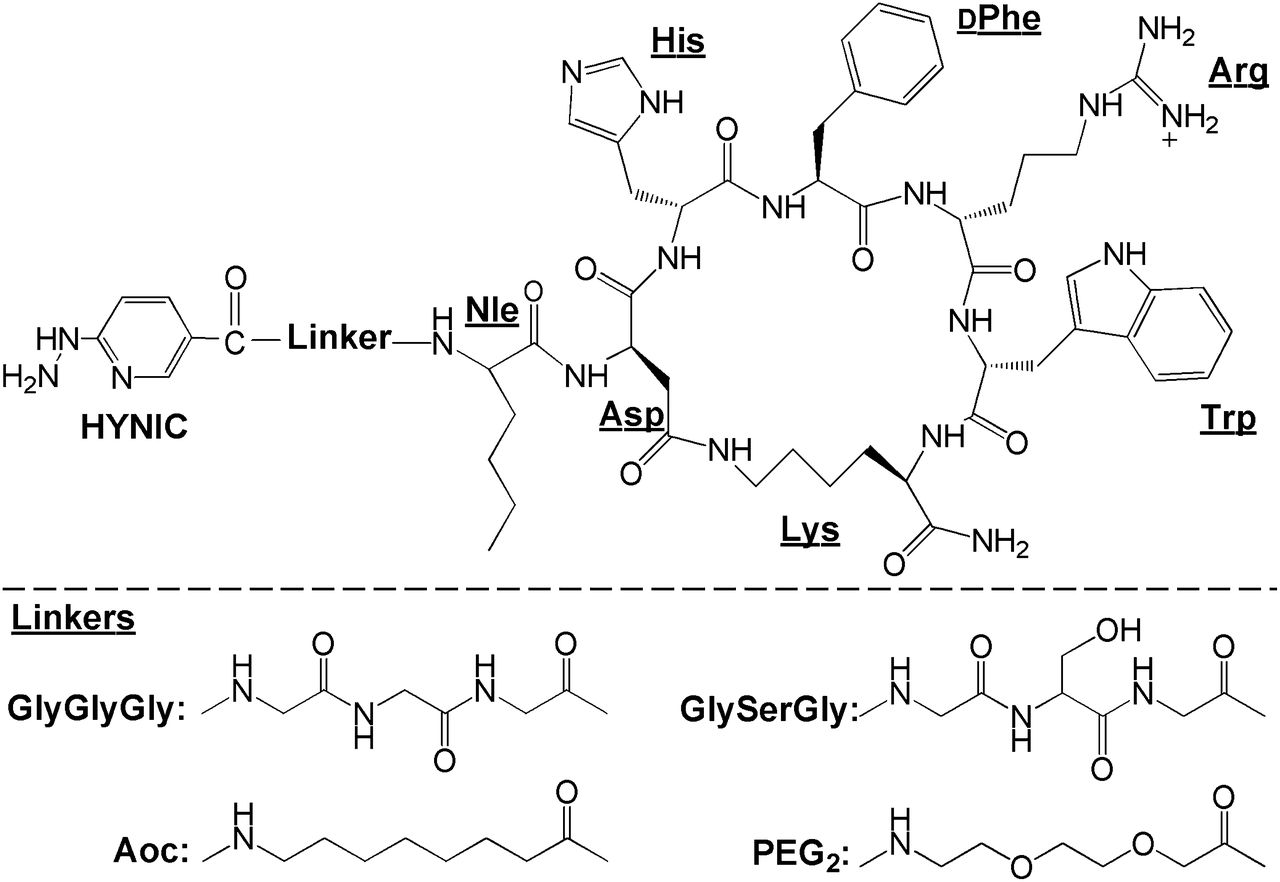
New HYNIC-GGGNle-CycMSHhex, HYNIC-GSGNle-CycMSHhex, HYNIC-AocNle-CycMSHhex, and HYNIC-PEG2Nle-CycMSHhex were synthesized and purified by RP-HPLC. All 4 peptides displayed greater than 95% purity after HPLC purification. The identities of HYNIC-GGGNle-CycMSHhex, HYNIC-GSGNle-CycMSHhex, HYNIC-AocNle-CycMSHhex, and HYNIC-PEG2Nle-CycMSHhex were confirmed by electrospray ionization mass spectrometry. The calculated and found molecular weights of HYNIC-GGGNle-CycMSHhex, HYNIC-GSGNle-CycMSHhex, HYNIC-AocNle-CycMSHhex, and HYNIC-PEG2Nle-CycMSHhex are presented in Table 1. The found molecular weights matched the calculated molecular weights. The schematic structures of HYNIC-GGGNle-CycMSHhex, HYNIC-GSGNle-CycMSHhex, HYNIC-AocNle-CycMSHhex, and HYNIC-PEG2Nle-CycMSHhex are shown in Figure 1. The IC50 values of HYNIC-GGGNle-CycMSHhex, HYNIC-GSGNle-CycMSHhex, HYNIC-AocNle-CycMSHhex, and HYNIC-PEG2Nle-CycMSHhex were 0.7 ± 0.1, 0.8 ± 0.09, 0.4 ± 0.08, and 0.3 ± 0.06 nM in B16/F1 melanoma cells, respectively (Fig. 2).
IC50 Values and Molecular Weights of 4 Peptides

Schematic structures of HYNIC-linker-Nle-CycMSHhex.

In vitro competitive binding curves of HYNIC-PEG2Nle-CycMSHhex (●, IC50 = 0.3 ± 0.06 nM), HYNIC-AocNle-CycMSHhex (▲, IC50 = 0.4 ± 0.08 nM), HYNIC-GGGNle-CycMSHhex (▪, IC50 = 0.7 ± 0.1 nM), and HYNIC-GSGNle-CycMSHhex (♦, IC50 = 0.8 ± 0.09 nM) in B16/F1 murine melanoma cells.
HYNIC-GGGNle-CycMSHhex, HYNIC-GSGNle-CycMSHhex, HYNIC-AocNle-CycMSHhex, and HYNIC-PEG2Nle-CycMSHhex were readily labeled with 99mTc with greater than 95% radiolabeling yields in EDDA/Tricine solution. Each radiolabeled peptide was completely separated from its excess nonlabeled peptide by RP-HPLC. All 99mTc-peptides displayed greater than 98% radiochemical purity after HPLC purification. The retention times of 99mTc(EDDA)-HYNIC-GGGNle-CycMSHhex, 99mTc(EDDA)-HYNIC-GSGNle-CycMSHhex, 99mTc(EDDA)-HYNIC-AocNle-CycMSHhex, and 99mTc(EDDA)-HYNIC-PEG2Nle-CycMSHhex were 12.7, 10.6, 24.0, and 16.8 min, respectively. The retention times of HYNIC-GGGNle-CycMSHhex, HYNIC-GSGNle-CycMSHhex, HYNIC-AocNle-CycMSHhex, and HYNIC-PEG2Nle-CycMSHhex were 9.5, 8.3, 17.9, and 14.6 min, respectively.
The melanoma-targeting and pharmacokinetic properties of 99mTc(EDDA)-HYNIC-GGGNle-CycMSHhex, 99mTc(EDDA)-HYNIC-GSGNle-CycMSHhex, 99mTc(EDDA)-HYNIC-AocNle-CycMSHhex, and 99mTc(EDDA)-HYNIC-PEG2Nle-CycMSHhex were determined in B16/F1 melanoma–bearing mice at 2 h after injection to select a lead radiolabeled peptide for further evaluation. The biodistribution results of these four 99mTc-peptides are shown in Table 2. 99mTc(EDDA)-HYNIC-GGGNle-CycMSHhex and 99mTc(EDDA)-HYNIC-GSGNle-CycMSHhex displayed substantial tumor uptake of 9.78 ± 3.40 percentage injected dose (%ID)/g and 7.41 ± 4.26 %ID/g at 2 h after injection. 99mTc(EDDA)-HYNIC-PEG2Nle-CycMSHhex showed higher tumor uptake of 14.32 ± 2.82 %ID/g at 2 h after injection. 99mTc(EDDA)-HYNIC-AocNle-CycMSHhex exhibited the highest tumor uptake of 22.3 ± 1.72 %ID/g at 2 h after injection among all four 99mTc-peptides. All 99mTc-peptides showed fast urinary clearance, approximately 89%–94% of injected activity had cleared the body at 2 h after injection. The accumulation in all normal organs except the kidney was lower than 0.81 %ID/g at 2 h after injection for all four 99mTc-peptides. The renal uptake values were in a similar range (3.9–6.5 %ID/g at 2 h after injection) for all four 99mTc-peptides. Thus, we selected 99mTc(EDDA)-HYNIC-AocNle-CycMSHhex as a lead peptide to further examine its full biodistribution and melanoma-imaging properties.
Biodistribution Comparison Among Peptides in B16/F1 Melanoma–Bearing C57 Mice 2 Hours After Injection
The full biodistribution results of 99mTc(EDDA)-HYNIC-AocNle-CycMSHhex are presented in Table 3. 99mTc(EDDA)-HYNIC-AocNle-CycMSHhex displayed high tumor uptake and prolonged tumor retention in B16/F1 melanoma–bearing C57 mice. The tumor uptake was 23.44 ± 3.37 %ID/g and 22.8 ± 1.71 %ID/g at 0.5 and 2 h after injection, respectively. As compared with the tumor uptake at 2 h after injection, 97.2% of the radioactivity remained in the tumor at 4 h after injection. In the melanoma uptake–blocking study, coinjection of NDP-MSH blocked 94.5% of tumor uptake (P < 0.05) at 2 h after injection, demonstrating that the tumor uptake was MC1 receptor-mediated. Normal-organ uptake of 99mTc(EDDA)-HYNIC-AocNle-CycMSHhex was lower than 1.26 %ID/g in normal tissues except for kidneys at 2, 4, and 24 h after injection. High tumor-to-blood and high tumor–to–normal-organ uptake ratios were achieved as early as 0.5 h after injection. As the major excretion pathway of 99mTc(EDDA)-HYNIC-AocNle-CycMSHhex, the kidney uptake was 19.65 ± 7.36 %ID/g at 0.5 h after injection and decreased to 1.05 ± 0.07 %ID/g at 24 h after injection. The tumor-to-kidney uptake ratios of 99mTc(EDDA)-HYNIC-AocNle-CycMSHhex were 3.29, 3.63, and 6.78 at 2, 4, and 24 h, respectively, after injection. Coinjection of NDP-MSH did not reduce the renal uptake of the 99mTc(EDDA)-HYNIC-AocNle-CycMSHhex activity at 2 h after injection, indicating that the renal uptake was not MC1 receptor–mediated. Meanwhile, l-lysine coinjection decreased 33% of the renal uptake (P < 0.05) without affecting the tumor uptake. Moreover, 99mTc(EDDA)-HYNIC-AocNle-CycMSHhex exhibited rapid urinary excretion. Approximately 88% of the activity cleared out of the body at 2 h after injection. The representative whole-body, coronal, and transversal SPECT/CT images are presented in Figure 3. The melanoma lesions were clearly visualized by SPECT/CT using 99mTc(EDDA)-HYNIC-AocNle-CycMSHhex as an imaging probe at 2 h after injection. 99mTc(EDDA)-HYNIC-AocNle-CycMSHhex exhibited high tumor–to–normal-organ uptake ratios except for the kidneys, which was consistent with the biodistribution results.
Biodistribution of 99mTc(EDDA)-HYNIC-AocNle-CycMSHhex in B16/F1 Melanoma–Bearing C57 Mice
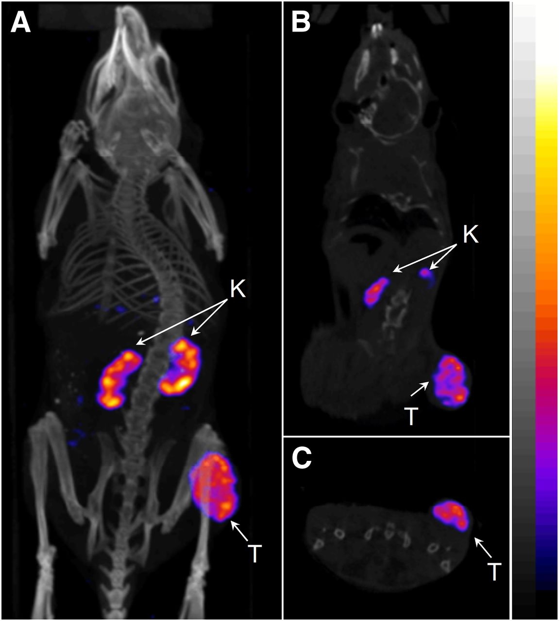
Representative whole-body (A), coronal (B), and transversal (C) SPECT/CT images of 99mTc(EDDA)-HYNIC-AocNle-CycMSHhex in B16/F1 melanoma–bearing C57 mouse at 2 h after injection. K = kidney; T = tumor.

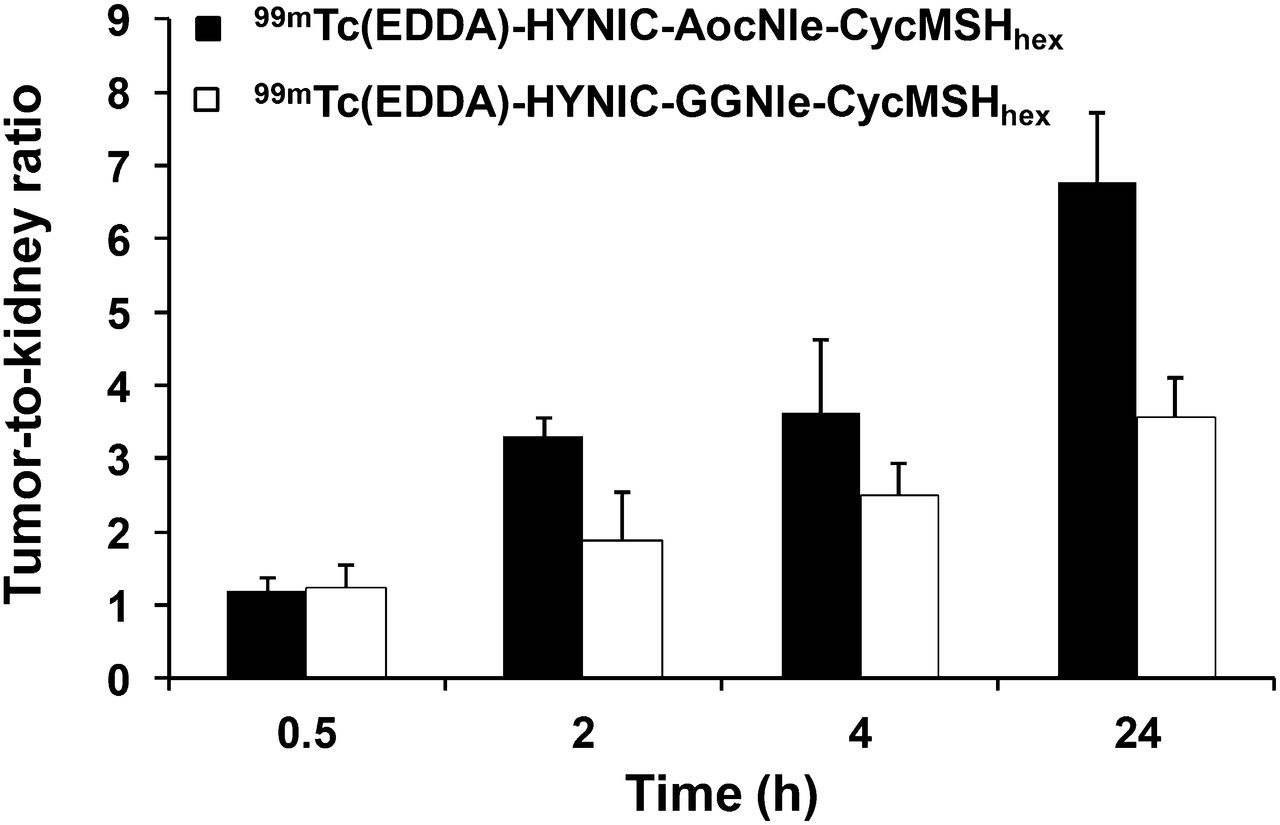
Comparison of tumor-to-kidney ratios between 99mTc(EDDA)-HYNIC-GGNle-CycMSHhex and 99mTc(EDDA)-HYNIC-AocNle-CycMSHhex at 0.5, 2, 4, and 24 h after injection. Data on 99mTc(EDDA)-HYNIC-GGNle-CycMSHhex (12) are shown for comparison.
DISCUSSION
Over the past several years, a number of research groups have reported 99mTc-labeled α-MSH peptides (2,5,8,12,22–24) to target the MC1 receptors for melanoma imaging, taking advantage of the ideal imaging properties of 99mTc. Recently, we have developed a novel class of radiolabeled lactam bridge–cyclized α-MSH peptides to target MC1 receptors for melanoma imaging. Specifically, we have radiolabeled HYNIC/DOTA/NOTA-GGNle-CycMSHhex with 99mTc, 111In, 67Ga, and 64Cu for SPECT and PET imaging of melanoma (9–12). In our previous report, we found that the Nle-CycMSHhex moiety was critical for nanomolar MC1 receptor binding affinity of the DOTA-conjugated peptide (9). Introduction of a -GlyGlu- linker between the Nle and CycMSHhex moiety sacrificed the receptor-binding affinity by 485-fold (9). Meanwhile, HYNIC was a better chelator for 99mTc than other N3S chelators in terms of melanoma uptake and urinary clearance (12). In this study, we managed to examine the effects of amino acid, hydrocarbon, and PEG linkers on the melanoma-targeting properties of 99mTc-labeled lactam bridge–cyclized α-MSH peptide. Specifically, we introduced -GGG-, -GSG-, -Aoc-, and -PEG2- linkers between the HYNIC and Nle-CycMSHhex moiety to generate new HYNIC-GGGNle-CycMSHhex, HYNIC-GSGNle-CycMSHhex, HYNIC-AocNle-CycMSHhex, and HYNIC-PEG2Nle-CycMSHhex peptides. The lengths of the -GGG-, -GSG-, -Aoc-, and -PEG2- linkers are the same, eliminating the effect of linker length on the melanoma-targeting properties.
The introduction of -GGG-, -GSG-, -Aoc-, and -PEG2- linkers retained the low-nanomolar MC1 receptor binding affinities of the peptides. The IC50 was 0.7 ± 0.1 nM for HYNIC-GGGNle-CycMSHhex, 0.8 ± 0.09 nM for HYNIC-GGGNle-CycMSHhex, 0.4 ± 0.08 nM for HYNIC-AocNle-CycMSHhex, and 0.3 ± 0.06 nM for HYNIC-PEG2Nle-CycMSHhex (Table 1), respectively. Furthermore, we radiolabeled these 4 peptides with 99mTc and determined their biodistribution properties in B16/F1 melanoma–bearing C57 mice at 2 h after injection to examine how the amino acid, hydrocarbon, and PEG linkers affected their melanoma-targeting and pharmacokinetic properties. Despite the slight difference in receptor-binding affinity among HYNIC-GGGNle-CycMSHhex, HYNIC-GSGNle-CycMSHhex, HYNIC-AocNle-CycMSHhex, and HYNIC-PEG2Nle-CycMSHhex peptides, we observed a dramatic difference in melanoma uptake among these four 99mTc-peptides. As shown in Table 2, 99mTc(EDDA)-HYNIC-AcoNle-CycMSHhex exhibited the highest tumor uptake among these four 99mTc-peptides at 2 h after injection in B16/F1 melanoma–bearing C57 mice. The tumor uptake of 99mTc(EDDA)-HYNIC-AcoNle-CycMSHhex was 2.3, 3.0, and 1.6 times the tumor uptake of 99mTc(EDDA)-HYNIC-GGGNle-CycMSHhex, 99mTc(EDDA)-HYNIC-GSGNle-CycMSHhex, and 99mTc(EDDA)-HYNIC-PEG2Nle-CycMSHhex, respectively, at 2 h after injection. Thus, we further examined the full biodistribution and melanoma-imaging properties of 99mTc(EDDA)-HYNIC-AocNle-CycMSHhex.
As shown in Table 3, 99mTc(EDDA)-HYNIC-AocNle-CycMSHhex displayed high tumor uptake and prolonged tumor retention in B16/F1 melanoma–bearing C57 mice. The tumor uptake was 23.44 ± 3.37 %ID/g and 22.8 ± 1.71 %ID/g at 0.5 and 2 h after injection, respectively. In comparison with the tumor uptake at 2 h after injection, 97.2% of the radioactivity remained in tumor at 4 h after injection. Coinjection of NDP-MSH blocked 94.5% of tumor uptake (P < 0.05) without affecting the renal uptake at 2 h after injection, demonstrating that the tumor uptake was MC1 receptor–mediated and that renal uptake was nonspecific. 99mTc(EDDA)-HYNIC-AocNle-CycMSHhex exhibited low accumulation in normal organs and rapid urinary excretion, resulting in high tumor–to–normal-organ uptake ratios. As we anticipated, the B16/F1 melanoma lesions were clearly visualized by SPECT/CT using 99mTc(EDDA)-HYNIC-AocNle-CycMSHhex as an imaging probe.
Recently, it has been reported that one more carboxylic group resulted in a dramatic decrease in the renal and liver uptake of 99mTc(CO)3-labeled lactam bridge–cyclized α-MSH peptides (8). Accordingly, we anticipate that the introduction of a negatively charged amino acid would further reduce the renal uptake. In this study, l-lysine coinjection decreased 33% of the renal uptake (P < 0.05) without affecting the tumor uptake, indicating that the overall positive charge of 99mTc(EDDA)-HYNIC-AocNle-CycMSHhex contributed to its nonspecific renal uptake. As shown in Figure 1, there was a positively charged side chain in Arg8, which contributed to the overall positive charge of 99mTc(EDDA)-HYNIC-AocNle-CycMSHhex. However, the Arg8 is critical for MC1 receptor binding. Thus, it would be better to introduce a negatively charged amino acid between the HYNIC and Aoc linker to decrease the overall positive charge of the peptide.
In this study, 99mTc(EDDA)-HYNIC-GGNle-CycMSHhex and 99mTc-(Arg11)CCMSH displayed comparably high melanoma uptake (13.23 ± 2.35 vs. 11.16 ± 1.77 %ID/g) at 4 h after injection. Remarkably, the tumor uptake of 99mTc(EDDA)-HYNIC-AocNle-CycMSHhex was 1.7 times the tumor uptake of 99mTc(EDDA)-HYNIC-GGNle-CycMSHhex at 4 h after injection. Meanwhile, 99mTc(EDDA)-HYNIC-AocNle-CycMSHhex displayed higher tumor-to-kidney uptake ratios than 99mTc(EDDA)-HYNIC-GGNle-CycMSHhex at 2, 4, and 24 h after injection (Fig. 4). The tumor-to-kidney uptake ratio of 99mTc(EDDA)-HYNIC-AocNle-CycMSHhex was 1.8, 1.5, and 1.9 times the tumor-to-kidney uptake ratio of 99mTc(EDDA)-HYNIC-GGNle-CycMSHhex at 2, 4, and 24 h after injection, respectively.
CONCLUSION
The biodistribution of 99mTc(EDDA)-HYNIC-GGGNle-CycMSHhex, 99mTc(EDDA)-HYNIC-GSGNle-CycMSHhex, 99mTc(EDDA)-HYNIC-AocNle-CycMSHhex, and 99mTc(EDDA)-HYNIC-PEG2Nle-CycMSHhex was determined in B16/F1 melanoma–bearing C57 mice in this study. Among these four 99mTc-peptides, 99mTc(EDDA)-HYNIC-AocNle-CycMSHhex exhibited the highest melanoma uptake (22.3 ± 1.72 %ID/g) and tumor-to-kidney uptake ratio at 2 h after injection. Overall, the properties of high melanoma uptake and fast urinary clearance of 99mTc(EDDA)-HYNIC-AocNle-CycMSHhex highlight its future clinical potential as an imaging probe for metastatic melanoma detection.
DISCLOSURE
The costs of publication of this article were defrayed in part by the payment of page charges. Therefore, and solely to indicate this fact, this article is hereby marked “advertisement” in accordance with 18 USC section 1734. This work was supported in part by the NIH grant NM-INBRE P20RR016480/P20GM103451 and University of New Mexico HSC RAC Award. The SPECT/CT image in this article was generated by the Keck-UNM Small Animal Imaging Resource established with funding from the W.M. Keck Foundation and the University of New Mexico Cancer Research and Treatment Center (NIH P30 CA118100). No other potential conflict of interest relevant to this article was reported.
Acknowledgments
We thank Drs. Jianquan Yang and Fabio Gallazzi for their technical assistance.
Footnotes
Published online Nov. 7, 2014.
- © 2014 by the Society of Nuclear Medicine and Molecular Imaging, Inc.
REFERENCES
- Received for publication July 18, 2014.
- Accepted for publication October 1, 2014.


