


Abstract
Overexpression, activation, and mutations of the epidermal growth factor receptor (EGFR) are commonly found in solid tumors. The aim of this study was to develop a PET-based method for detecting the constitutively active mutant de2-7 EGFR, which is associated with disease progression and resistance to chemotherapy and radiotherapy in glioma. Methods: The chimeric antibody ch806, which selectively binds an epitope of the EGFR that is exposed only on overexpressed, mutant, or ligand-activated forms of the receptor, was conjugated to the radiohalogen 124I via the residualizing ligand IMP-R4, and in vitro properties were characterized. In vivo biodistribution and small-animal PET studies were performed in BALB/c nude mice bearing U87MG.de2-7 glioma xenografts. Imaging results were correlated with measured tumor uptake of the radioconjugate. Results: 124I-IMP-R4-ch806 had an immunoreactivity of 78.3% and was stable for 7 d when incubated in serum in vitro. The biodistribution analysis of 124I-IMP-R4-ch806 demonstrated a maximal uptake of 30.95 ± 6.01 percentage injected dose per gram (%ID/g) in U87MG.de2-7 xenografts at 48 h after injection, with prolonged tumor retention (6.07 ± 0.80 %ID/g at 216 h after injection). The tumor-to-blood ratio increased from 0.44 at 4 h after injection to a maximum of 4.70 at 168 h after injection. PET of 124I-IMP-R4-ch806 biodistribution was able to clearly detect the U87MG.de2-7 tumors at 24 h after injection and for at least 168 h after injection. Correlation between tumor PET image quantitation of 124I-IMP-R4-ch806 and %ID/g determined from resected tissues (r = 0.9350) was excellent. Conclusion: These results show that immuno-PET with 124I-IMP-R4-ch806 is feasible and allows noninvasive quantitation of de2-7 EGFR expression in vivo.
Monoclonal antibodies (mAbs) are an established therapeutic tool in oncology for immunotherapy and signaling abrogation of tumor cells and as carrier molecules to deliver a toxic load (e.g., radioimmunotherapy) (1). More recently, these antibodies have been labeled with positron emitters for use as a diagnostic tool in combination with PET scanning (immuno-PET) (1–4). This technique has been developed to allow the noninvasive, high-resolution, quantitative imaging of tumors using mAbs raised against tumor-specific or -prevalent antigens. Potential applications of immuno-PET include aiding in antibody development and in patient selection for antibody therapy either by confirming the presence of antibody binding in patients planned for immunotherapy or by determining the radiation dosimetry in patients planned for radioimmunotherapy (1,2).
The epidermal growth factor receptor (EGFR) is an attractive target for tumor-targeted antibody therapy because it is overexpressed in many types of epithelial tumors and is associated with poor prognosis in several tumor types (5). Overexpression of the receptor is often caused by amplification of the EGFR gene, an event also linked with EGFR mutation (6). The de2-7 EGFR (or EGFRvIII) extracellular truncation of the EGFR is the most common EGFR mutation and is frequently expressed in glioblastoma and some other tumor types, including prostate and breast cancer (7). Inhibition of the EGFR by mAbs and tyrosine kinase is a rational strategy for the development of new cancer therapeutics, because of the high expression on epithelial tumors and the role of EGFR signaling in maintaining the neoplastic phenotype of cancer cells. Consequently, several anti-EGFR therapeutics have been reported in the literature, with several undergoing clinical evaluation (5).
We have previously described ch806, a chimeric form of a novel anti-EGFR antibody that binds the EGFR deletion variant de2-7 EGFR and wild-type EGFR expressed in cells overexpressing the receptor. ch806 has negligible binding to normal tissues—including the skin and liver—that express physiologic levels of EGFR (8–15). We have shown in a Phase I trial that ch806 has impressive therapeutic activity in preclinical models and specifically targets tumors but not normal tissues (10,16). In previous studies, we have shown that ch806 directly labeled with 125I shows minimal tumor retention, most likely a result of this internalized antibody being catabolized (10). To develop a noninvasive method for targeting and quantitating de2-7 EGFR expression in glioma, we have developed an immuno-PET technique using ch806 conjugated with 124I via the residualizing ligand IMP-R4 (IMP-R4 is MCC-Lys(MCC)-Lys(X)-d-Tyr-d-Lys(X)-OH, wherein MCC is 4-(N-maleimidomethyl)-cyclohexane-1-carbonyl and X is 1-((4-thiocarbonylamino)benzyl)-diethylenetriaminepentaacetic acid) (17), resulting in specific tumor localization and high-resolution PET images of glioma xenografts.
MATERIALS AND METHODS
All analytic-grade reagents, except where stated, were obtained from Merck Pty. Ltd. 124I in 0.02 N NaOH (370 MBq/mL [10 mCi/mL]) was purchased from Advanced Nuclide Technologies. The residualizing peptide IMP-R4 was provided by Immunomedics, Inc. Radioactivity was measured either with a dose calibrator (Atomlab-100; Biodex) or an automated γ-counter (Cobra II; Canberra-Packard).
Antibodies and Cells
The ch806 and huA33 mAbs (IgG1) were produced (Biologic Production Facility, LICR) as previously described (3,10,18). The A33-expressing colorectal cancer cell line SW1222 and the transfected human glioblastoma cell line U87MG.de2-7 that expresses the de2-7 EGFR have been described previously (3,11).
Radiolabeling
Detailed radiolabeling conditions using IMP-R4 have been reported previously (17). In brief, 0.53 mg of ch806 antibody (5.3 mg/mL) was reduced with a 60-fold excess of dithiothreitol for 40 min at room temperature. The treated antibody was then purified twice using a centrifugal chromatographic technique (19) with two 3.0-mL columns of Sephadex G50 equilibrated with deaerated 0.1 M sodium phosphate buffer, pH 6.6, containing 5 mM ethylenediaminetetraacetic acid. Before antibody purification, the IMP-R4 peptide was radiolabeled by neutralizing the pH of 0.1 mL of 124I-NaOH using 0.1 mL of 0.5 M potassium phosphate buffer, pH 7.0. An aliquot of IMP-R4 (31 μg) was added to the mixture, followed by IODO-GEN (Pierce) precoated glass beads (a 0.5 mg/mL concentration of IODO-GEN in chloroform). After 20 min, the mixture was transferred to an Eppendorf tube containing 9.0 μL of 20 mM 4-hydroxyphenylacetic acid dissolved in 40 mM sodium phosphate buffer, pH 7.4. The reduced antibody was then added to the radioactive mixture and incubated for a further 30 min at room temperature. The radiolabeled antibody was subsequently purified under gravity using a 5-mL Sephadex G50 column equilibrated in saline.
Immunoreactivity Assays
The immunoreactivity of antibody 124I-IMP-R4-ch806 was determined using the Lindmo assay. This cell-based assay comprised incubation of 20 ng of radiolabeled antibody with increasing concentrations of antigen-expressing U87MG.de2-7 cells (ranging from 0 × 106 cells to 5.0 × 10⁶ cells in 1.0 mL of cell culture medium) and continuous mixing on a rotation device. After 45 min at room temperature, the cells were centrifuged and washed 3 times with medium before radioactive counting to determine the extent of binding in comparison to known standards. To demonstrate the specificity of binding, 20 μg of unlabeled ch806 were added to the assays and extent of binding determined similarly. Scatchard assays were also performed. Serial dilutions starting from 10 μg/mL of unlabeled ch806 antibody were added to 20 ng of labeled antibody, followed by the addition of 1.0 × 10⁶ U87MG.de2-7 cells.
The stability of 124I-IMP-R4-ch806 was determined by incubating 10 μg in 80 μL of human serum (final volume, 106 μL) for periods up to 7 d at 37°C. An aliquot of the mixture was diluted for analysis of radiochemical purity on instant thin-layer chromatography silica gel–impregnated glass fibers developed with 10% trichloroacetic acid and immunoreactivity assays (single point with 5 × 10⁶ U87MG.de2-7 cells/mL) on days 3 and 7. The immunoreactivities of 124I-IMP-R4-huA33 (5.0 × 10⁶ LIM 1215 cells/mL) and directly labeled 124I-ch806 (5 × 10⁶ U87MG.de2–7 cells/mL) were determined similarly.
Animals and Tumors
The U87MG.de2-7 cell line and the A33 antigen–expressing control cell line SW1222 were grown in RPMI 1640 with standard additives and 5% fetal calf serum. Cells were harvested at the point of confluence using phosphate-buffered saline/0.05% ethylenediaminetetraacetic acid (w/v) and resuspended in medium. Approximately 3.0 × 10⁶ U87MG.de2-7 cells in 0.1 mL of phosphate-buffered saline were injected subcutaneously into the flanks of 3- to 4-wk-old female BALB/c nude mice. Tumors were measured using the formula (L × W2) × 0.5, where length (L) was the maximal measurement of the tumor and width (W) was the measurement perpendicular to it (3).
Biodistribution Studies and Measurement of Tumor Weight
Two weeks after tumor inoculation, mice received tail vein injections of a sterile filtered mixture of 8.6 μg of 124I-IMP-R4-ch806 (159.1 kBq [4.3 μCi]) in 0.1 mL of 0.01% w/v human serum albumin in saline. Biodistribution was studied and tumors were weighed at 4, 24, 48, 72, 120, 168, and 216 h after injection of 124I-IMP-R4-ch806. At each time point, 5 mice were sacrificed by isoflurane (Forane; Baxter) overinhalation, followed by cervical dislocation. The total blood volume was collected by exsanguination via cardiac puncture. Tumors and organs (skin, liver, spleen, intestine, stomach, kidneys, brain, femoral bone, lungs, and heart) were then harvested, blotted dry, and weighed (Sartorius Basic Balance). All samples were counted in a γ-scintillation counter (Cobra II; Canberra-Packard) set for the 540-keV window. Calibrated standards of a known radioactive concentration prepared from the injected material were subsequently scanned with each mouse measurement, thereby enabling tumor uptake to be corrected for the physical decay of the isotope.
Two controls were included in this study. An IgG1 isotype control antibody, humanized huA33, was similarly radiolabeled with 124I via the residualizing ligand IMP-R4. The resultant 124I-IMP-R4-huA33 (40.7 kBq [1.1 μCi]/5.5 μg) was also injected into U87MG.de2-7 tumor–bearing mice, and its biodistribution was assessed at 4 h and at day 3 after injection. The second control consisted of injecting mice with directly labeled 124I-ch806 (125.8 kBq [3.4 μCi]/5.2 μg) and assessing its biodistribution at day 3 after injection.
Results of labeled antibody distribution over time were expressed as percentage injected dose per gram (%ID/g) ([counts per minute for tissue sample/counts per minute for standard] × 100/weight in grams) and as tumor-to-blood ratios. Blood clearance kinetics were determined using a curve-fitting program (WinNonlin, version 5.2; Pharsight), assuming a 2-compartment model. Cumulative retention of radiolabeled ch806 by tumor was determined by integrating the area under the time–activity curve for localization in xenografts, and the results were expressed as %ID/g/h.
PET/CT Camera Imaging
PET/CT was performed at 4, 24, 48, and 168 h after injection. Two mice were examined by PET/CT at each of these time points. Mice were anesthetized with a mixture of ketamine and xylaxine. If necessary, parenteral anesthesia was supplemented with inhalational anesthesia during scanning using a nose cone containing cotton wool saturated with isoflurane under an approved animal ethics protocol (Austin Health Animal Ethics Committee). PET/CT images were acquired on a Philips Gemini-2 camera. The CT tube voltage was reduced to the minimum of 90 kV to improve contrast and to allow a higher anode current for better signal-to-noise ratio. Mice were scanned simultaneously, with scan durations varying from 900 s on day 0 to 2,700 s on day 7. The PET emission scans were corrected for attenuation using the 137Cs singles–acquired transmission scans. The scans were then reconstructed using the row-action maximization-likelihood algorithm with 3-dimensional reconstruction, with standardized uptake values (SUVs) based on the mean mouse weight and the injected activity. In addition, an imaging standard comprising 10% of the injected dose was placed within the field of view for reference.
PET/CT and Biodistribution Comparison
The results of tumor volume and CT-derived tumor volume were determined and compared by linear regression analysis. The mouse PET scans were reconstructed to give SUV images, and the mouse CT images were used to define the xenograft region of interest (ROI). This ROI was then used with the coregistered PET images to determine the tumor volume and uptake of 124I-IMP-R4-ch806 in units of SUV. The resultant maximum SUV for the ROI was corrected for the effects of partial volume from a recovery coefficient calibration and then compared with the resected xenograft-determined %ID/g.
As a result of the finite spatial resolution of the reconstructed PET images, the SUV required correction for recovery loss. The recovery coefficient was quantified by measuring the SUV for a series of spheres with volumes ranging from 0.59 to 20.19 mL. These spheres were filled with a 100 kBq/mL concentration of 124I-IMP-R4-ch806 and then scanned and reconstructed. The PET/CT-derived tumor concentration of 124I-IMP-R4-ch806 was compared with biodistribution data by linear regression analysis.
RESULTS
In Vitro Properties of 124I-Radioconjugated Antibodies
Ch806 was radiolabeled to a specific activity of 19.61 MBq/mg (0.53 mCi/mg) of protein, with a labeling efficiency of 51.2%. Analysis of radioactivity showed that 96.6% of 124I was bound to protein, as determined by thin-layer chromatography. Radiolabeled antibody retained immunoreactivity to de2-7 EGFR–expressing cells with 78.3% binding using the Lindmo assay. The apparent association constant determined by Scatchard analysis was 5.6 × 10⁸ M−1, and the number of antibodies bound per cell was 1.12 × 10⁶ (data not shown). The stability of the radioconjugate was demonstrated by immunoreactivities of 52.1% and 34.3% on days 3 and 7, respectively, and high-performance liquid chromatography analysis of samples confirmed the integrity of labeled antibody over time.
For controls, the 124I-IMP-R4-huA33 was prepared at a specific radioactivity of 7.4 MBq/mg (0.2 mCi/mg), with 95.4% of 124I being bound to protein, and with an immunoreactivity of 55.6%. The directly labeled 124I-ch806 was prepared at 22.2 MBq/mg (0.6 mCi/mg), 97.1% of 124I was bound to protein, and the immunoreactivity was 77.2%.
Biodistribution
The biodistribution properties of 124I-IMP-R4-ch806 in xenografted mice are shown in Figure 1A. The mean uptake of 124I-IMP-R4-ch806 by U87MG.de2-7 xenografts reached a maximum of 30.95 ± 6.01 %ID/g at 48 h after injection, when the mean tumor weight was 0.29 ± 0.13 g (range, 0.13–0.45 g). Tumor uptake was 14.64 ± 2.52 %ID/g by 120 h after injection. This apparent decrease may be due in part to the rapid growth of these aggressive tumor xenografts between the 2 time points; they had almost tripled in weight between the 48- and 120-h measurements (mean weight, 0.83 ± 0.29 g at 120 h; range, 0.49–1.16 g). The tumor-to-blood ratios increased from 0.45 ± 0.07 (at 4 h after injection) to 5.25 ± 2.45 (120 h after injection). Uptake in normal tissues (e.g., skin and liver) was low, with a time-dependent decrease paralleling that in the blood. The blood α-half-life (t1/2) was 3.47 ± 0.34 h, β-t1/2 was 56.63 ± 14.53 h, and area under the curve was 159.18 h/ng/mL, with a clearance value of 62.82 ± 14.5 mL/h.

Biodistribution properties of 124I-IMP-R4-ch806 and control constructs in BALB/c nude mice bearing U87MG.de2-7 xenografts. Results of biodistribution of 24I-IMP-R4-ch806 (left to right) at 4, 24, 48, 72, and 120 h (A), 124I-IMP-R4-huA33 (4 and 72 h) (B), and 124I-ch806 direct conjugate (72 h) (C) in BALB/c nude mice bearing U87MG.de2-7 xenografts. Results are shown for each tissue as mean (±SD) %ID/g values.
The specificity of tumor targeting was demonstrated using an isotype control IgG1-humanized mAb huA33. The biodistribution of 124I-IMP-R4-huA33 at 4 and 72 h is shown in Figure 1B. At 4 h after injection, the amount of radioconjugate in U87MG.de2-7 tumors was 6.96 ± 0.67 %ID/g. By 72 h after injection, the corresponding value was 7.85 ± 0.56 %ID/g tumor. The tumor-to-blood ratio was low (0.68 ± 0.08), indicating a lack of specific uptake. A second biodistribution study with directly labeled 124I-ch806 at 72 h after injection (Fig. 1C) also showed low de2-7 EGFR tumor uptake at 72 h (7.14 ± 0.62 %ID/g), with a tumor-to-blood ratio of 0.96 ± 0.08.
PET/CT
Tumor uptake of 124I-IMP-R4-ch806 was evident by 24 h after injection and increased with time. No normal tissue uptake was evident, consistent with the biodistribution studies. Uptake of 124I-IMP-R4-ch806 in de2-7 EGFR–expressing tumor increased over time, and prolonged retention was evident even up to 168 h after injection (Fig. 2).

Immuno-PET of de2-7 EGFR expression in glioma xenografts. Small-animal PET image at 168 h after injection (A), 3-dimensional surface–rendered CT image (B), and fused immuno-PET and CT image (C).
Correlative Analyses
Comparison of the directly measured xenograft mass (Mxenograft) and CT-determined volume (CTvol) showed a strong linear correlation between the mass and volume parameters (Mxenograft = 1.40 × CTvol, r = 0.9708). The corrected maximum SUV for the mouse tumor ROI was compared with the %ID/g determined in resected xenografts, the results of which are displayed in Figure 3. An excellent correlation (r = 0.9350) was obtained after recovery coefficient correction.
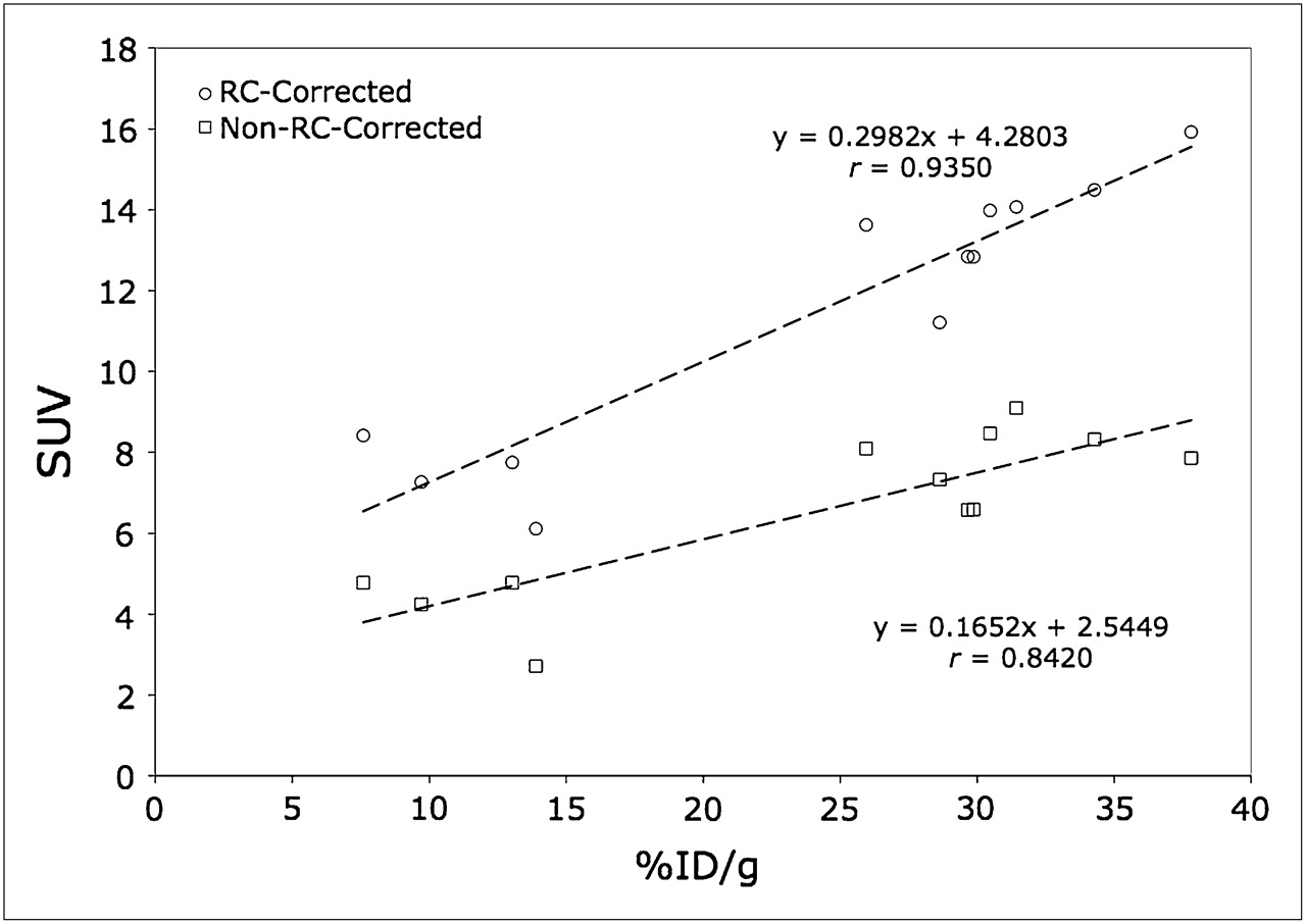
Recovery coefficient (RC)–corrected and noncorrected maximum SUV vs. measured 124I-ch806 %ID/g illustrating time distribution of xenograft samples.
DISCUSSION
We report on the successful use of immuno-PET with ch806 labeled with 124I via the residualizing ligand IMP-R4 to detect xenografts expressing de2-7 EGFR. We selected 124I as our radiolabel because it is readily available and has a sufficiently long t1/2 (4.2 d) to make it compatible with the biodistribution properties of intact antibodies such as ch806. The quantitation of 124I-ch806 uptake was significantly correlated with well counter–based quantitation, despite the small volume of tumors. Although several studies have been published regarding immuno-PET using short-lived (t1/2, 1–2 h) positron emitters such as 18F-FDG (20) or 68Ga (21–23), these are suitable only for antibody fragments or smaller antibody constructs because of their short t1/2. Although the medium-lived (t1/2, 12–16 h) isotopes such as 64Cu (24–27), 86Y (28,29), and 76Br (30,31) have been used to label both intact antibodies and partial antibody constructs, the most favorable isotopes for labeling whole antibodies are long-lived isotopes such as 124I or 89Zr (3,4,32). Both 124I (33,34) and 89Zr (32,35–38) have been conjugated to mAbs in preclinical (1,3,32,35–37) and clinical studies (33,34,37,38).
It was necessary to conjugate 124I to ch806 using the residualizing peptide IMP-R4 to increase tumor retention of the labeled antibody, because ch806 is internalized by de2-7 EGFR–expressing cells, which leads to dehalogenation of radioiodine to antibody with standard tyrosine linkage (15). The residualizing peptide IMP-R4 provides a conjugation method that can be successfully applied to animal studies and potentially to human trials. Conjugation with IMP-R4 did not alter the functional characteristic of ch806 in vitro, with the affinity, number of receptors bound, and immunoreactivity of 124I-IMP-R4-ch806 being highly comparable to the murine antibody (13) and similar to the same antibody labeled with 111In (10,39). Although the immunoreactivity of 124I-IMP-R4-ch806 did decline with time, the results are consistent with other immunoconjugates successfully applied in the clinic (16,18).
124I-IMP-R4-ch806 also retained specificity for tumor-expressed de2-7 EGFR, with no significant binding to normal tissues observed. This specificity is consistent with that observed for ch806 for tumor-expressed EGFR seen in patients (16) and in marked contrast to the high liver uptake seen with 111In-C225 anti-EGFR antibody in clinical trials (40). 124I-IMP-R4-ch806 was also an excellent molecular imaging agent for PET/CT. The ability to capture high-resolution images was evident from 24 h after injection and persisted until 168 h after injection, when the xenografts remained clearly visible on PET. There was also an excellent correlation between the assessment of tumor volume determined by CT and the tumor weight determined at necropsy. The volumes of the lesions determined from CT were used subsequently to correct the tumor uptake for partial-volume effect. Immuno-PET was able to detect tumors as small as 230 mm3 in this experiment, showing the high resolution and specificity of this method. More important, there was good correlation between the quantification of 124I-IMP-R4-ch806 uptake by PET and by direct measurement at necropsy (r2 = 0.8743).
CONCLUSION
We have shown that the radiohalogen 124I can be used for indirect labeling of ch806, allowing high-resolution imaging and quantification of de2-7 EGFR expression in gliomas. We also confirm the high tumor specificity of ch806, indicating that it has therapeutic potential both as a naked antibody and as a carrier antibody for payloads (e.g., radioisotopes, toxins). Because the conditions that allow ch806 binding (EGFR overexpression, mutation, and ligand activation) are essentially restricted to tumors (8,39), immuno-PET with ch806 may have an important role in the noninvasive molecular characterization of EGFR-expressing tumors in patients.
Acknowledgments
This work was supported in part by NHMRC Program grants 280916 and 487922.
Footnotes
-
COPYRIGHT © 2010 by the Society of Nuclear Medicine, Inc.
References
- Received for publication July 13, 2009.
- Accepted for publication February 22, 2010.


