


Article Figures & Data
Figures
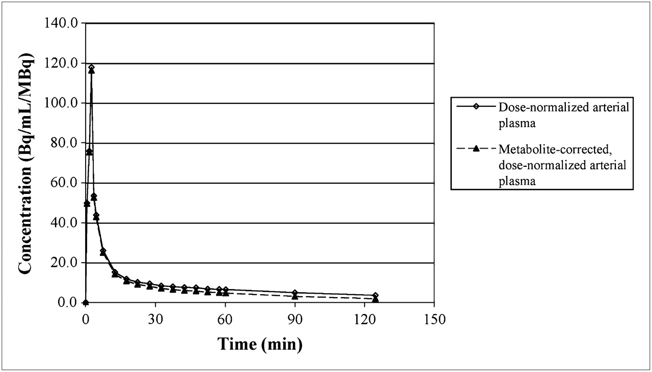
- FIGURE 1.
Standard 18F-FLT plasma curves presented as value normalized by administered dose (Bq/mL/MBq). Solid line represents measured curve, and dashed line is metabolite-corrected curve. Individual patient data and table of plotted values are available in supplemental material.
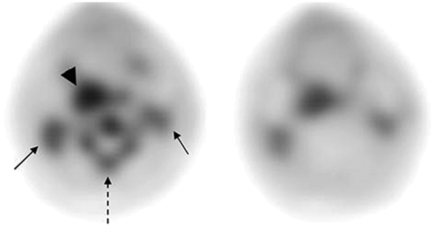
- FIGURE 2.
Pretherapy (left) and midtherapy (right) 18F-FLT PET images of patient with right tonsillar neoplasm (arrowhead), with bilateral cervical nodal metastases (solid arrows). Note interval decrease in 18F-FLT uptake in tumor and marked reduction in cervical bone marrow activity (dashed arrow).
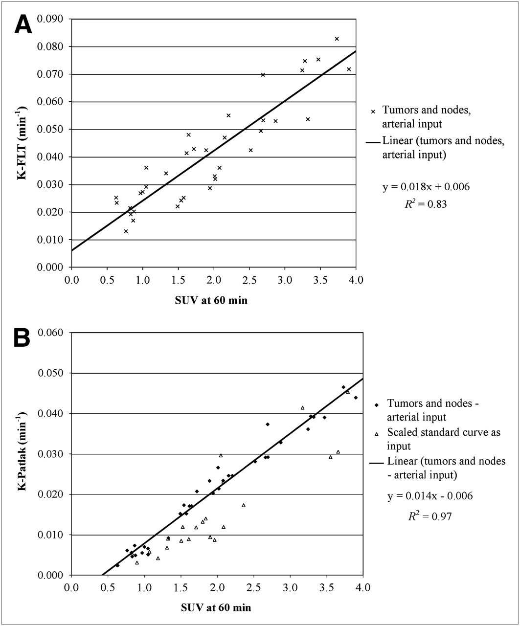
- FIGURE 3.
Comparison of SUV60 with K-FLT derived from 4-parameter fits in 4 patients with arterial blood sampling (A) and with K-Patlak in all patients (B), including pretherapy and midtherapy scans. B includes values determined in patients without arterial sampling through use of scaled standard input function.
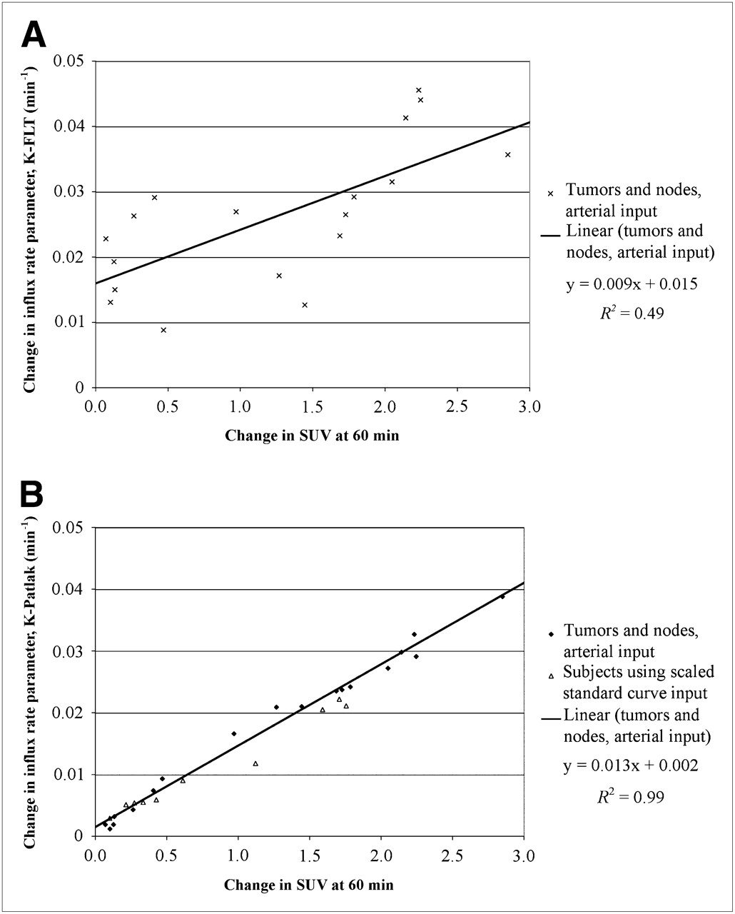
- FIGURE 4.
Comparison of change in SUV60 between pretherapy and midtherapy scans with change in K-FLT in patients with arterial blood sampling (A) and comparison of SUV and K-Patlak in all patients (B). B includes values determined in patients without arterial sampling through use of scaled standard input function.
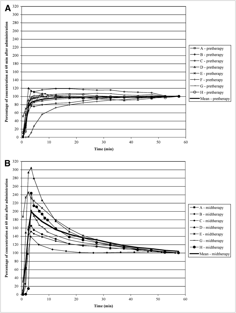
- FIGURE 5.
18F-FLT uptake in primary tumor at different time points expressed as percentage of the concentration at 60 min after administration at pretherapy (A) and midtherapy (B) scans. Heavy solid line represents mean value at each time point. At 45 min, uptake is within ±10% of 60-min concentration in all patients for both pretherapy and midtherapy scans.
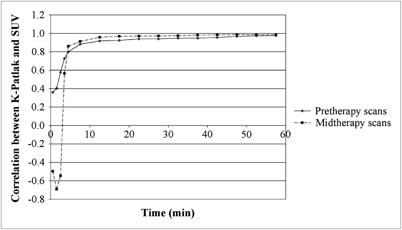
- FIGURE 6.
Correlation between K-Patlak and SUV at particular time point vs. midpoint time (min) of imaging interval for pretherapy (solid curve) and midtherapy (dashed curve) scans.


Tables
Parameter Tissue K-FLT-4p (1/min) K-FLT-3p (1/min) K-Patlak (1/min) SUV-60 SUV-60 max Primary tumor Before therapy 0.075 ± 0.006 0.054 ± 0.006 0.042 ± 0.004 3.4 ± 0.5 4.8 ± 0.8 Midtherapy 0.040 ± 0.010 0.029 ± 0.015 0.018 ± 0.016 1.8 ± 1.1 2.6 ± 1.6 18F-FLT–avid neck nodes Before therapy 0.047 ± 0.015 0.031 ± 0.008 0.026 ± 0.008 2.3 ± 0.7 3.2 ± 1.2 Midtherapy 0.024 ± 0.007 0.017 ± 0.011 0.010 ± 0.007 1.2 ± 0.5 1.7 ± 0.8 K-FLT-4p = influx parameter as defined in text derived from 4-parameter model fit; K-FLT-3p = influx parameter derived from fit holding k4 = 0; SUV-60 = mean SUV from 55 to 60 min after initiation of 18F-FLT infusion; max = maximum value for same region of interest.
Data are mean ± SD for patients with full arterial and venous sampling (n = 4 on 2 occasions).
Parameter Primary tumor and neck node Primary tumor only Neck node only K-Patlak vs. K-FLT-4p 0.896 0.931 0.862 K-Patlak vs. K-FLT-3p 0.987 0.983 0.987 K-Patlak vs. SUV-60 0.987 0.980 0.988 K-Patlak vs. SUV-60 max 0.966 0.972 0.958 K-FLT-4p vs. K-FLT-3p 0.916 0.953 0.873 K-FLT-4p vs. SUV-60 0.910 0.904 0.895 K-FLT-4p vs. SUV-60 max 0.889 0.875 0.873 K-FLT-3p vs. SUV-60 0.969 0.961 0.975 K-FLT-3p vs. SUV-60 max 0.944 0.951 0.939 SUV-60 vs. SUV-60 max 0.987 0.990 0.983 K-FLT-4p = influx parameter as defined in text derived from 4-parameter model fit; K-FLT-3p = influx parameter derived from fit holding k4 = 0; SUV-60 = mean SUV from 55 to 60 min after initiation of 18F-FLT infusion; max = maximum value for same region of interest.
Supplemental Data
Files in this Data Supplement:


Jump to section
Related Articles
Cited By...
- Hybrid Imaging PET/CT with Application of 18F-Fluorothymidine in Patients with Head and Neck Carcinoma Undergoing Radiotherapy
- 18F-FLT PET During Radiotherapy or Chemoradiotherapy in Head and Neck Squamous Cell Carcinoma Is an Early Predictor of Outcome
- PET Imaging During Radiotherapy of Head and Neck Cancer
- Usefulness of 3'-Deoxy-3'-18F-Fluorothymidine PET for Predicting Early Response to Chemoradiotherapy in Head and Neck Cancer
- The future of imaging: developing the tools for monitoring response to therapy in oncology: the 2009 Sir James MacKenzie Davidson Memorial lecture
- 18F-FLT PET/CT for Early Response Monitoring and Dose Escalation in Oropharyngeal Tumors
- Histopathologic Validation of 3'-Deoxy-3'-18F-Fluorothymidine PET in Squamous Cell Carcinoma of the Oral Cavity