


Abstract
Patients with pancreatic cancer continue to have a poor prognosis, with a 5-y survival rate of less than 5%. Surgery is the only treatment that offers a potential cure. Determining resectability is the principal goal of staging in pancreatic cancer patients. Our objective was to evaluate the value of combined contrast-enhanced 18F-FDG PET/CT in assessing the resectability of pancreatic cancer and to compare enhanced PET/CT with the performance of PET alone and unenhanced PET/CT. Methods: Fifty patients (25 women and 25 men; mean age, 64.3 y; range, 39–84 y) with biopsy-proven pancreatic adenocarcinoma underwent enhanced 18F-FDG PET/CT for the evaluation of resectability. Criteria for unresectability were distant metastases, peritoneal carcinomatosis, arterial infiltration, or invasion of neighboring organs other than the duodenum. The performance of enhanced PET/CT regarding resectability was compared with that of PET alone and unenhanced PET/CT. Histology, intraoperative findings, and follow-up CT with clinical investigations were used as the reference standard. Results: According to the reference standard, 27 patients had disease that was not resectable because of distant metastases (n = 17), peritoneal carcinomatosis (n = 5), or local infiltration (n = 5). In the assessment of resectability, PET alone had a sensitivity of 100%, specificity of 44%, accuracy of 70%, positive predictive value of 61%, and negative predictive value of 100%; unenhanced PET/CT had respective values of 100%, 56%, 76%, 66%, and 100%; and enhanced PET/CT, 96%, 82%, 88%, 82%, and 96%. In 5 patients, unresectability was missed by all imaging methods and was diagnosed intraoperatively. Enhanced PET/CT was significantly superior to PET alone (P = 0.035), and there was a trend for enhanced PET/CT to be superior to unenhanced PET/CT (P = 0.070). Conclusion: The use of enhanced PET/CT as a 1-stop-shop imaging protocol for assessing the resectability of pancreatic cancer is feasible and accurate. Enhanced PET/CT is significantly superior to PET alone.
Patients with pancreatic cancer continue to have a poor prognosis, with a 5-y survival rate of less than 5%. Surgery is the only treatment that offers a potential cure, but only 15%−20% of the patients are candidates for surgery (1,2). Determining resectability is the principal goal of staging in pancreatic cancer patients. The current preoperative imaging standard for pancreatic cancer staging is contrast-enhanced multidetector CT (3,4). Endoscopic ultrasound is also routinely used in many centers for local staging and biopsy guidance (5,6). 18F-FDG PET/CT is a powerful imaging method for the staging of many cancers and also has been shown to affect the oncologic management of pancreatic cancer patients (7). However, because of the limited information available in the literature, the role of 18F-FDG PET/CT in the management of pancreatic cancer remains undefined. The 1-stop-shop imaging approach with whole-body 18F-FDG PET combined with enhanced multidetector CT in a single investigation is feasible with the current generation of scanners, is convenient for patients, and appears to be an attractive staging tool for pancreatic cancer. The aim of this study was to evaluate the value of combined enhanced 18F-FDG PET/CT in determining the resectability of pancreatic cancer and to compare enhanced PET/CT with PET alone and with unenhanced PET/CT.
MATERIALS AND METHODS
Eligibility Criteria
Patients with biopsy-proven pancreatic cancer who underwent staging PET/CT at the University Hospital of Zurich were eligible for this retrospective analysis. The disease was considered resectable in the absence of distant metastases, arterial infiltration, and infiltration of organs other than the duodenum or stomach. The study was conducted in accordance with the local guidelines established by the ethics committee for retrospective evaluation, and written informed consent was waived for all patients.
PET/CT
All data were acquired on a combined PET/CT in-line system (Discovery ST; GE Healthcare). This dedicated system integrates a PET scanner (Advance Nxi; GE Healthcare) with a multislice helical CT scanner (Lightspeed 16; GE Healthcare) and permits the acquisition of coregistered CT and PET images in a single session.
The patients fasted for at least 4 h before scanning, which started approximately 60 min after the injection of 370–400 MBq of 18F-FDG. All patients were tested for a normal glucose level (range, 80–120 mg/dL [4.4–6.7 mmol/L]) before scanning. Patients with elevated glucose levels were rescheduled, prepared with insulin, and scanned when they had normal glucose levels. Patients were examined in the supine position. Initially, a low-dose CT scan was acquired starting from the level of the head using the following parameters: 40 mAs, 140 kV, 0.5 s/tube rotation, a slice thickness of 4.25 mm, a scan length of 867 mm, and a data acquisition time of 22.5 s. The CT scan was acquired during breath holding in the normal expiratory position. The low-dose CT data were used for attenuation correction and lesion localization, and the images were reconstructed using a standard iterative algorithm. Immediately after the CT acquisition, a PET emission scan was acquired with a time of 3 min per cradle position with a 1-slice overlap in 2-dimensional mode (matrix, 128 × 128). The 8–9 cradle positions starting from the head and continuing to the knees resulted in an acquisition time of approximately 24–27 min. Afterward, enhanced CT of the abdomen was performed on the same scanner using a dual-phase pancreatic protocol. The contrast agent (150 mL, Ultravist 300; Schering) was injected with a power injector at a rate of 3.0–4.0 mL/s through a 21-gauge catheter placed in the antecubital vein. A bolus-tracking program (SmartPrep; GE Healthcare) was used to monitor contrast enhancement after injection and before initiation of the diagnostic scans. The region-of-interest cursor for bolus tracking was placed in the aorta at the level of the diaphragmatic dome. Real-time low-dose serial monitoring scanning was initiated 5 s after the start of the contrast injection. Sections 1.25 mm in nominal thickness were obtained from the diaphragm to the inferior part of the duodenum after triggering of 60 Hounsfield units in the aortic region of interest. After a 70-s delay, a portovenous phase from the diaphragm to the symphysis pubis was obtained with a 2.5-mm thickness. In patients for whom high-quality dual-phase abdominal CT had already been performed at an outside institution less than 2 wk before the PET/CT scan, enhanced CT was not repeated and these patients were excluded from the study. The acquired images were reviewed with software providing multiplanar reformatted images of PET alone, CT alone, and fused unenhanced PET/CT and enhanced PET/CT imaged with linked cursors using a Xeleris workstation (GE Healthcare). PET/CT was performed according to the recently published Procedure Guideline for Tumor Imaging with 18F-FDG PET/CT 1.0 (8).
Image Interpretation
The PET/CT images were analyzed by 2 dual–board-certified nuclear radiology physicians with 7 and 3 y of experience in PET/CT reading, with specialization in tumor staging and abdominal imaging. Images were evaluated by consensus. The only information the readers had was that the investigation was being done for pancreatic cancer staging. They were unaware of other clinical information and the results of other imaging modalities (e.g., endoscopic ultrasound or MRI). First, the reader interpreted the PET images alone; in a second step, the reader interpreted the unenhanced PET/CT images; and in a third step, the reader interpreted the enhanced PET/CT images. There was at least a 2-wk delay between the readings. The PET images were analyzed for the presence and nature of lesions with focally increased 18F-FDG uptake. For all patients, the attenuation-corrected PET images were used for analysis. Lesions were interpreted as metastases if the uptake was higher than the uptake of the surrounding background tissue so that a focal lesion was clearly depicted. 18F-FDG uptake thought to be physiologic or due to benign variants such as uptake in muscles or brown fat or uptake caused by pulmonary infiltration was considered nonmalignant. 18F-FDG–negative pulmonary nodules without calcifications or fatty content were diagnosed as pulmonary metastases (Fig. 1). The enhanced-CT part was analyzed using the established criteria for the assessment of the primary tumor: vessel involvement (>180° of circumferential contiguity of tumor to vessel), organ infiltration, and distant metastases (9,10).

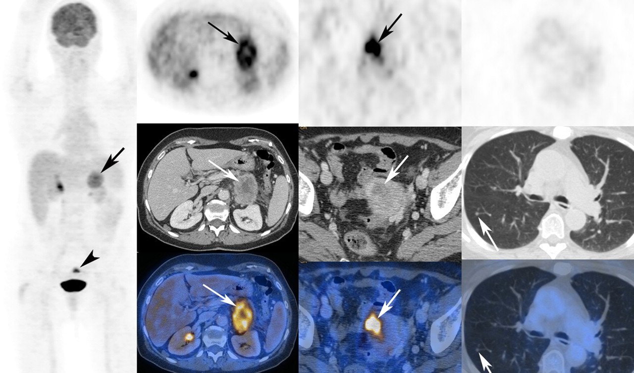
A 73-y-old woman with cancer of pancreatic tail. (First column) Primary tumor (arrow) and incidentally found small uterine cancer (arrowhead) are seen on maximum-intensity projection. (Second column) Primary tumor is seen on axial PET, enhanced CT, and fused enhanced PET/CT (arrows). (Third column) Incidentally found small uterine cancer is seen on axial PET, enhanced CT, and fused enhanced PET/CT (arrows). (Fourth column) Small 18F-FDG–negative right lung metastasis is seen (arrows).
Reference Standard
The reference standard was intraoperative findings, histologic findings, or clinical and imaging follow-up of at least 6 mo if biopsies were judged too invasive to be performed.
Statistical Analysis
Data were analyzed on a patient basis using SPSS 15 for Windows (SPSS Inc.). Statistical significance was assessed with the sign test. A P value of less than 0.05 was considered to indicate a significant difference. Bonferroni correction was not possible because of the small number of patients.
RESULTS
Fifty consecutive patients (25 women and 25 men; mean age, 64.3 y; range, 39–84 y) with biopsy-proven adenocarcinoma of the pancreas underwent enhanced 18F-FDG PET/CT for evaluation of resectability between April 2004 and December 2006. All patients had biopsy-proven adenocarcinoma of the pancreas. Eighteen patients had distant metastases. Of these, metastases were histologically proven in 10 patients. In 8 patients, metastases were confirmed by the radiologic appearance, with progression on follow-up PET/CT or CT scans and clinical investigations including CA 19-9 measurements. Thirty-five patients underwent surgical exploration, of which 28 (56% of the 50 total) underwent tumor resection (24 Whipple operations and 4 left resections) with a curative intent.
According to the reference standard, 27 patients had disease that was not resectable because of distant metastases (n = 17), peritoneal carcinomatosis (n = 2), or local infiltration (n = 5); a combination of peritoneal carcinomatosis and metastasis (n = 1); or a combination of peritoneal carcinomatosis and arterial infiltration (n = 2). Twenty-three patients had resectable disease. The discrepancy that 5 patients underwent tumor resection despite unresectability regarding the reference standard is explained by an aggressive surgical approach in unclear cases: tumor resection was performed in 2 patients with unclear lung nodules, which turned out to be lung metastases during follow-up; in 1 patient with a liver metastasis discovered during the Whipple procedure; and in 2 patients with arterial infiltration, 1 of whom underwent arterial grafting. The sensitivity, specificity, accuracy, NPV, and PPV of the various imaging methods were calculated on the basis of the reference standard (Table 1).
Value of PET, Unenhanced PET/CT, and Enhanced PET/CT in Assessment of Overall Resectability in 50 Patients with Pancreatic Cancer
Distant Metastases
Liver Metastases.
Eleven patients had liver metastases. In 6 patients, liver metastases were 18F-FDG–negative; in 5 patients, liver metastases were 18F-FDG–positive (mean maximal standardized uptake value, 5.6; range, 3.8–6.9). PET alone and unenhanced PET/CT detected liver metastases in 5 of these 11 patients (sensitivity, 46%; specificity, 100%), whereas enhanced PET/CT detected liver metastases in 9 (82%/97%). In 4 patients, all 3 imaging modalities (PET, unenhanced PET/CT, and enhanced PET/CT) detected the liver metastases. In 2 patients, all imaging modalities failed to detect the liver metastases and they were detected intraoperatively. In 1 patient, a liver metastasis was diagnosed with enhanced PET/CT but was missed intraoperatively. Follow-up imaging clearly showed progressive liver metastases in this patient.
Lung Metastases.
Seven patients had lung metastases. In 6 patients, lung metastases were 18F-FDG–negative (mean size, 4.6 mm; range, 3–7 mm). One patient had multiple 18F-FDG–positive lung metastases (mean maximal standardized uptake value, 6.5; maximal size, 20 mm) With PET alone, lung metastases were diagnosed in only 1 patient (14%/100%), but all lung metastases were diagnosed with unenhanced PET/CT and enhanced PET/CT (100%/100%). There was no case of false-positive diagnosis of lung metastases.
Bone Metastases.
Three patients had bone metastases. All bone metastases were 18F-FDG–positive (mean maximal standardized uptake value, 6.2; range, 4.5–6.9). PET, unenhanced PET/CT, and enhanced PET/CT detected the bone metastases in all 3 patients (100%/100%).
Peritoneal Carcinomatosis
Five patients had peritoneal carcinomatosis, which was detected by PET in 1 patient (20%/100%), by unenhanced PET/CT in 3 (60%/100%), and by enhanced PET/CT in 4 (80%/100%). In 1 patient, the peritoneal carcinomatosis was diagnosed only intraoperatively.
Arterial Infiltration
Five patients had arterial infiltration of the celiac trunk or the superior mesenteric artery. Enhanced PET/CT diagnosed arterial infiltration in all 5 patients (100%/100%). PET and unenhanced PET/CT failed to detect arterial infiltration in all 5 cases (0%/100%).
No patient had disease that was unresectable because of infiltration of organs other than the duodenum or stomach.
Table 2 summarizes the results for detection of metastases, peritoneal carcinomatosis, and arterial infiltration.
Value of PET, Unenhanced PET/CT, and Enhanced PET/CT in Detection of Metastases, Peritoneal Carcinomatosis, and Arterial Infiltration in 50 Patients with Pancreatic Cancer
Overall Resectability Assessment
Comparing Accuracy of Different Imaging Modalities.
Although we did not find a significant difference between unenhanced PET/CT and PET alone, a trend toward superiority for enhanced PET/CT over unenhanced PET/CT was found (P = 0.070). Furthermore, enhanced PET/CT was significantly superior to PET alone in our analysis (P = 0.035) (Table 1): 12 patients (24%) with disease judged to be resectable on PET demonstrated local unresectability or distant metastases on enhanced PET/CT.
Despite the improved accuracy of enhanced PET/CT over PET, 5 patients (10%) were judged to have resectable disease on enhanced PET/CT but had surgically unresectable disease at laparotomy: 2 patients had liver metastases, 2 had infiltration of the mesenteric root, and 1 had peritoneal carcinomatosis (Table 3).
Detection of Reasons for Unresectability with Different Imaging Methods in 27 Patients with Unresectable Disease
Incidental Detection of Simultaneous Other Cancers.
PET/CT also detected simultaneous cancer in 2 patients (4%). One patient had a non–small cell lung cancer (T1 stage), and the other patient had cancer of the uterus (T1 stage), both of which were successfully resected.
DISCUSSION
Although the morbidity and mortality of pancreas surgery has decreased during recent years, the long-term outcome of patients with pancreatic cancer remains poor. This poor outcome is generally attributed to a relatively chemoresistant disease and undetected metastases at the time of surgery (2) A multimodality regimen including adjuvant chemotherapy can improve survival after a curative resection (11). Furthermore, accurate staging is imperative for optimal patient selection, which can be improved by PET/CT, compared with standard staging (7).
Our results show that the 1-stop-shop protocol with enhanced multislice 18F-FDG PET/CT is feasible and accurate for preoperative pancreatic cancer staging.
In our patients, the liver was the organ most frequently affected by metastases, showing distant metastases in 11 patients. Other authors compared PET and CT in detecting liver metastases from pancreatic cancer and described an accuracy of about 90% for PET alone, which was comparable to the accuracy of enhanced CT or ultrasound (12). For hepatic metastasis, Diederichs et al. described a PET sensitivity of 70% and specificity of 95% caused by some metastases smaller than 1 cm being missed by PET (13). The additional use of contrast material increased the sensitivity for liver metastases from 46% to 82% in our patients. We also observed 1 patient with a false-positive diagnosis of liver metastases on enhanced PET/CT. This patient underlines the necessity of not only relying on imaging results but also obtaining histologic confirmation of suspected liver metastases or at least performing adequate imaging follow-up in selected cases. In 2 patients, all imaging modalities failed to detect small liver metastases, which were discovered during operative exploration together with intraoperative ultrasound.
It is known that PET alone is not sensitive enough to find lung metastases smaller than 1 cm, especially if they are in the lower parts of the lungs, where respiratory motion decreases their detectability. Therefore, some authors recommend additional diagnostic lung CT for tumors that tend to metastasize to the lungs, such as sarcomas (14). Our results emphasize that preoperative imaging should include a lung CT scan and that suggestive pulmonary nodules should be biopsied before a pancreatic resection is performed.
PET alone is clearly limited in the detection of peritoneal carcinomatosis, especially in cases of diffuse infiltration without formation of larger nodules. PET/CT can partially overcome the limitation of PET alone in diagnosing peritoneal carcinomatosis by showing stranding, peritoneal nodules, or ascites. Exploration remains the gold standard for the diagnosis of peritoneal carcinomatosis, considering the limited resolution of all established imaging methods (15,16).
Although infiltration of the portal vein is not generally considered a contraindication for surgery, infiltration of the superior mesenteric artery or celiac trunk precludes surgery in most centers. Therefore, the diagnosis of infiltration of the superior mesenteric artery or celiac trunk preoperatively is crucial for surgical planning. Arterial infiltration cannot be detected by PET alone or unenhanced PET/CT because the vessels cannot be delineated. Thin-section helical CT is reliable in assessing local resectability, with a sensitivity and specificity of 84% and 98%, respectively, if criteria such as the >180° circumferential contiguity of tumor to vessel are used (9). The ability to create high-resolution 2-dimensional and 3-dimensional maximum-intensity projections and volume-rendered images is an advantage of the thin-slice multidetector CT technique that has replaced conventional angiography (17,18). The 1-stop-shop enhanced-PET/CT approach offers the advantage of clear visualization of the relationship between the important vessels and the pancreatic tumor in a volume-rendered 3-dimensional CT angiography/PET combination as demonstrated in Figure 2.
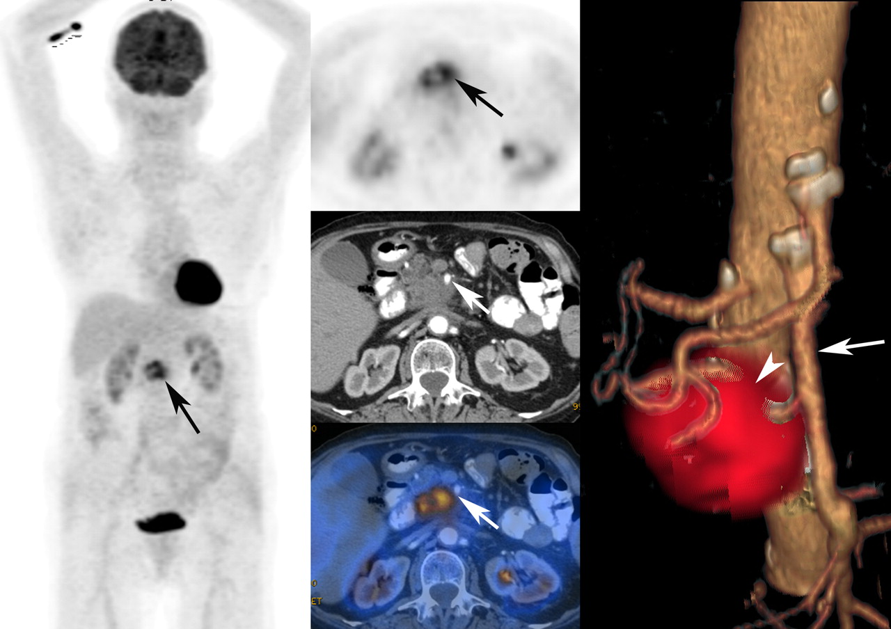
A 72-y-old woman with cancer of pancreatic head. (First column) Maximum-intensity-projection image shows 18F-FDG–active primary (arrow) without 18F-FDG–active distant metastases. (Second column) Axial PET, enhanced CT, and enhanced PET/CT in arterial phase show that tumor is growing around superior mesenteric artery (arrows). (Third column) Software-fused volume-rendered CT angiography and PET illustrate normal variant, with additional branch (arrowhead) from superior mesenteric artery (long arrow) supplying liver. Additional branch is infiltrated by pancreatic-head cancer.
In addition to improved staging of pancreatic cancer, enhanced PET/CT also detects simultaneous cancers, as reported previously (7). These incidental findings affect the oncologic treatment of the patients, since undetected primary tumors may metastasize before their detection.
Because the intravenous contrast protocol is used only for the abdominal part of the CT study, radiation exposure remains acceptable, at approximately 12 mSv. Also, enhanced CT is not repeated in our daily routine if high-quality enhanced CT has been performed recently.
The higher resectability rate in our population (46%), compared with previous publications (10%−20%), is presumably related to a referral bias and due to the aggressiveness of the pancreas surgeons at our institution (1): only patients whose disease was deemed resectable on standard staging were referred for PET/CT; those with clear evidence of metastatic disease on ultrasound or enhanced CT were not. This circumstance strengthens the value of enhanced PET/CT, which revealed mainly previously undetected metastases.
Experience with other tracers, such as 18F-fluorothymidine for pancreatic adenocarcinomas, is limited (19), and comparative studies with 18F-FDG are missing. It is known that in neuroendocrine pancreatic tumors, alternative tracers such as 68Ga-DOTA-NOC and 18F-DOPA PET work better than 18F-FDG (20).
CONCLUSION
We have demonstrated for, what is to our knowledge, the first time the feasibility and accuracy of a 1-stop-shop imaging approach by combining enhanced CT and PET in a single investigation in this analysis of 50 patients with histologically proven pancreatic cancer. Enhanced PET/CT was significantly superior to PET alone for the preoperative assessment of respectability.
Footnotes
-
COPYRIGHT © 2008 by the Society of Nuclear Medicine, Inc.
References
- Received for publication February 7, 2008.
- Accepted for publication May 16, 2008.


