
Main menu

  • Home
  • Content
    • Current
    • Ahead of print
    • Past Issues
    • JNM Supplement
    • SNMMI Annual Meeting Abstracts
  • Subscriptions
    • Subscribers
    • Institutional and Non-member
    • Rates
    • Corporate & Special Sales
    • Journal Claims
  • Authors
    • Submit to JNM
    • Information for Authors
    • Assignment of Copyright
    • AQARA requirements
  • Info
    • Permissions
    • Advertisers
    • Continuing Education
  • About
    • About Us
    • Editorial Board
    • Contact Information
  • More
    • Alerts
    • Feedback
    • Help
    • SNMMI Journals
  • SNMMI
    • JNM
    • JNMT
    • SNMMI Journals
    • SNMMI

User menu

  • Subscribe
  • My alerts
  • Log in
  • My Cart

Search

  • Advanced search
Journal of Nuclear Medicine
  • SNMMI
    • JNM
    • JNMT
    • SNMMI Journals
    • SNMMI
  • Subscribe
  • My alerts
  • Log in
  • My Cart
Journal of Nuclear Medicine

Advanced Search

  • Home
  • Content
    • Current
    • Ahead of print
    • Past Issues
    • JNM Supplement
    • SNMMI Annual Meeting Abstracts
  • Subscriptions
    • Subscribers
    • Institutional and Non-member
    • Rates
    • Corporate & Special Sales
    • Journal Claims
  • Authors
    • Submit to JNM
    • Information for Authors
    • Assignment of Copyright
    • AQARA requirements
  • Info
    • Permissions
    • Advertisers
    • Continuing Education
  • About
    • About Us
    • Editorial Board
    • Contact Information
  • More
    • Alerts
    • Feedback
    • Help
    • SNMMI Journals
  • Follow JNM on Twitter
  • Visit JNM on Facebook
  • Join JNM on LinkedIn
  • Subscribe to our RSS feeds
OtherBASIC SCIENCE INVESTIGATIONS

Epidermal Growth Factor Receptor Inhibition Modulates the Nuclear Localization and Cytotoxicity of the Auger Electron–Emitting Radiopharmaceutical 111In-DTPA–Human Epidermal Growth Factor

Kristy E. Bailey, Danny L. Costantini, Zhongli Cai, Deborah A. Scollard, Zhuo Chen, Raymond M. Reilly and Katherine A. Vallis
Journal of Nuclear Medicine September 2007, 48 (9) 1562-1570; DOI: https://doi.org/10.2967/jnumed.107.044073
Kristy E. Bailey
  • Find this author on Google Scholar
  • Find this author on PubMed
  • Search for this author on this site
Danny L. Costantini
  • Find this author on Google Scholar
  • Find this author on PubMed
  • Search for this author on this site
Zhongli Cai
  • Find this author on Google Scholar
  • Find this author on PubMed
  • Search for this author on this site
Deborah A. Scollard
  • Find this author on Google Scholar
  • Find this author on PubMed
  • Search for this author on this site
Zhuo Chen
  • Find this author on Google Scholar
  • Find this author on PubMed
  • Search for this author on this site
Raymond M. Reilly
  • Find this author on Google Scholar
  • Find this author on PubMed
  • Search for this author on this site
Katherine A. Vallis
  • Find this author on Google Scholar
  • Find this author on PubMed
  • Search for this author on this site
  • Article
  • Figures & Data
  • Info & Metrics
  • PDF
Loading

Article Figures & Data

Figures

  • Tables
  • FIGURE 1. 
    • Download figure
    • Open in new tab
    • Download powerpoint
    FIGURE 1. 

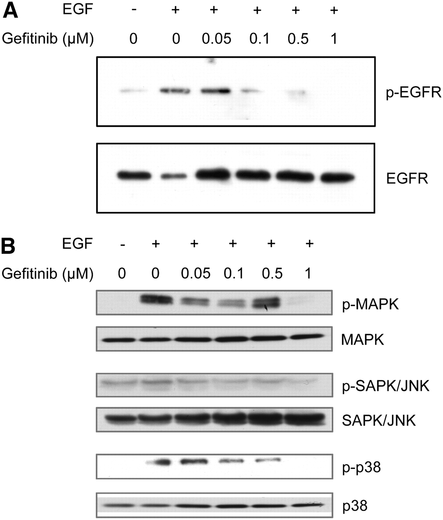
    EGF-induced EGFR phosphorylation and activation is blocked by 1 μM gefitinib in MDA-MB-468 cells. Western blot analyses of MDA-MB-468 cells without (−) or stimulated with 20 ng/mL EGF (+) for 15 min, following incubation with 0.05–1 μM gefitinib for 3 h at 37°C. (A) Total protein samples were probed with antiphospho-EGFR Tyr-1173 and anti-EGFR antibodies. (B) Anti-MAPK, antiphospho-MAPK (p42/p44), anti-SAPK/JNK, antiphospho-SAPK/JNK, anti-p38, and antiphospho-p38 antibodies were used to determine phosphorylation status of downstream proteins in EGFR signaling cascade. Phosphorylation of EGFR and activation of downstream proteins was completely blocked by 1 μM gefitinib.

  • FIGURE 2. 
    • Download figure
    • Open in new tab
    • Download powerpoint
    FIGURE 2. 

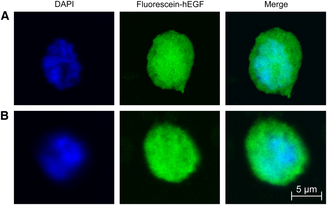
    Visualization of nuclear fluorescein-labeled-hEGF in presence of gefitinib using confocal microscopy. (A) MDA-MB-468 cells treated with fluorescein-hEGF for 1 h at 37°C. (B) MDA-MB-468 cells pretreated with gefitinib (1 μM) for 3 h followed by fluorescein-hEGF and gefitinib for 1 h at 37°C. Cells were also incubated with DAPI to visualize the cell nucleus. Images represent a 1-μm slice through center of cell. Fluorescein-hEGF was observed in the nucleus in absence and presence of 1 μM gefitinib.
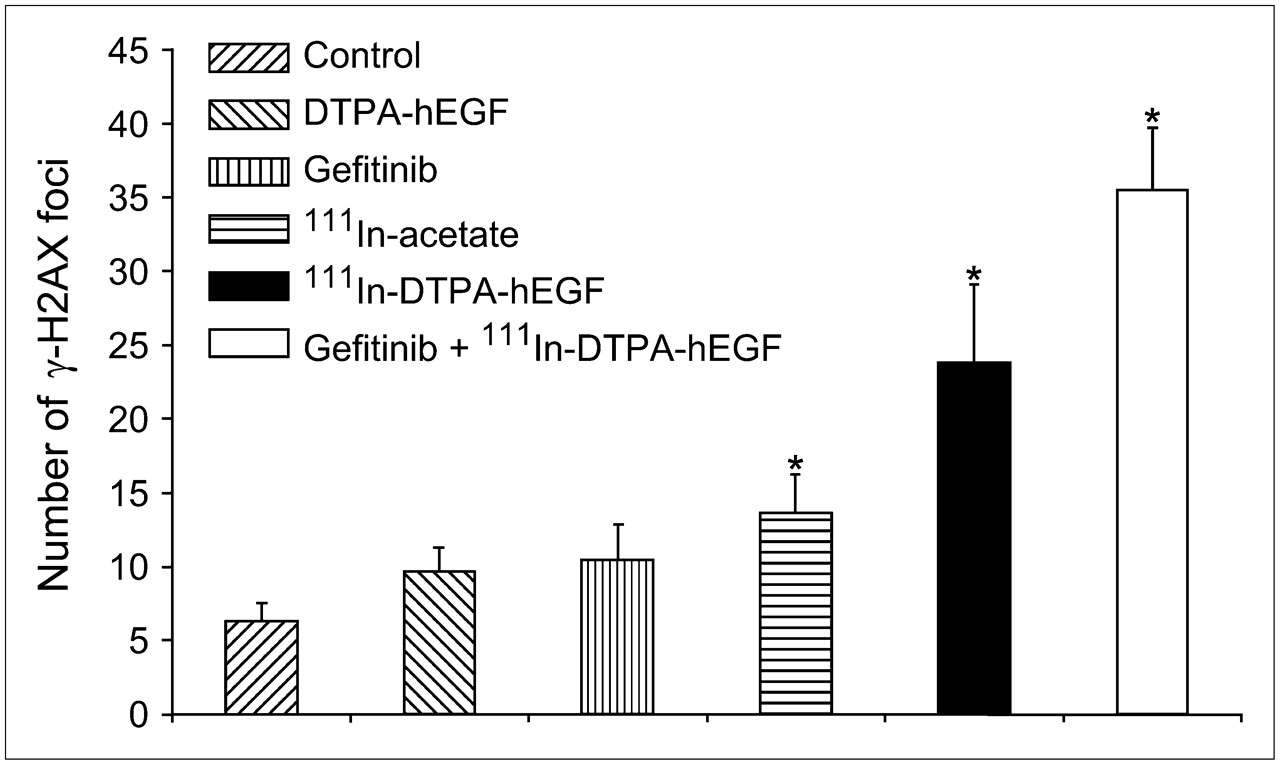
  • FIGURE 3. 
    • Download figure
    • Open in new tab
    • Download powerpoint
    FIGURE 3. 

    Effect of gefitinib and 111In-DTPA-hEGF on induction of γ-H2AX foci in MDA-MB-468 cells. γ-H2AX assay was performed after exposure of MDA-MB-468 cells to DTPA-hEGF (250 ng/mL), gefitinib (1 μM), 111In-acetate (1.5 MBq/mL), 111In-DTPA-hEGF (250 ng/mL, 1.5 MBq/mL), gefitinib (1 μM) plus 111In-DTPA-hEGF (250 ng/mL, 1.5 MBq/mL), or DMEM alone (control) for 20 h. Optical sections (1.2 μm) through the cells were obtained using a confocal microscope and were processed using ImageJ (U.S. National Institutes of Health). Error bars represent SEM number of γ-H2AX foci from 3 separate experiments. Thirty cells were counted to generate each data point for each experiment. *P < 0.05 (compared with control).

  • FIGURE 4. 
    • Download figure
    • Open in new tab
    • Download powerpoint
    FIGURE 4. 

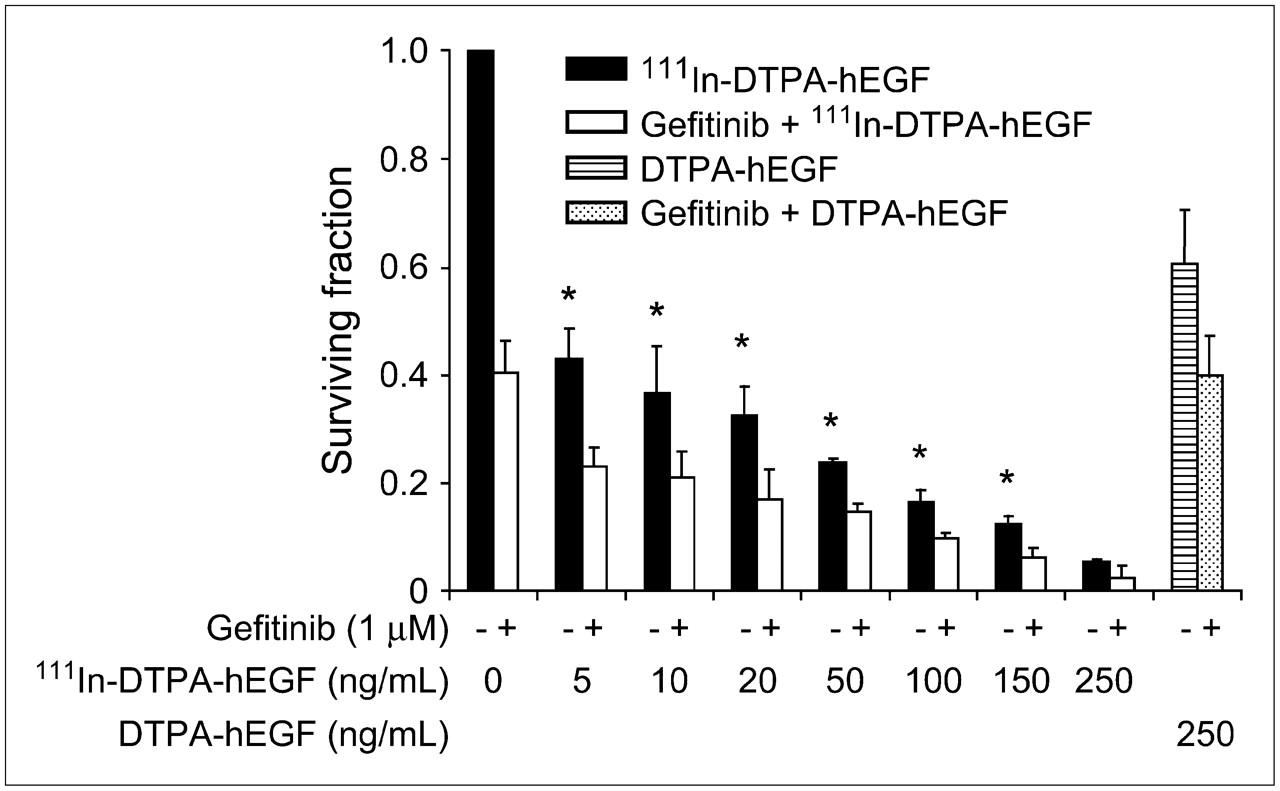
    Gefitinib enhances 111In-DTPA-hEGF–mediated cytotoxicity. Clonogenic assay was performed using MDA-MB-468 cells treated with increasing concentrations of 111In-DTPA-hEGF (5–250 ng/mL, 6 MBq/μg) for 24 h at 37°C (black bars) or pretreated with gefitinib (1 μM) for 3 h followed by treatment with a range of concentrations of 111In-DTPA-hEGF plus gefitinib (1 μM) for 24 h at 37°C (white bars). At each concentration of 111In-DTPA-hEGF tested, addition of gefitinib resulted in enhanced cytotoxicity. The SF of cells exposed to DTPA-hEGF (250 ng/mL) in the absence or presence of gefitinib (1 μM) was also measured (lined and speckled bars, respectively). 111In-DTPA-hEGF (250 ng/mL) was ∼10-fold more cytotoxic than unlabeled DTPA-hEGF (SF, 6.1% ± 1.9% vs. 60.6% ± 9.9%, respectively; P < 0.01). The SF of cells exposed to DTPA-hEGF (250 ng/mL) plus gefitinib (1 μM) was not statistically significant from the SF of cells exposed to 1 μM gefitinib alone (39.9% ± 7.4% vs. 40.3% ± 6.1%, respectively; P = 0.5). Error bars represent SD of the mean SF calculated from 3 separate experiments. *P < 0.05 (compared with control).

Tables

  • Figures
    • View popup
    TABLE 1

    Effect of Gefitinib on Binding, Internalization, and Nuclear Localization of 111In-DTPA-hEGF in MDA-MB-468 Human Breast Cancer Cells

    Proportion (%)Proportion of cell-bound 111In-DTPA-hEGF (%)
    TreatmentRadioactivity bound to cellsCell-bound radioactivity internalized by cellsRadioactivity within cytoplasmRadioactivity within nucleus
    111In-DTPA-hEGF54.7 ± 8.676.9 ± 5.962.3 ± 9.514.6 ± 4.0
    111In-DTPA-hEGF + gefitinib58.1 ± 2.881.1 ± 1.055.0 ± 6.226.0 ± 5.5*
    • ↵* P < 0.05.

    • Data are expressed as mean ± SD of 3 experiments.

    • View popup
    TABLE 2

    Effect of Gefitinib on Radiation-Absorbed Dose to Cell Nucleus from 111In-DTPA-hEGF Localized at Cell Membrane, Cytoplasm, or Nucleus of MDA-MB-468 EGFR-Overexpressing Human Breast Cancer Cells*

    TreatmentCell compartmentĀ (Bq × s)†S ([Gy/Bq × s] × 10−4)Radiation dose to nucleus Embedded Image, (Gy)‡
    111In-DTPA-hEGFMembrane4,2401.780.75
    Cytoplasm11,3073.183.4
    Nucleus2,65160.316.0
    Total20.15
    111In-DTPA-hEGF + gefitinibMembrane3,4321.780.61
    Cytoplasm9,8943.183.15
    Nucleus4,73560.328.55
    Total32.31
    • ↵* Radiation-absorbed dose (Embedded Image) to cell nucleus was estimated using cellular radiation dosimetry model of Goddu et al. (22): Embedded Image = Ā × S, where S is radiation-absorbed dose in nucleus (Gy) per unit of cumulated radioactivity in source compartment, Ā (Bq × s).

    • ↵† Ā = A0/λ, where A0 is amount of radioactivity localized in the compartment at time 0, and λ is radioactive decay constant for 111In (2.83 × 10⁻⁶/s). Rapid localization of 111In-DTPA-hEGF in the compartment and rate of elimination corresponding to radioactive decay of radionuclide 111In are assumed.

    • ↵‡ Based on each MDA-MB-468 cell having a diameter of 10 μm and a nuclear diameter of 6 μm. Assumes targeting of a single cell with 111In-DTPA-hEGF to receptor saturation. At receptor saturation, ∼48 mBq 111In-DTPA-hEGF would be bound to each MDA-MB-468 cell at a specific activity of 3.7 MBq/μg.

PreviousNext
Back to top

In this issue

Journal of Nuclear Medicine: 48 (9)
Journal of Nuclear Medicine
Vol. 48, Issue 9
September 2007
  • Table of Contents
  • Table of Contents (PDF)
  • About the Cover
  • Index by author
Print
Download PDF
Article Alerts
Sign In to Email Alerts with your Email Address
Email Article

Thank you for your interest in spreading the word on Journal of Nuclear Medicine.

NOTE: We only request your email address so that the person you are recommending the page to knows that you wanted them to see it, and that it is not junk mail. We do not capture any email address.

Enter multiple addresses on separate lines or separate them with commas.
Epidermal Growth Factor Receptor Inhibition Modulates the Nuclear Localization and Cytotoxicity of the Auger Electron–Emitting Radiopharmaceutical 111In-DTPA–Human Epidermal Growth Factor
(Your Name) has sent you a message from Journal of Nuclear Medicine
(Your Name) thought you would like to see the Journal of Nuclear Medicine web site.
Citation Tools
Epidermal Growth Factor Receptor Inhibition Modulates the Nuclear Localization and Cytotoxicity of the Auger Electron–Emitting Radiopharmaceutical 111In-DTPA–Human Epidermal Growth Factor
Kristy E. Bailey, Danny L. Costantini, Zhongli Cai, Deborah A. Scollard, Zhuo Chen, Raymond M. Reilly, Katherine A. Vallis
Journal of Nuclear Medicine Sep 2007, 48 (9) 1562-1570; DOI: 10.2967/jnumed.107.044073

Citation Manager Formats

  • BibTeX
  • Bookends
  • EasyBib
  • EndNote (tagged)
  • EndNote 8 (xml)
  • Medlars
  • Mendeley
  • Papers
  • RefWorks Tagged
  • Ref Manager
  • RIS
  • Zotero
Share
Epidermal Growth Factor Receptor Inhibition Modulates the Nuclear Localization and Cytotoxicity of the Auger Electron–Emitting Radiopharmaceutical 111In-DTPA–Human Epidermal Growth Factor
Kristy E. Bailey, Danny L. Costantini, Zhongli Cai, Deborah A. Scollard, Zhuo Chen, Raymond M. Reilly, Katherine A. Vallis
Journal of Nuclear Medicine Sep 2007, 48 (9) 1562-1570; DOI: 10.2967/jnumed.107.044073
Twitter logo Facebook logo LinkedIn logo Mendeley logo
  • Tweet Widget
  • Facebook Like
  • Google Plus One
Bookmark this article

Jump to section

  • Article
    • Abstract
    • MATERIALS AND METHODS
    • RESULTS
    • DISCUSSION
    • CONCLUSION
    • Acknowledgments
    • Footnotes
    • References
  • Figures & Data
  • Info & Metrics
  • PDF

Related Articles

  • This Month in JNM
  • PubMed
  • Google Scholar

Cited By...

  • Evaluation of Cobalt-Labeled Octreotide Analogs for Molecular Imaging and Auger Electron-Based Radionuclide Therapy
  • ErbB-2 Blockade and Prenyltransferase Inhibition Alter Epidermal Growth Factor and Epidermal Growth Factor Receptor Trafficking and Enhance 111In-DTPA-hEGF Auger Electron Radiation Therapy
  • RNA helicase A is a DNA-binding partner for EGFR-mediated transcriptional activation in the nucleus
  • The Monte Carlo Method in Nuclear Medicine: Current Uses and Future Potential
  • Cellular Dosimetry of 111In Using Monte Carlo N-Particle Computer Code: Comparison with Analytic Methods and Correlation with In Vitro Cytotoxicity
  • Relationship Between Induction of Phosphorylated H2AX and Survival in Breast Cancer Cells Exposed to 111In-DTPA-hEGF
  • Google Scholar

More in this TOC Section

  • Tumor Uptake of Anti-CD20 Fabs Depends on Tumor Perfusion
  • How Sensitive Is the Upper Gastrointestinal Tract to 90Y Radioembolization? A Histologic and Dosimetric Analysis in a Porcine Model
  • 11C-Methionine PET of Myocardial Inflammation in a Rat Model of Experimental Autoimmune Myocarditis
Show more Basic Science Investigations

Similar Articles

SNMMI

© 2023 Journal of Nuclear Medicine

Powered by HighWire