


Article Figures & Data
Figures
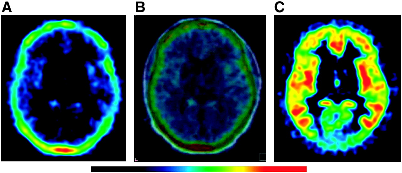
- FIGURE 1.
Horizontal 18F-FCWAY images before and after administration of disulfiram. (A) Baseline image at 2 h shows high activity in skull, consistent with metabolism of 18F-FCWAY to 18F-fluoride ion. (B) Coregistered baseline PET and MR images show skull activity spilling into adjacent brain—for example, into occipital cortex and cerebellum at bottom of image. (C) Repeated PET scan in same subject after disulfiram (500 mg on prior night) shows marked reduction of skull activity and better visualization of brain.
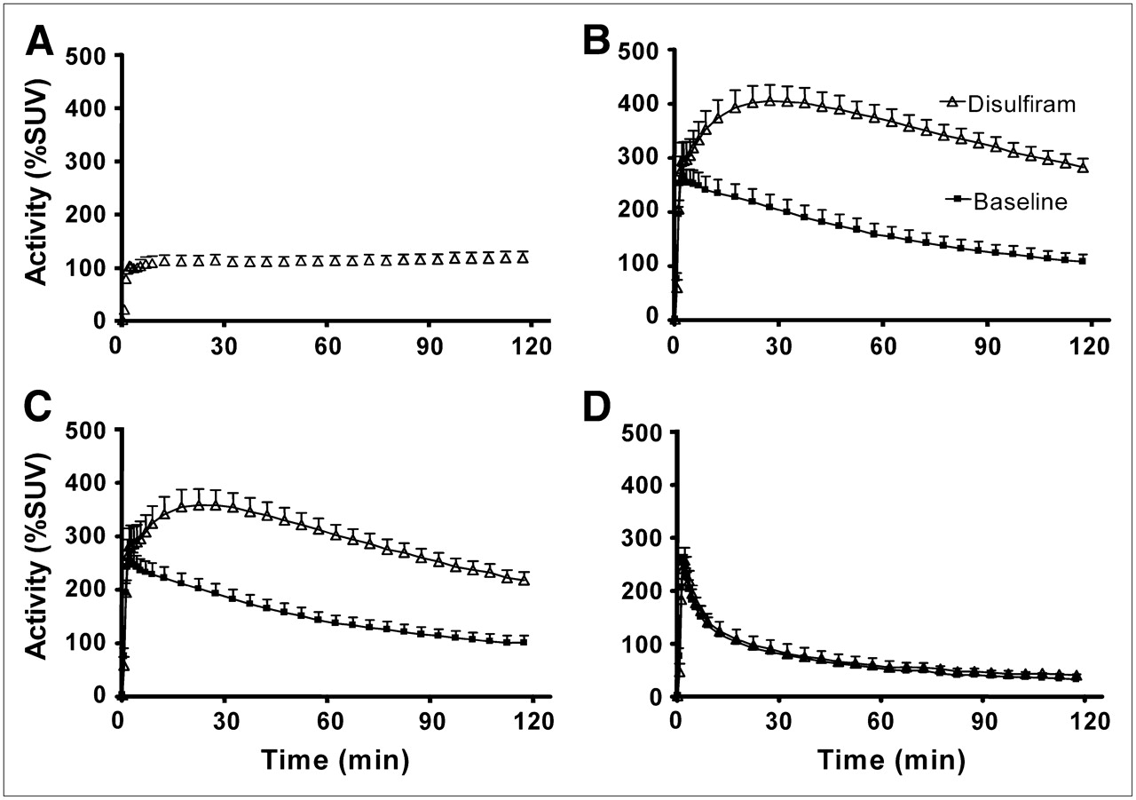
- FIGURE 2.
Time–activity curves for 18F-FCWAY in brain and skull. (A) Disulfiram had no effect on initial uptake of radioactivity (∼100% SUV) into skull within first few minutes but later blocked continued accumulation seen in baseline conditions. (B and C) In 2 regions (temporal and frontal cortices) with high 5-HT1A receptor densities, disulfiram increased brain uptake and delayed time of peak radioactivity to approximately 30 min. (D) Disulfiram had insignificant effects on time–activity curve in cerebellum, region with few 5-HT1A receptors. Symbols represent mean ± SD (n = 5).
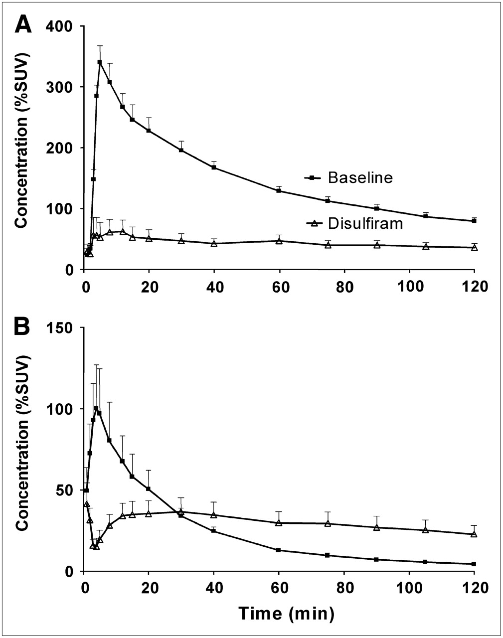
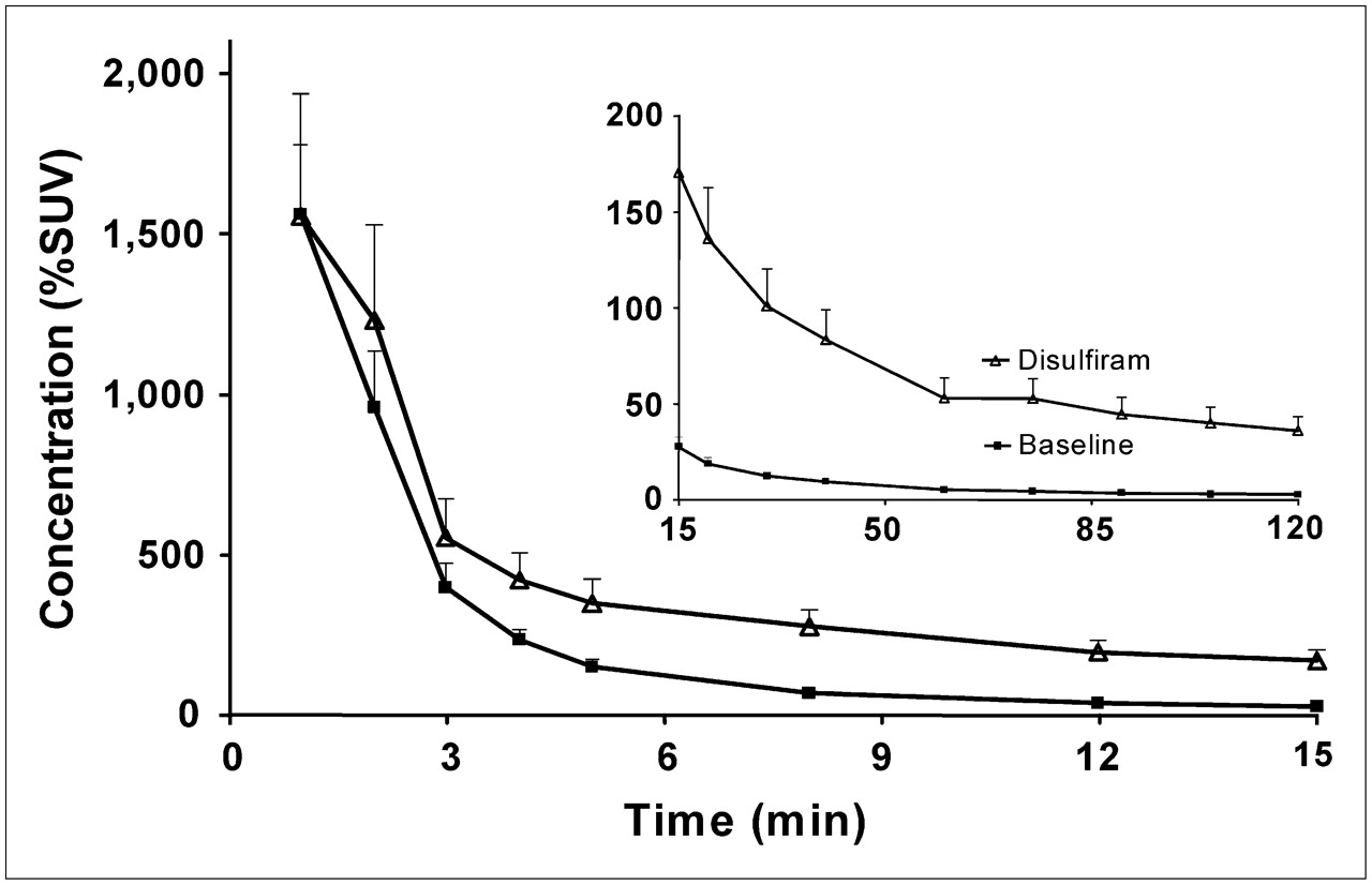
- FIGURE 3.
Plasma concentration of 18F-FCWAY, separated from radiometabolites. Disulfiram increased concentration of 18F-FCWAY and, thereby, decreased its removal from plasma. Disulfiram significantly decreased clearance of 18F-FCWAY by 47%. Symbols represent mean of 5 subjects, with SD bars only partially visible because some were less than size of symbol itself.
- FIGURE 4.
Plasma concentration of 2 radiometabolites of 18F-FCWAY. (A) Disulfiram decreased plasma concentration of 18F-fluoride ion. Expressed relative to AUC from time 0 to 120 min, disulfiram decreased plasma 18F-fluoride ion by 69%. (B) Disulfiram had time-dependent effects on plasma concentration of 18F-FC, initially increasing but later decreasing concentration of this radiometabolite. Symbols represent mean of 5 subjects, with SD bars only partially visible because some were less than size of symbol itself.
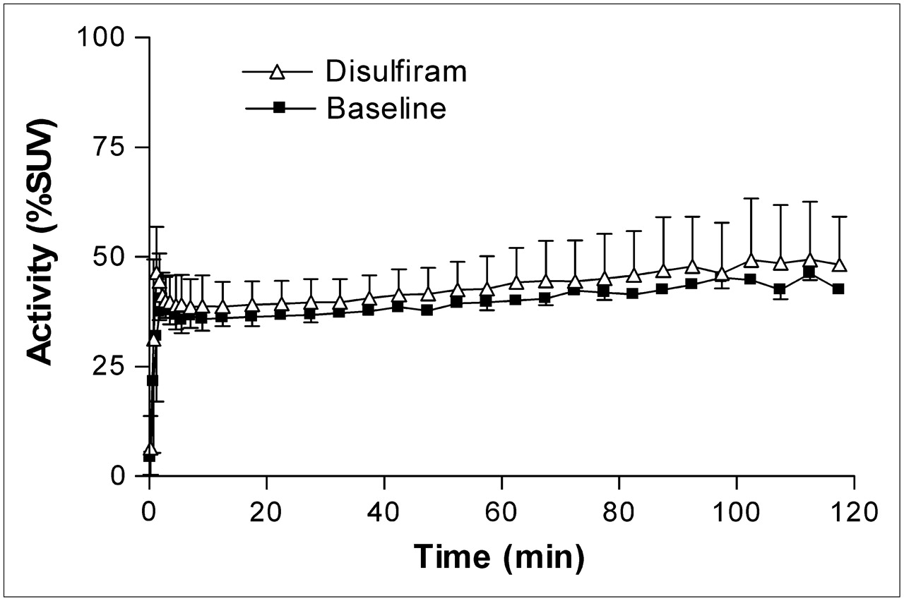
- FIGURE 5.
11C-Loperamide brain uptake in rhesus monkeys before and after administration of disulfiram (1,000-mg cumulative dose by mouth). Maximal brain uptake was low (∼50% SUV), consistent with loperamide being a substrate for P-gp efflux. Disulfiram had statistically insignificant effects on brain uptake. Symbols represent mean ± SD of 3 animals studied at baseline and after disulfiram.


Tables
VT VS VS corrected for fP Site Baseline Disulfiram Baseline Disulfiram Baseline Disulfiram Frontal cortex Distribution volume 7.5 ± 1.1 4.0 ± 15.1 6.9 ± 1.1 3.5 ± 13.4 56.7 ± 9.7 26.2 ± 7.6 COV (%) 3.5 ± 0.4 1.1 ± 0.4 3.7 ± 0.4 1.6 ± 0.6 Parietal cortex Distribution volume 7.3 ± 1.2 4.0 ± 15.5 6.8 ± 1.3 3.5 ± 13.5 56.0 ± 12.7 26.1 ± 7.6 COV (%) 3.3 ± 0.2 1.2 ± 0.4 3.5 ± 0.2 1.6 ± 0.4 Temporal cortex Distribution volume 8.3 ± 1.8 5.1 ± 18.8 7.7 ± 1.8 4.6 ± 16.9 62.9 ± 12.9 33.5 ± 8.6 COV (%) 3.4 ± 0.7 1.2 ± 0.4 3.6 ± 0.7 1.5 ± 0.4 Cerebellum Distribution volume 2.4 ± 1.7 0.7 ± 2.7 1.9 ± 1.6 0.4 ± 1.7 14.6 ± 11.2 2.7 ± 1.6 COV (%) 9.9 ± 9.3 4.7 ± 2.9 14.7 ± 16.9 11.3 ± 7.2 Putamen Distribution volume 2.2 ± 1.3 0.9 ± 3.5 1.8 ± 1.2 0.6 ± 2.4 13.0 ± 7.4 4.5 ± 1.5 COV (%) 10.7 ± 2.4 4.9 ± 1.4 13.3 ± 2.3 7.5 ± 1.3


Jump to section
Related Articles
Cited By...
- Common anesthetic used in preclinical PET imaging inhibits metabolism of the PET tracer [18F]3F4AP
- High-Contrast PET imaging with [18F]-NT160, a Class-IIa Histone Deacetylase (Class-IIa HDAC) Probe for In Vivo Imaging of Epigenetic Machinery in the Central Nervous System
- Biodistribution, Tumor Detection, and Radiation Dosimetry of 18F-5-Fluoro-2'-Deoxycytidine with Tetrahydrouridine in Solid Tumors
- Structure-Activity Relationship of 2-Arylquinolines as PET Imaging Tracers for Tau Pathology in Alzheimer Disease
- Using Cerebral White Matter for Estimation of Nondisplaceable Binding of 5-HT1A Receptors in Temporal Lobe Epilepsy
- Radiodefluorination of 3-Fluoro-5-(2-(2-[18F](fluoromethyl)-thiazol-4-yl)ethynyl)benzonitrile ([18F]SP203), a Radioligand for Imaging Brain Metabotropic Glutamate Subtype-5 Receptors with Positron Emission Tomography, Occurs by Glutathionylation in Rat Brain