


Article Figures & Data
Figures
- FIGURE 1.
DAT imaging in PD patient followed for 4 y. Visually, there is decreased uptake of radiotracer in striatum, indicative of ongoing loss of presynaptic dopaminergic function. Although the loss is easily noted, it is difficult to determine clinical meaningfulness of this extent of signal change without quantitation.
- FIGURE 2.
Display showing example patient's brain scan (right half of figure) being registered to standard average scan (left half of figure).
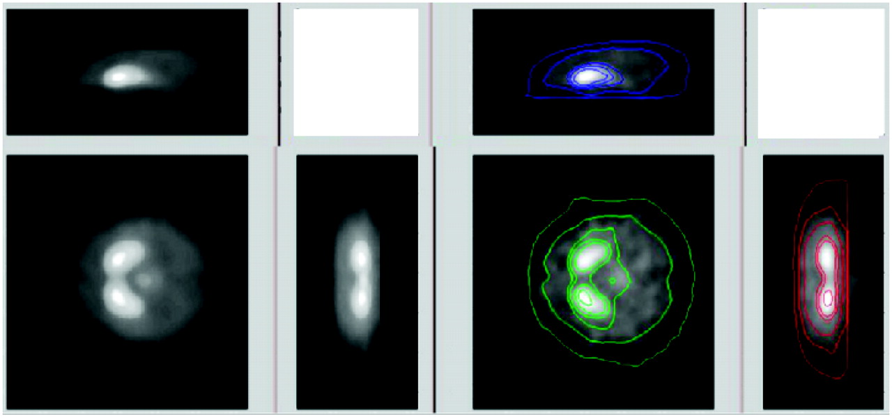
- FIGURE 3.
Display showing selection of horizontal striatal midplane (left half of figure) and subsequent localization of left and right striatum (right half of figure), both based on location of maxima in either sagittal or coronal profiles.
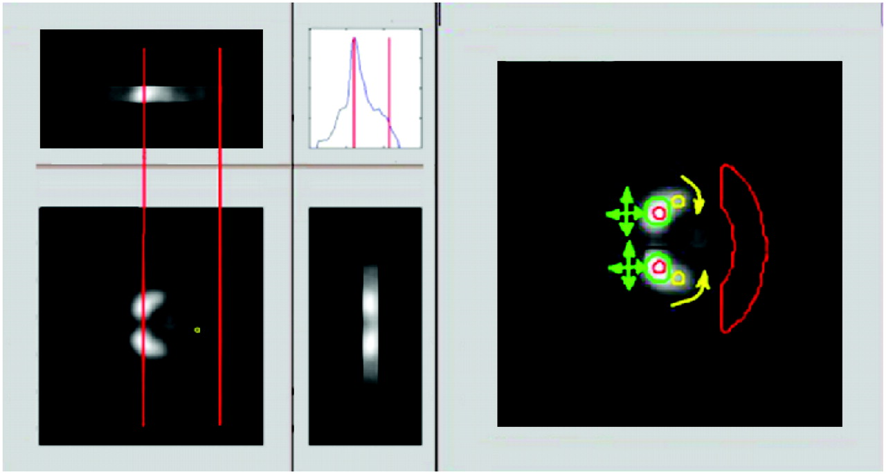
- FIGURE 4.
Display showing automatic measurement of head size (left half of figure), whereby distance from midpoint between the 2 striata to edge of occipital lobes is used to scale subsequent ROI placement. Right half of figure shows placement of caudate ROIs (small red circles), putamen ROIs (small yellow circles), and occipital ROI (large truncated annulus).
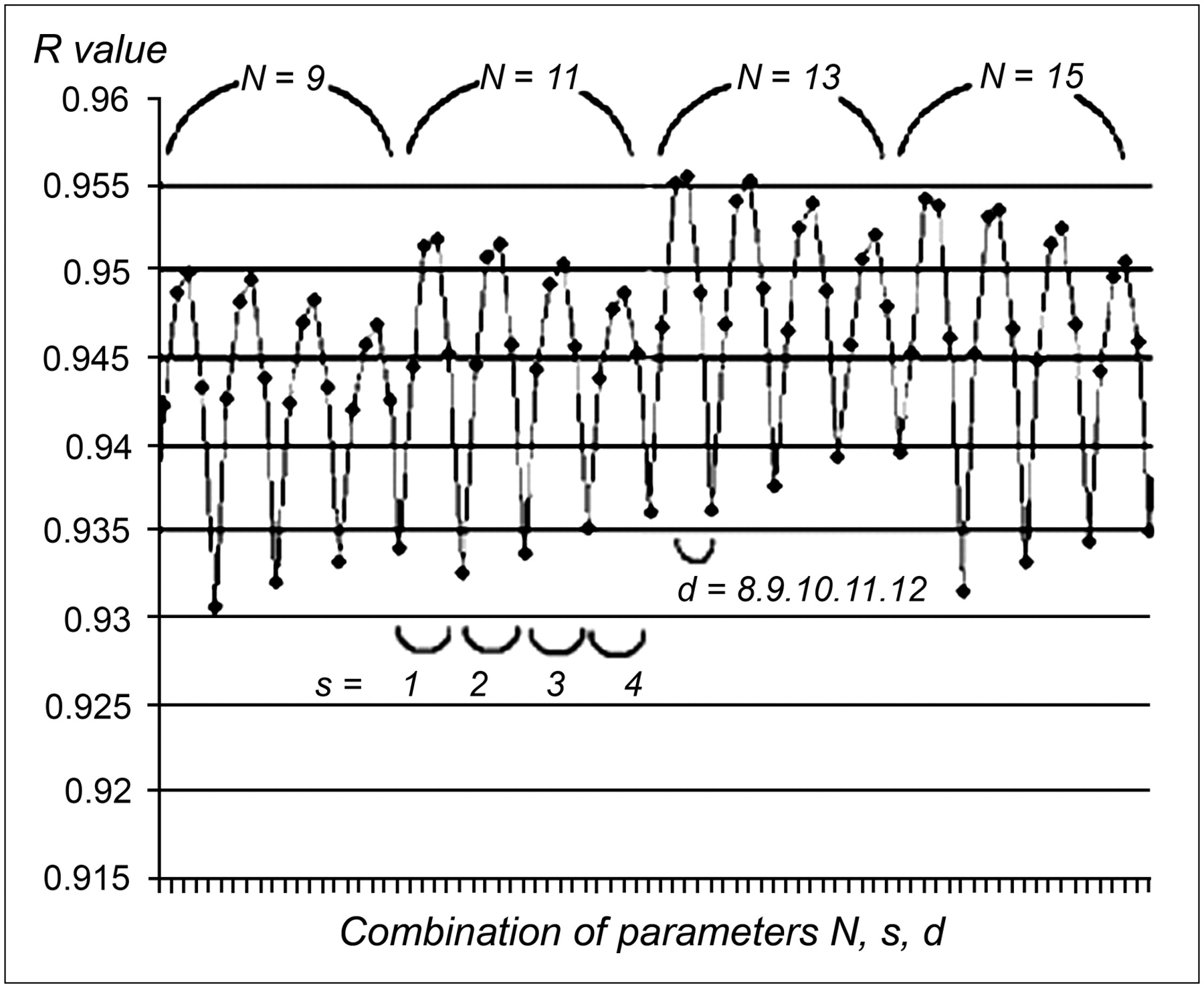
- FIGURE 5.
Graph of R value versus combination of N, S, and d parameters used by OSA. R value comes from comparison of OSA V3″ values to those obtained by experienced image processing researcher. Values of N vary from 9 to 15 from left to right along axis; values of s change from 1 to 4 and cycle more slowly from left to right than do values of d, which cycles more quickly through values of 8 to 12.
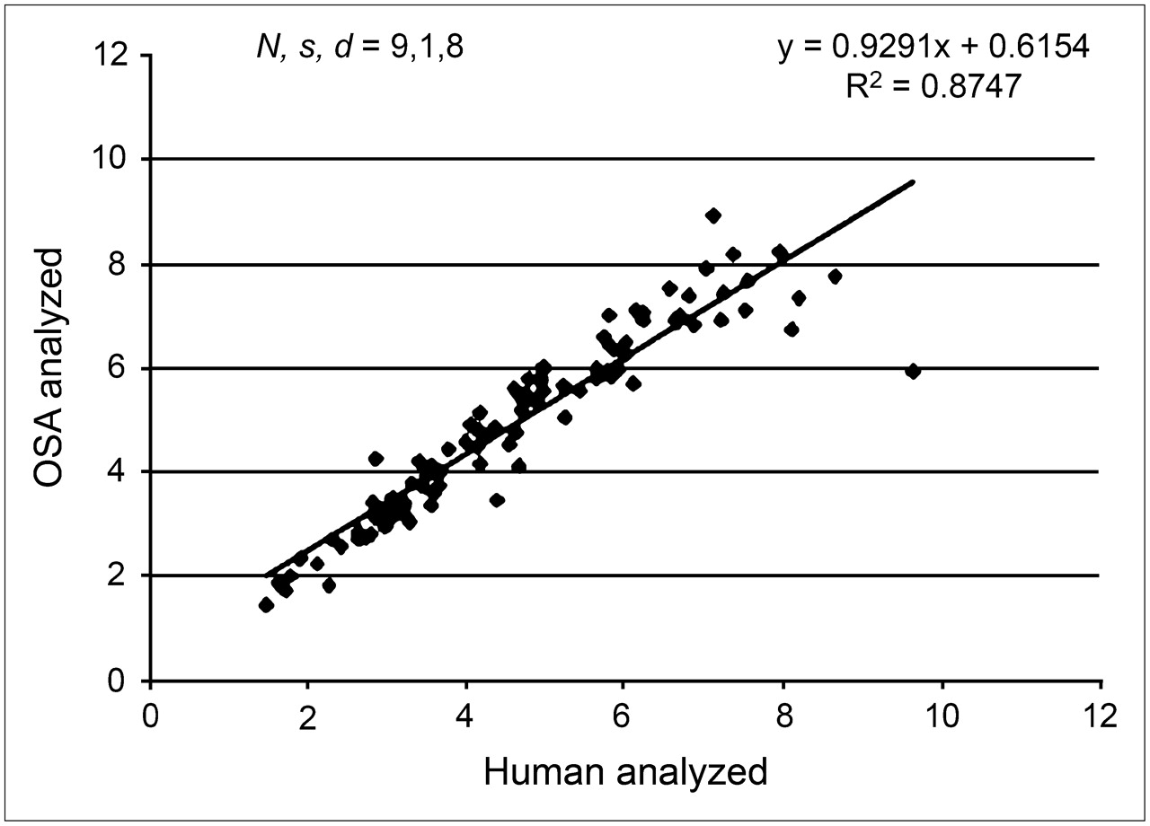
- FIGURE 6.
Scatter plot of OSA V3″ evaluations compared with those of experienced researcher for OSA parameters N, s, and d set to 9, 1, and 8, respectively. These parameters yielded optimal slope of 0.93 and demonstrated best accuracy for OSA analysis.
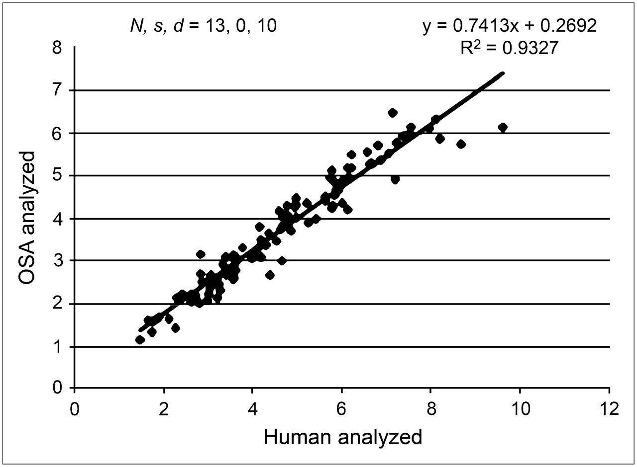
- FIGURE 7.
Scatter plot of OSA V3″ evaluations compared with those of experienced researcher for OSA parameters N, s, and d set to 13, 0, and 10, respectively. These parameters yielded optimal R value of 0.96 (R2 = 0.93) and demonstrated best precision for OSA analysis.
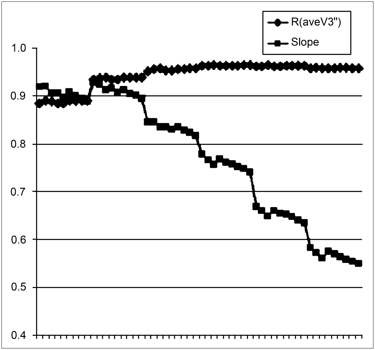
- FIGURE 8.
Plot of both R value and regression slope vs. combination of N, S, and d parameters used by OSA. Parameters along horizontal axis are similar to those in Figure 5 in that, here, values of N vary from 3 to 17 from left to right along axis; values of s change from 1 to 4 and cycle more slowly from left to right than do values of d, which cycles more quickly through values of 8 to 12.


Jump to section
Related Articles
Cited By...
- Optimization of Parameters for Quantitative Analysis of 123I-Ioflupane SPECT Images for Monitoring Progression of Parkinson Disease
- The Semicolon Sign: Dopamine Transporter Imaging Artifact from Head Tilt
- Value of Semiquantitative Analysis for Clinical Reporting of 123I-2-{beta}-Carbomethoxy-3{beta}-(4-Iodophenyl)-N-(3-Fluoropropyl)Nortropane SPECT Studies
- Receiver-Operating-Characteristic Analysis of an Automated Program for Analyzing Striatal Uptake of 123I-Ioflupane SPECT Images: Calibration Using Visual Reads
- SNM Practice Guideline for Dopamine Transporter Imaging with 123I-Ioflupane SPECT 1.0