


Article Figures & Data
Figures
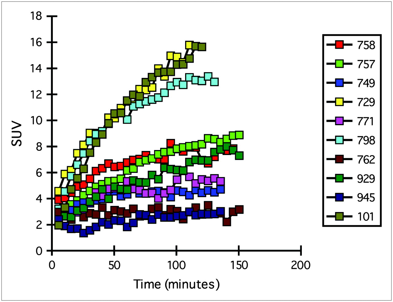
- FIGURE 1.
Tissue time–activity curves for 10 patients with solitary pulmonary nodules imaged over time with dynamic emission PET (23). Lesions were identified, ROI analysis performed, and SUV determined. 18F-FDG uptake plateaued at various times after injection. Reprinted with permission from the Society of Nuclear Medicine.
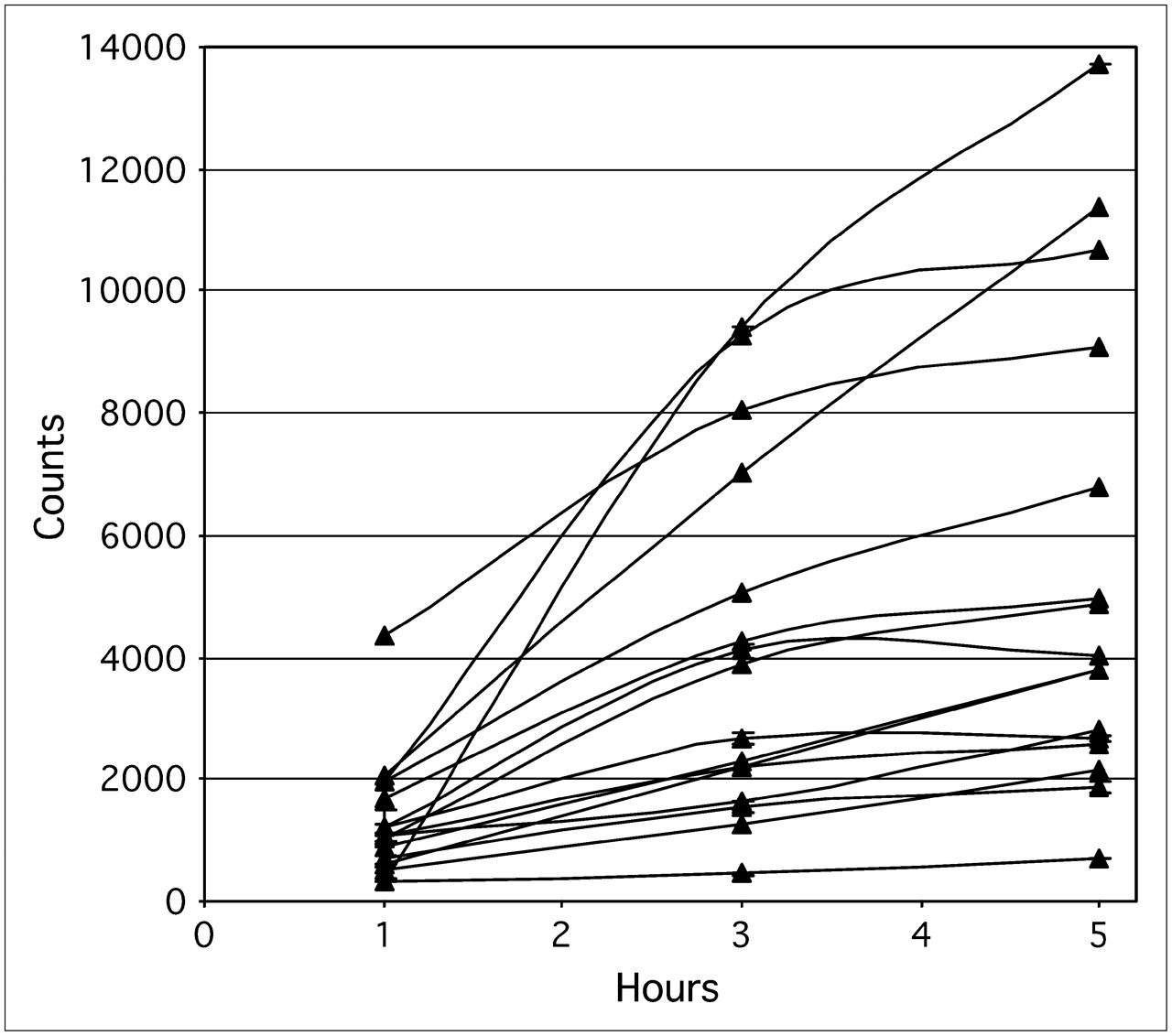
- FIGURE 2.
Tissue time–activity curves for 16 patients. Static PET was performed at 1, 3, and 5 h after injection of 18F-FDG, and activity in lesions was determined. 18F-FDG uptake plateaued at various times after injection and, in several lesions, was still increasing even at 5 h after injection. (Courtesy of Karen Kurdziel.)

Tables
Method Advantage Disadvantage Dependency Visual Static/whole-body imaging Subjectivity Uptake time No need for blood sampling Chance of threshold variation between readers Blood glucose concentration Short scan times Low statistics Partial-volume effects ± Attenuation correction Single snapshot of dynamic process Dependency on background activity SUV Static/whole-body imaging Numerous methods of calculation Uptake time Semiquantitative analysis Low statistics Blood glucose concentration No need for blood sampling Single snapshot of dynamic process Body weight Ease of computation Need for attenuation correction Partial-volume effects Inaccuracy in detecting small changes Kinetic Dynamic data acquisition Need for input function (arterial preferred) Partial-volume effects Quantitative analysis Complexity of computation Quality of input function Low dependency on imaging time Parameter Recommendation Patient preparation Patients fast overnight for morning scan or 4 h for afternoon scan. Venous serum glucose concentration is measured before injection (<120 mg/dL for nondiabetic patients and 150–200 mg/dL for diabetic patients). Diabetic patients are scanned in morning after overnight fast and before first use of medication. Patients are well hydrated and, if possible, drink 500 mL of water after injection and before scanning. For renal/pelvic imaging, furosemide (20–40 mg) may be given 10–15 min after 18F-FDG injection, or urinary catheter may be used. All medications being taken by patients are recorded. Diazepam or other mild sedative may be used at clinician's discretion to decrease uptake in muscle. PET timing Pretreatment and posttreatment scans are acquired. Pretreatment scans are acquired as close to start of therapy as possible (preferably <2 wk). Posttreatment scans are acquired no sooner than 2 wk after end of chemotherapy to avoid transient increases or decreases. Timing is determined by endpoint being assessed. Timing of scans after changes due to radiotherapy needs further investigation. Whole-body imaging begins 60 ± 10 min after injection of 18F-FDG. Attenuation correction Attenuation correction is used. No standard procedure has yet been recommended. Procedure chosen is documented. 18F-FDG dose No standard dose has yet been recommended. Doses of 370–740 MBq (10–20 mCi) are appropriate. Dose injected is documented.


Jump to section
Related Articles
Cited By...
- Principles of Tracer Kinetic Analysis in Oncology, Part II: Examples and Future Directions
- Whole-Body Parametric Imaging of 18F-FDG PET Using uEXPLORER with Reduced Scanning Time
- Performance of Digital PET Compared with High-Resolution Conventional PET in Patients with Cancer
- Does 2-FDG PET Accurately Reflect Quantitative In Vivo Glucose Utilization?
- Linsitinib (OSI-906) for the Treatment of Adult and Pediatric Wild-Type Gastrointestinal Stromal Tumors, a SARC Phase II Study
- First-in-Human Phase I Study to Evaluate the Brain-Penetrant PI3K/mTOR Inhibitor GDC-0084 in Patients with Progressive or Recurrent High-Grade Glioma
- Use of a Qualification Phantom for PET Brain Imaging in a Multicenter Consortium: A Collaboration Between the Pediatric Brain Tumor Consortium and the SNMMI Clinical Trials Network
- Prospective Study of Serial 18F-FDG PET and 18F-Fluoride PET to Predict Time to Skeletal-Related Events, Time to Progression, and Survival in Patients with Bone-Dominant Metastatic Breast Cancer
- Early prediction of lenvatinib treatment efficacy by using 18F-FDG PET/CT in patients with unresectable or advanced thyroid carcinoma that is refractory to radioiodine treatment: a protocol for a non-randomized single-arm multicenter observational study
- Linker engineering in anti-TAG-72 antibody fragments optimizes biophysical properties, serum half-life, and high-specificity tumor imaging
- 89Zr-anti-{gamma}H2AX-TAT but not 18F-FDG Allows Early Monitoring of Response to Chemotherapy in a Mouse Model of Pancreatic Ductal Adenocarcinoma
- Test-Retest Variability in Lesion SUV and Lesion SUR in 18F-FDG PET: An Analysis of Data from Two Prospective Multicenter Trials
- Qualification of National Cancer Institute-Designated Cancer Centers for Quantitative PET/CT Imaging in Clinical Trials
- Repeatability of SUV in Oncologic 18F-FDG PET
- Reliability of PET/CT Shape and Heterogeneity Features in Functional and Morphologic Components of Non-Small Cell Lung Cancer Tumors: A Repeatability Analysis in a Prospective Multicenter Cohort
- Getting the Most out of 18F-FDG PET Scans: The Predictive Value of 18F-FDG PET-Derived Blood Flow Estimates for Breast Cancer
- Accuracy and Precision of Partial-Volume Correction in Oncological PET/CT Studies
- Multicenter Clinical Trials Using 18F-FDG PET to Measure Early Response to Oncologic Therapy: Effects of Injection-to-Acquisition Time Variability on Required Sample Size
- Dose-Finding Quantitative 18F-FDG PET Imaging Study with the Oral Pan-AKT Inhibitor GSK2141795 in Patients with Gynecologic Malignancies
- Molecular Imaging to Plan Radiotherapy and Evaluate Its Efficacy
- Complexity of Response Evaluation During Primary Systemic Therapy of Breast Cancer: Scoring Systems and Beyond-Preliminary Results
- Phase II Study of Lapatinib in Combination With Trastuzumab in Patients With Human Epidermal Growth Factor Receptor 2-Positive Metastatic Breast Cancer: Clinical Outcomes and Predictive Value of Early [18F]Fluorodeoxyglucose Positron Emission Tomography Imaging (TBCRC 003)
- Repeatability of 18F-FDG PET/CT in Advanced Non-Small Cell Lung Cancer: Prospective Assessment in 2 Multicenter Trials
- Summary of the UPICT Protocol for 18F-FDG PET/CT Imaging in Oncology Clinical Trials
- 18F-FLT PET Evaluation of Radiation Response
- FDG PET-CT in follicular lymphoma: a case-based evidence review
- TBCRC 008: Early Change in 18F-FDG Uptake on PET Predicts Response to Preoperative Systemic Therapy in Human Epidermal Growth Factor Receptor 2-Negative Primary Operable Breast Cancer
- Quantitative PET/CT Scanner Performance Characterization Based Upon the Society of Nuclear Medicine and Molecular Imaging Clinical Trials Network Oncology Clinical Simulator Phantom
- Role of Imaging in the Staging and Response Assessment of Lymphoma: Consensus of the International Conference on Malignant Lymphomas Imaging Working Group
- The predictive role of interim positron emission tomography for Hodgkin lymphoma treatment outcome is confirmed using the interpretation criteria of the Deauville five-point scale
- Sarcoma Mid-Therapy [F-18]Fluorodeoxyglucose Positron Emission Tomography (FDG PET) and Patient Outcome
- Early Predictive Value of Non-response to Docetaxel in Neoadjuvant Chemotherapy in Breast Cancer Using 18F-FDG-PET
- Impact of Different Standardized Uptake Value Measures on PET-Based Quantification of Treatment Response
- Comparison of EORTC Criteria and PERCIST for PET/CT Response Evaluation of Patients with Metastatic Colorectal Cancer Treated with Irinotecan and Cetuximab
- Interim 18F-FDG PET in Hodgkin Lymphoma: Would PET-Adapted Clinical Trials Lead to a Paradigm Shift?
- Reporting Guidance for Oncologic 18F-FDG PET/CT Imaging
- Differences in the Biologic Activity of 2 Novel MEK Inhibitors Revealed by 18F-FDG PET: Analysis of Imaging Data from 2 Phase I Trials
- Sepantronium Bromide (YM155) Enhances Response of Human B-Cell Non-Hodgkin Lymphoma to Rituximab
- Noise Considerations for PET Quantification Using Maximum and Peak Standardized Uptake Value
- 18F-FDG PET as a Surrogate Biomarker in Non-Small Cell Lung Cancer Treated with Erlotinib: Newly Identified Lesions Are More Informative Than Standardized Uptake Value
- 18F-FDG-PET/CT Imaging as an Early Survival Predictor in Patients with Primary High-Grade Soft Tissue Sarcomas Undergoing Neoadjuvant Therapy
- Assessment of Valvular Calcification and Inflammation by Positron Emission Tomography in Patients With Aortic Stenosis
- Mutatis Mutandis: Harmonize the Standard!
- Need for Standardization of 18F-FDG PET/CT for Treatment Response Assessments
- Imaging in Sarcoma
- 18F-FDG PET/CT for Monitoring Treatment Responses to the Epidermal Growth Factor Receptor Inhibitor Erlotinib
- Prevalence and Risk Factors of Carotid Vessel Wall Inflammation in Coronary Artery Disease Patients: FDG-PET and CT Imaging Study
- Practical Approach for Comparative Analysis of Multilesion Molecular Imaging Using a Semiautomated Program for PET/CT
- Positron Emission Tomography-Computed Tomography (PET-CT) After Induction Therapy Is Highly Predictive of Patient Outcome in Follicular Lymphoma: Analysis of PET-CT in a Subset of PRIMA Trial Participants
- FDG-PET as a Potential Tool for Selecting Patients with Advanced Non-Small Cell Lung Cancer Who May Be Spared Maintenance Therapy after First-Line Chemotherapy
- Changes in 18F-Fluorodeoxyglucose and 18F-Fluorodeoxythymidine Positron Emission Tomography Imaging in Patients with Non-Small Cell Lung Cancer Treated with Erlotinib
- Standardization of Quantitative Imaging: The Time Is Right, and 18F-FDG PET/CT Is a Good Place to Start
- Variations in Clinical PET/CT Operations: Results of an International Survey of Active PET/CT Users
- PET and PET/CT of the Neck Lymph Nodes Improves Risk Prediction in Patients with Squamous Cell Carcinoma of the Oral Cavity
- Variations in PET/CT Methodology for Oncologic Imaging at U.S. Academic Medical Centers: An Imaging Response Assessment Team Survey
- Monitoring Response to Antiangiogenic Therapy in Non-Small Cell Lung Cancer Using Imaging Markers Derived from PET and Dynamic Contrast-Enhanced MRI
- Phase II Study of Daily Sunitinib in FDG-PET-Positive, Iodine-Refractory Differentiated Thyroid Cancer and Metastatic Medullary Carcinoma of the Thyroid with Functional Imaging Correlation
- A functional dynamic scoring model to elucidate the significance of post-induction interim fluorine-18-fluorodeoxyglucose positron emission tomography findings in patients with Hodgkin's lymphoma
- Imaging Atherosclerotic Plaque Inflammation by Fluorodeoxyglucose With Positron Emission Tomography: Ready for Prime Time?
- Imaging in drug development: will it deliver on the promise of personalised medicine?
- SUVs: Always a Good Choice?
- Guidelines for the Development and Incorporation of Biomarker Studies in Early Clinical Trials of Novel Agents
- A Phase I Clinical Trial of CHT-25 a 131I-Labeled Chimeric Anti-CD25 Antibody Showing Efficacy in Patients with Refractory Lymphoma
- Phase I Biodistribution and Pharmacokinetic Study of Lewis Y-Targeting Immunoconjugate CMD-193 in Patients with Advanced Epithelial Cancers
- Frontiers of Biomedical Imaging Science 2009: Workshop Report and Research Opportunities
- 18F-FDG PET/CT for Image-Guided and Intensity-Modulated Radiotherapy
- Repeatability of 18F-FDG PET in a Multicenter Phase I Study of Patients with Advanced Gastrointestinal Malignancies
- Expert Opinions on Positron Emission Tomography and Computed Tomography Imaging in Lymphoma
- Chemotherapy Response Evaluation in Metastatic Colorectal Cancer with FDG PET/CT and CT Scans
- Qualification of PET Scanners for Use in Multicenter Cancer Clinical Trials: The American College of Radiology Imaging Network Experience
- Comparison of Imaging Protocols for 18F-FDG PET/CT in Overweight Patients: Optimizing Scan Duration Versus Administered Dose
- NCCN Task Force: Clinical Utility of PET in a Variety of Tumor Types
- Role of 18F-FDG PET in Assessment of Response in Non-Small Cell Lung Cancer
- From RECIST to PERCIST: Evolving Considerations for PET Response Criteria in Solid Tumors
- Standards for PET Image Acquisition and Quantitative Data Analysis
- Monitoring Predominantly Cytostatic Treatment Response with 18F-FDG PET
- FDG-PET/CT Imaging Predicts Histopathologic Treatment Responses after the Initial Cycle of Neoadjuvant Chemotherapy in High-Grade Soft-Tissue Sarcomas
- Measuring Response with FDG-PET: Methodological Aspects
- Considerations for the Use of Imaging Tools for Phase II Treatment Trials in Oncology
- 18F-FDG PET/CT as an Indicator of Progression-Free and Overall Survival in Osteosarcoma
- Relationships Among Regional Arterial Inflammation, Calcification, Risk Factors, and Biomarkers: A Prospective Fluorodeoxyglucose Positron-Emission Tomography/Computed Tomography Imaging Study
- In Reply
- Standardization of Scanning for [18F]Fluorodeoxyglucose Positron Emission Tomography
- Imaging Surrogates of Tumor Response to Therapy: Anatomic and Functional Biomarkers
- Early Prediction of Response to Sunitinib After Imatinib Failure by 18F-Fluorodeoxyglucose Positron Emission Tomography in Patients With Gastrointestinal Stromal Tumor
- 18F-FDG PET and PET/CT in the Evaluation of Cancer Treatment Response
- Spatial Heterogeneity in Sarcoma 18F-FDG Uptake as a Predictor of Patient Outcome
- [18F]Fluorothymidine Positron Emission Tomography before and 7 Days after Gefitinib Treatment Predicts Response in Patients with Advanced Adenocarcinoma of the Lung
- Combined Assessment of Metabolic and Volumetric Changes for Assessment of Tumor Response in Patients with Soft-Tissue Sarcomas
- Tumor Metabolism and Blood Flow Changes by Positron Emission Tomography: Relation to Survival in Patients Treated With Neoadjuvant Chemotherapy for Locally Advanced Breast Cancer
- The role of clinical imaging in oncological drug development
- Analysis and Reproducibility of 3'-Deoxy-3'-[18F]Fluorothymidine Positron Emission Tomography Imaging in Patients with Non-Small Cell Lung Cancer
- Treatment Monitoring by 18F-FDG PET/CT in Patients with Sarcomas: Interobserver Variability of Quantitative Parameters in Treatment-Induced Changes in Histopathologically Responding and Nonresponding Tumors
- Atherosclerosis Inflammation Imaging with 18F-FDG PET: Carotid, Iliac, and Femoral Uptake Reproducibility, Quantification Methods, and Recommendations
- Molecular Imaging of Metastatic Potential
- Reduction of Glucose Metabolic Activity Is More Accurate than Change in Size at Predicting Histopathologic Response to Neoadjuvant Therapy in High-Grade Soft-Tissue Sarcomas
- The role of FDG-PET scans in patients with lymphoma
- Early Prediction of Response to Chemotherapy and Survival in Malignant Pleural Mesothelioma Using a Novel Semiautomated 3-Dimensional Volume-Based Analysis of Serial 18F-FDG PET Scans
- Prospective Assessment of Discontinuation and Reinitiation of Erlotinib or Gefitinib in Patients with Acquired Resistance to Erlotinib or Gefitinib Followed by the Addition of Everolimus