


Article Figures & Data
Figures
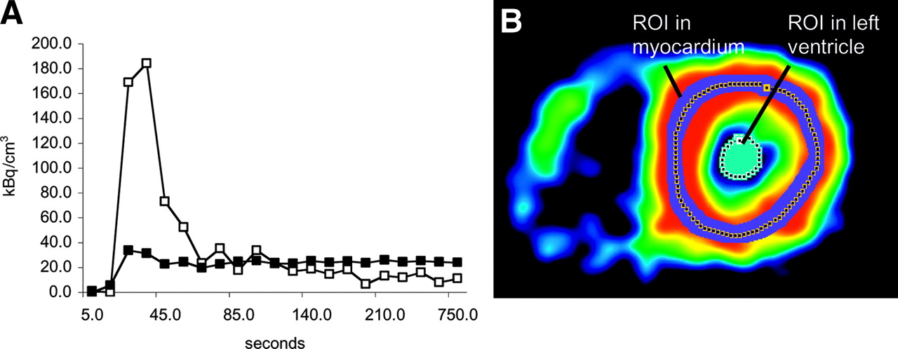
- FIGURE 1.
(A) Time–activity curves of left ventricular blood pool (□) (input function) and left ventricular myocardium (▪). (B) ROIs for left ventricular cavity and myocardial tissue drawn on a short-axis cut of the heart.
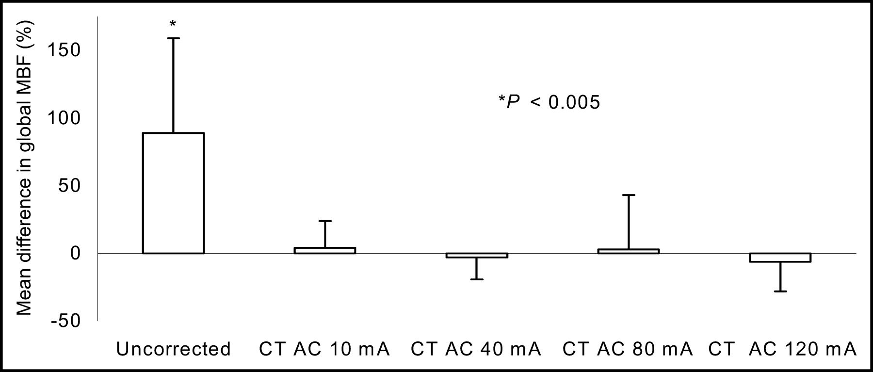
- FIGURE 2.
Percentage difference of CT AC at different tube currents vs. standard 68Ge AC.
- FIGURE 3.
Repeated MBF measurements using CT AC (10 mA). Coefficients of variance (COV) are given as percentages.
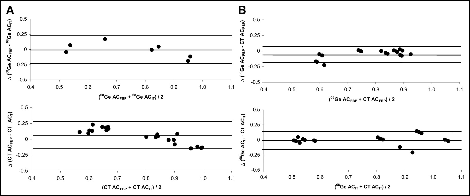
- FIGURE 4.
Bland–Altman plots of MBF (mL/min/g) in group 3. (A) Comparison of reconstruction algorithms: FBP vs. IT. Top graph shows 68Ge AC (RC = 0.22 mL/min/g [29%]). Bottom graph shows CT AC (RC = 0.23 mL/min/g [29%]). (B) Comparison of AC sources: 68Ge vs. CT. Top graph shows FBP (RC = 0.13 mL/min/g [17%]). Bottom graph shows IT (RC = 0.15 mL/min/g [19%]).


Tables
Region Patient 1 Patient 2 Patient 3 Rest Stress Rest Stress Rest Stress MBF COV MBF COV MBF COV MBF COV MBF COV MBF COV Septal 0.49 ± 0.03 6 0.87 ± 0.10 12 0.55 ± 0.02 4 1.15 ± 0.07 6 0.88 ± 0.01 1 2.21 ± 0.04 2 Anterior 0.28 ± 0.01 3 0.38 ± 0.01 4 0.35 ± 0.02 7 0.77 ± 0.02 3 0.75 ± 0.04 5 1.03 ± 0.07 7 Lateral 0.28 ± 0.02 9 0.44 ± 0.04 9 0.48 ± 0.04 9 0.93 ± 0.03 4 0.75 ± 0.04 6 1.26 ± 0.06 5 Inferior 0.34 ± 0.03 9 0.40 ± 0.03 9 0.51 ± 0.02 4 0.60 ± 0.03 5 0.78 ± 0.02 2 2.18 ± 0.10 5 Regional myocardial blood flow is in mL/min/g (mean ± SD) of repeated measurements using CT AC (10 mA), and respective coefficients of variance (COV) are given as percentages.
In this issue


Jump to section
Related Articles
Cited By...
- Absolute Myocardial Blood Flow and Flow Reserve Assessed by Gated SPECT with Cadmium-Zinc-Telluride Detectors Using 99mTc-Tetrofosmin: Head-to-Head Comparison with 13N-Ammonia PET
- The Effect of Misregistration Between CT-Attenuation and PET-Emission Images in 13N-Ammonia Myocardial PET/CT
- SNMMI/ASNC/SCCT Guideline for Cardiac SPECT/CT and PET/CT 1.0
- Hybrid PET/MR Imaging of the Heart: Potential, Initial Experiences, and Future Prospects
- Evaluation of a Mitochondrial Voltage Sensor, (18F-Fluoropentyl)Triphenylphosphonium Cation, in a Rat Myocardial Infarction Model
- Diagnostic Value of 13N-Ammonia Myocardial Perfusion PET: Added Value of Myocardial Flow Reserve
- Structural Abnormalities of the Coronary Arterial Wall--in Addition to Luminal Narrowing--Affect Myocardial Blood Flow Reserve
- Improved Outcome Prediction by SPECT Myocardial Perfusion Imaging After CT Attenuation Correction
- Agreement of Visual Estimation of Coronary Artery Calcium From Low-Dose CT Attenuation Correction Scans in Hybrid PET/CT and SPECT/CT With Standard Agatston Score
- Validation of CT Attenuation Correction for High-Speed Myocardial Perfusion Imaging Using a Novel Cadmium-Zinc-Telluride Detector Technique
- Decreased Perfusion in the Lateral Wall of the Left Ventricle in PET/CT Studies with 13N-Ammonia: Evaluation in Healthy Adults
- Long-Term Prognostic Value of 13N-Ammonia Myocardial Perfusion Positron Emission Tomography: Added Value of Coronary Flow Reserve
- Comparison of Myocardial Perfusion 82Rb PET Performed with CT- and Transmission CT-Based Attenuation Correction
- SPECT/CT
- Reducing Radiation Dose in Rest-Stress Cardiac PET/CT by Single Poststress Cine CT for Attenuation Correction: Quantitative Validation
- Absolute Quantification of Myocardial Blood Flow with 13N-Ammonia and 3-Dimensional PET
- Radiation Dose to Patients From Cardiac Diagnostic Imaging
- Added Value of Coronary Artery Calcium Score as an Adjunct to Gated SPECT for the Evaluation of Coronary Artery Disease in an Intermediate-Risk Population
- Frequent Diagnostic Errors in Cardiac PET/CT Due to Misregistration of CT Attenuation and Emission PET Images: A Definitive Analysis of Causes, Consequences, and Corrections
- PET/CT Attenuation Correction: Breathing Lessons
- Cardiac Image Fusion from Stand-Alone SPECT and CT: Clinical Experience
- Respiration-Averaged CT for Attenuation Correction in Canine Cardiac PET/CT
- Artifacts from Misaligned CT in Cardiac Perfusion PET/CT Studies: Frequency, Effects, and Potential Solutions
- Noninvasive Characterization of Myocardial Molecular Interventions by Integrated Positron Emission Tomography and Computed Tomography
- Comparison of 64-Slice CT with Gated SPECT for Evaluation of Left Ventricular Function
- 18F-Choline Images Murine Atherosclerotic Plaques Ex Vivo
- Caffeine Decreases Exercise-Induced Myocardial Flow Reserve
- PET/CT: Challenge for Nuclear Cardiology
- Novel Doppler Assessment of Intracoronary Volumetric Flow Reserve: Validation Against PET in Patients With or Without Flow-Dependent Vasodilation
- Integrated PET/CT for the Assessment of Coronary Artery Disease: A Feasibility Study
- Myocardial Blood Flow Measurement by PET: Technical Aspects and Clinical Applications
- {beta}-Adrenergic Blockade and Myocardial Perfusion in Coronary Artery Disease: Differential Effects in Stenotic Versus Remote Myocardial Segments