


Abstract
Increased 18F-FDG activity in fatty tissue has previously been reported with PET/CT. We previously named this activity uptake in supraclavicular area fat (“USA-Fat”). We and others have speculated that this uptake exists in metabolically active brown adipose tissue (BAT). Such tissue might be expected to have varying metabolic activity depending on the ambient temperature. The purpose of this study was to evaluate the frequency of USA-Fat and its relationship to the outdoor temperature. Methods: Between July 2001 and June 2002, 1,017 consecutive whole-body scans were obtained with a PET/CT scanner and 18F-FDG for clinical patients. PET images were reviewed for the presence of USA-Fat. Results: USA-Fat was observed in 68 scans obtained from 62 patients (51 female and 11 male). The incidence of USA-Fat was highest, at 13.7%, in January through March, while outside temperatures were low, and was significantly lower, at 4.1%, during the rest of the year. Conclusion: The incidence of USA-Fat is clearly increased during the cooler period of the year. This finding suggests that stimulation by cold temperatures increases the frequency with which USA-Fat occurs, supporting underlying BAT as the etiology for this activity.
The recent introduction of in-line PET/CT scanners in clinical oncologic imaging allows the combination of anatomic and metabolic studies. The presence of increased 18F-FDG metabolic activity occurring in fat tissue, previously attributed to muscular uptake, has recently been reported on images obtained with PET/CT scanners (1,2). Because it was described in the supraclavicular region, this activity has been called uptake in supraclavicular area fat (“USA-Fat”). Brown adipose tissue (BAT) has been hypothesized to be the cause of this increased metabolic activity in fat tissue (1,2). BAT is known to be stimulated by several factors, including exposure to cold (3). An increase in the incidence of USA-Fat during periods of cold outdoor temperatures might be observed and would support the BAT hypothesis. The purpose of this study was to assess the relationship between the monthly incidence of USA-Fat and the outdoor temperature in patients imaged with a PET/CT scanner. Further, we sought to evaluate the sequential behavior of this uptake in patients for whom serial scans were available.
MATERIALS AND METHODS
For 905 patients (443 female and 462 male), 1,017 consecutive clinical whole-body scans were obtained with 18F-FDG and a PET/CT scanner from July 2001 to June 2002. Mean patient age (±SD) was 58.1 ± 15.1 y (range, 1–93 y). Forty-two scans were obtained for 21 patients 18 y old or less. The scans were retrospectively assessed for the presence of USA-Fat. All patients had been referred for the evaluation of known or suspected cancer.
Scanning was performed using the Discovery LS PET/CT scanner (General Electric Medical Systems). Patients fasted at least 4 h before the PET acquisition. Intravenous injection of 555–740 MBq of 18F-FDG was followed by a tracer uptake phase of about 60 min, during which the patients sat in a quiet room without talking.
CT Scanning
The CT portion of the Discovery LS consists of a multidetector helical scanner (LightSpeed Plus; General Electric Medical Systems). Imaging parameters were as follows for an acquisition at 5 bed positions: 140 kV(p), 0.8 s per CT rotation, a pitch of 6, a table speed of 22.5 mm/s, 722.5-mm coverage, and a 31.9-s acquisition time. CT was performed before emission PET. The current of the CT tube was adjusted according to patient weight. CT data were resized from a 512 × 512 matrix to a 128 × 128 matrix to match the PET data in order to generate a CT transmission map and to fuse images.
PET Scanning
Emission data were acquired for 5–7 bed positions, typically from the base of the skull through the mid thigh. Emission data were acquired for 5 min for each bed position. Each bed had 35 scanning planes with a 14.6-cm longitudinal field of view and a 1-slice overlap. PET images were reconstructed using CT for attenuation correction with the ordered-subsets expectation maximization algorithm (2 iterations, 28 subsets) and an 8-mm gaussian filter using a 128 × 128 matrix.
Image Analysis
All PET/CT scans were examined retrospectively by 1 observer on an interactive computer display using fusion software (eNTEGRA; General Electric Medical Systems). This software allows the review of PET, CT, and fused data using transaxial, sagittal, and coronal displays. PET images of the supraclavicular area were evaluated for the presence of obvious abnormally increased 18F-FDG uptake (i.e., more intense than background activity) with corresponding fat-density tissue in the fused and CT images (USA-Fat). Monthly outdoor temperature (minimum, maximum, and average) from the northern hemisphere city where the PET/CT scanner was installed was obtained from the web site of the state meteorology office (http://www.meto.umd.edu). Patients with at least 1 study demonstrating USA-Fat and with serial scans available were analyzed subsequently.
Statistical Analysis
Results were expressed as mean ± SD. Differences in the incidence of USA-Fat were tested using the Fisher exact test. Differences in age and body mass index (BMI) between the groups with and without USA-Fat were tested by a t test. Two-tailed P values less than 0.05 were considered significant.
RESULTS
General Parameters
Sixty-eight of the 1,017 scans (6.7%) demonstrated USA-Fat (Fig. 1). The 68 scans with USA-Fat were obtained from 62 patients (11 male and 51 female). The incidence of USA-Fat was 10.5% in scans of female patients and 2.9% in scans of male patients (P < 0.0001). Among patients with USA-Fat, male patients were younger than female patients (32.8 ± 20 y vs. 50 ± 13 y, P = 0.02). Patients with USA-Fat were younger than patients without (46.9 ± 15 y vs. 58.9 ± 15 y, P < 0.0001). The incidence of USA-Fat was 23.8% in scans of patients aged 18 y or less and 5.9% in scans of patients older than 18 y (P = 0.0002).
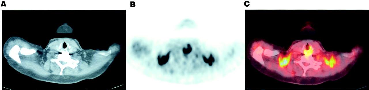
Example of USA-Fat in 56-y-old woman with lung cancer, evaluated for suspected tumor recurrence. 18F-FDG PET was performed in January. Transverse images are displayed. (A) CT shows no pathologic lymph nodes in supraclavicular regions. (B and C) Intense bilateral 18F-FDG uptake fusing in fat is seen on PET image (B) and fused image (C).
There was no significant difference in BMI between patients with USA-Fat and patients without, although patients with USA-Fat tended to be thinner (BMI, 23.6 ± 5 vs. 31 ± 127, P = 0.09). BMI was 21.6 ± 5 for male patients and 24 ± 5 for female patients with USA-Fat (P = 0.17).
Patients with USA-Fat were referred because of breast cancer (9 patients), gynecologic cancer (12 patients), lymphoma (9 patients), digestive tract cancer (13 patients), lung cancer or nodule (10 patients), thyroid cancer (2 patients), head and neck cancer (4 patients), melanoma (1 patients), or sarcoma (2 patients).
Relationship of USA-Fat to Temperature
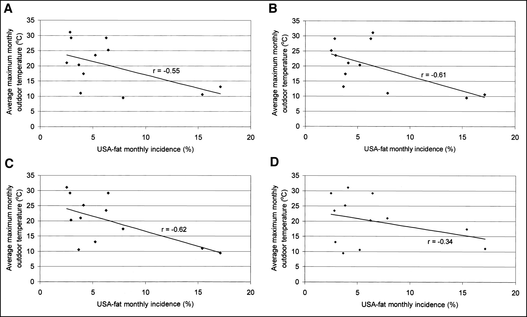
Figure 2 demonstrates the monthly incidence of USA-Fat and its temporal relationship to the average monthly maximum temperature. Details are given in Table 1. When compared with the incidence from April to December, the incidence was highest from January to March in the whole group of patients (13.7% vs. 4.1%, P < 0.0001), in male patients (6.6% vs. 1.3%, P = 0.032), in female patients (20% vs. 6.9%, P < 0.0001), and in patients older than 18 y (12.5% vs. 3.5%, P < 0.0001). The incidence from January to March was not significantly higher in patients aged 18 y or less (35.7% vs. 17.9%, P = 0.26), but the number of patients was limited. A negative correlation was observed between outdoor temperature and the monthly incidence of USA-Fat and was strongest (r = −0.62) when the incidence in 1 mo was compared with the temperature occurring 2 mo earlier (Fig. 3).
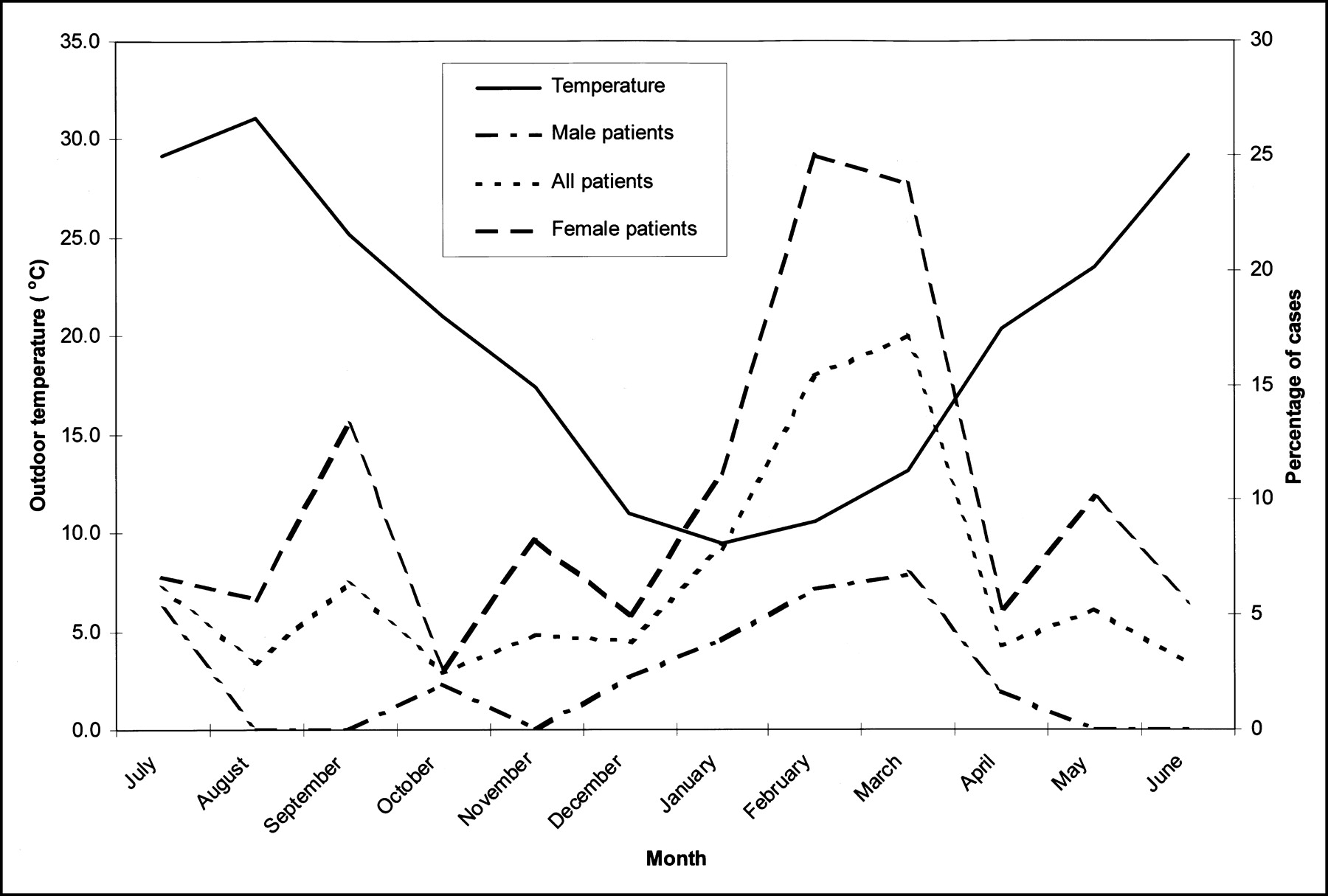
Monthly incidence of USA-fat and its relationship to outdoor temperature. Monthly average maximum outdoor temperature is displayed in degrees Celsius. Incidence of USA-Fat is displayed as percentage of cases for female patients, male patients, and all patients.

Correlation between incidence of USA-Fat and average monthly outdoor maximum temperature. Incidence is compared with temperature of same month (A) and temperature of 1 mo (B), 2 mo (C), and 3 mo (D) before.
Monthly Incidence of USA-Fat in Male Patients, Female Patients, and All Patients
Repeated Scans
Among the 62 patients with USA-Fat, 12 underwent 35 studies during the survey period. The results are shown in Table 2. The studies of patients 5, 6, 8, and 9 showed a pattern of activity in fat during cold months. All studies of patients 1, 3, and 10 showed USA-Fat.
Evaluation of Patients with at Least 1 PET/CT Scan Showing 18F-FDG Activity in Fat Tissue and Serial Scans
DISCUSSION
Increased 18F-FDG uptake occurring in fat tissue (USA-Fat) has been described with 18F-FDG and PET/CT and has recently been attributed, without biopsy confirmation, to brown fat metabolic activity (1,2). A similar phenomenon of increased tracer activity in fatty tissue has recently been described with 131I-metaiodobenzylguanidine in rodents (4), with documentation by autoradiography that BAT was the underlying target for this activity. BAT has been reported in humans on necropsy series of outdoor workers exposed to cold (5). Cold stimulation causes overexpression of glucose transporter 4 in brown fat (6,7). We showed a clear temporal relationship between USA-Fat and the outdoor temperature. There is a clear rise in the monthly incidence of USA-Fat during the winter, providing indirect evidence that BAT is responsible for this phenomenon. Interestingly, the phenomenon is delayed by a few months in regard to the beginning of a decrease in outside temperature, suggesting that, even if cold can activate brown fat rapidly, a longer stimulation is necessary before it can be imaged with 18F-FDG and PET. Accordingly, the negative correlation between temperature and USA-Fat incidence was strongest when the incidence was compared with the temperature occurring 2 mo earlier, suggesting that a relatively prolonged period of cold is required to activate BAT sufficiently.
The increased prevalence of USA-Fat in women was again found, as it was observed in the previous studies (1,2). The male patients with this phenomenon were younger and leaner than the female patients. The increased mass of BAT in female rats in comparison with male rats (8), as well as the higher level of BAT activation in cold-exposed female rats in comparison with male, is known (9).
The evaluation of patients with serial scans also indicated that USA-Fat more frequently occurs in colder than in warmer months, but not necessarily in all patients. We do not have specific information on our patients’ individual behavior. Indeed, in summer, some patients might have spent much time in an air-conditioned environment. This suggests that, although BAT is stimulated by exposure to cold, other important factors not yet identified are involved in increased BAT metabolism in patients.
CONCLUSION
USA-Fat has a clear temporal relationship to outdoor temperature, showing an increased frequency during months with “cold” outdoor temperatures. This supports the hypothesis that BAT is the underlying substratum of this phenomenon and that BAT demonstrates increased 18F-FDG uptake when stimulated by cold.
Footnotes
Received Dec. 26, 2002; revision accepted Apr. 21, 2003.
For correspondence or reprints contact: Richard L. Wahl, MD, Division of Nuclear Medicine, Department of Radiology, Johns Hopkins Medical Institutions, 601 N. Caroline St., Baltimore, MD 21287-0817.
E-mail: rwahl{at}jhmi.edu
In this issue


Jump to section
Related Articles
Cited By...
- Brown Adipose Tissue: A Protective Mechanism Against "Preprediabetes"?
- Perspectives on Brown Adipose Tissue Imaging: Insights from Preclinical and Clinical Observations from the Last and Current Century
- Brown Adipose Tissue is Associated with Improved Cardiometabolic Health and Regulates Blood Pressure
- Brown adipocyte glucose metabolism: a heated subject
- The significance of beige and brown fat in humans
- Pediatric Brown Adipose Tissue on 18F-FDG PET: Diazepam Intervention
- Human Brown Adipose Tissue: What We Have Learned So Far
- Infrared thermography in the detection of brown adipose tissue in humans
- The prevalence and predictors of active brown adipose tissue in Chinese adults
- Quantification of Human and Rodent Brown Adipose Tissue Function Using 99mTc-Methoxyisobutylisonitrile SPECT/CT and 18F-FDG PET/CT
- Adipose Tissue Biology and Cardiomyopathy: Translational Implications
- Brown Adipose Tissue: Mechanisms and Potential Therapeutic Targets
- Thermal Control of Brown Adipose Tissue in 18F-FDG PET
- Inverse association between brown adipose tissue activation and white adipose tissue accumulation in successfully treated pediatric malignancy
- 18F-Fluorobenzyl Triphenyl Phosphonium: A Noninvasive Sensor of Brown Adipose Tissue Thermogenesis
- Prevention of Brown Adipose Tissue Activation in 18F-FDG PET/CT of Breast Cancer Patients Receiving Neoadjuvant Systemic Therapy
- CT Hounsfield Units of Brown Adipose Tissue Increase with Activation: Preclinical and Clinical Studies
- Brown Adipose Tissue and Seasonal Variation in Humans
- Early and Late Therapy Response Assessment With [18F]Fluorodeoxyglucose Positron Emission Tomography in Pediatric Hodgkin's Lymphoma: Analysis of a Prospective Multicenter Trial
- A novel approach for reduction of brown fat uptake on FDG PET
- High Incidence of Metabolically Active Brown Adipose Tissue in Healthy Adult Humans: Effects of Cold Exposure and Adiposity
- Prospective Evaluation of Physiologic Uptake Detected with True Whole-Body 18F-FDG PET/CT in Healthy Subjects
- Preliminary PET/CT Study of 18F-FDG Uptake in Cervical and Supraclavicular Brown Adipose Tissue
- Comparison of Uptake of Multiple Clinical Radiotracers into Brown Adipose Tissue Under Cold-Stimulated and Nonstimulated Conditions
- Brown Fat Imaging with 18F-6-Fluorodopamine PET/CT, 18F-FDG PET/CT, and 123I-MIBG SPECT: A Study of Patients Being Evaluated for Pheochromocytoma
- Effect of Nicotine and Ephedrine on the Accumulation of 18F-FDG in Brown Adipose Tissue
- 18FDG Uptake in Brown Fat: Potential for False Positives
- Improvements in Cancer Staging with PET/CT: Literature-Based Evidence as of September 2006
- Impact of Animal Handling on the Results of 18F-FDG PET Studies in Mice
- 18F-FDG Imaging: Pitfalls and Artifacts
- Intense 18F-FDG Uptake in Brown Fat Can Be Reduced Pharmacologically
- Brown Adipose Tissue and Nuclear Medicine Imaging