


Abstract
This study used PET to measure the time course of the brain concentration of 18F-labeled N-(4-acetyl-1-piperazinyl)-p-fluorobenzamide monohydrate (FK960), a novel antidementia drug, after oral administration to conscious rhesus monkeys. Methods: Three young-adult male rhesus monkeys were tested. FK960 (0.1 mg/kg) containing about 370 MBq of 18F-FK960 was administered orally to each monkey. Dynamic PET images were acquired for 4 h from 5 min after the administration. Arterial blood samples were withdrawn during PET scanning and were analyzed by an automatic well γ-counter and thin-layer chromatography to determine the time course of authentic 18F-FK960 activity concentration in plasma. FK960 concentrations in brain and plasma were calculated in units of mol/L using the specific activity of FK960 preparations. Results: 18F-FK960 penetrated the blood–brain barrier and underwent perfusion-dependent distribution in the entire brain. Maximal concentrations in the brain and plasma were 1.11 ± 0.30 × 10⁻⁷ mol/L (at 3.0 ± 0.6 h after administration) and 4.04 ± 1.29 × 10⁻⁷ mol/L (at 2.0 ± 1.1 h after administration), respectively. Conclusion: We succeeded in measuring the FK960 concentration in the brains of conscious monkeys and in plasma after oral administration at a dose of 0.1 mg/kg. The results suggested that this method can measure the FK960 concentration in the human brain, and a potential use of the PET technique in drug development was demonstrated.
- N-(4-acetyl-1-piperazinyl)-p-fluorobenzamide monohydrate
- antidementia drug
- PET
- rhesus monkey
- pharmacokinetics
To succeed in new drug development, dose setting in a clinical trial is important. Because each drug has its corresponding target site, achievement of the most suitable drug concentration in the target organ or tissue becomes critical to allow the drug to exhibit maximal effect. Although pharmacokinetic data in animal experiments are usually used to estimate the clinical dose, discrepancies in absorption, distribution, and metabolism of drugs between experimental animals and humans still remains in most cases, and these make the dose estimation difficult. Especially when a drug acts in the brain, further difficulty is caused by the presence of a defense system, the blood–brain barrier.
N-(4-acetyl-1-piperazinyl)-p-fluorobenzamide monohydrate (FK960) (Fig. 1) is a novel potential antidementia drug that has improved memory impairment in several kinds of rodent and nonhuman primate animal models (1–3). However, the dose–response relationships of FK960 in memory improvement in animal models of amnesia and in the enhancement of long-term potentiation (LTP) in hippocampal slices were bell shaped (1,2). The dose–response relationship of somatostatin in enhancement of LTP in hippocampal slices was also bell shaped (4). Therefore, suitable dose setting is still more critical for FK960 to exhibit efficacy in patients.
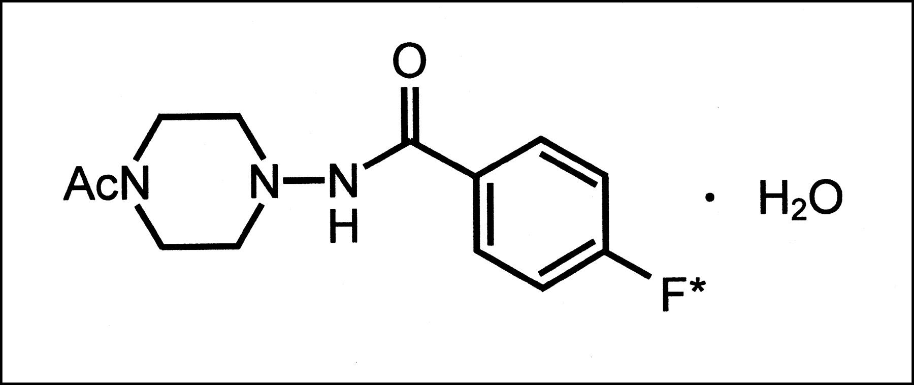
Chemical structure of FK960 (N-(4-acetyl-1-piperazinyl)-p-fluorobenzamide monohydrate). Asterisk denotes position of 18F labeling.
Recently, we succeeded in synthesizing 18F-labeled FK960 using an automated apparatus for 18F labeling to serve in a PET study (5). Because PET can measure radioactivity concentration dynamically in a target organ in a living subject with minimal invasion, the acquisition of bridging data between animals and humans may be expected. FK960 is also a drug to be administered perorally; therefore, in the present study, we measured the brain concentration of FK960 after oral administration in conscious monkeys using PET with 18F-FK960, which may reduce any discrepancies in the obtained data between animals and humans. A human PET study similar to the present study will need to be performed with the subjects conscious.
MATERIALS AND METHODS
Animals
Studies were performed on 3 male rhesus monkeys (Macaca mulatta; age, 7 y; body weight range, 8.08–9.08 kg). The monkeys were maintained and handled in accordance with the recommendations of the United States National Institutes of Health, and all animal experiments were performed in compliance with the animal ethics committee of The Medical and Pharmacological Research Center Foundation (Hakui, Japan). Each monkey had a specially designed head holder (6,7), and this was used to stereotactically fix each monkey’s head to a chair during PET scanning. The monkeys were allowed to recover from the procedure for more than 1 mo. They had previously been acclimatized to the chair restraint during repeated training sessions several times a week for more than 1 mo before the PET study began.
Drug Testing
FK960 was provided by Fujisawa Pharmaceutical Co., Ltd. (Osaka, Japan), and was dissolved in sterile physiologic saline at a concentration of 0.4 mg/mL before each study. 18F-FK960 (mean radiochemical purity, 97.4%; mean specific activity, 954 GBq/mg) was synthesized by an automated apparatus before each study, and the product obtained from the apparatus was an 18F-FK960 solution in 2 mL of saline. Then, about 370 MBq of the 18F-FK960 solution was mixed with the solution of unlabeled FK960. With the addition of saline, 0.1 mg of FK960 per kilogram of body weight, containing about 370 MBq (mean ± SD, 366 ± 14 MBq) of 18F-FK960 solution (a final volume of 5 mL), was prepared.
PET Experiment
PET scans were obtained with a high-resolution animal PET scanner (SHR-7700; Hamamatsu Photonics K.K., Hamamatsu, Japan) with a transaxial resolution of 2.6 mm in full width at half maximum in the center of the scan field and a center-to-center distance of 3.6 mm (8). 68Ga–68Ge blank scanning (120 min) was performed before each study. During catheterization of the femoral artery for arterial blood sampling, each monkey was transiently anesthetized with about 2% sevoflurane in a N2O:O2 gas mixture (7:3). After catheterization, anesthesia was immediately discontinued. For PET scans, each monkey’s head was fixed to a chair with a head holder and was stereotactically aligned parallel to the orbitomeatal plane with a laser marker; 68Ga–68Ge transmission scanning (30 min) was then performed.
After the transmission scan, a probe was inserted from the mouth to the stomach of each monkey. Through the probe, the 5 mL of prepared 18F-FK960 solution were administered, and another 5 mL of saline were administered to wash the inside of the probe. The remaining radioactivity in the probe was also measured by a radioisotope calibrator (CRC-127R; Capintec Instruments Inc., Ramsey, NJ) to calculate a net dose of 18F-FK960. Starting 5 min after oral administration of 18F-FK960, PET emission scanning was performed for 4 h; the scan consisted of 48 frames of 5 min each. During emission scanning, each monkey’s ears were unplugged, its eyes were open, and it was under a dimmed light.
Arterial blood samples were drawn (at 0, 10, 20, 30, 40, 50, 60, 80, and 100 min and at 2, 2.5, 3, 3.5, and 4 h) and were centrifuged to obtain plasma. They were analyzed for radioactivity by a γ-counter (1480 Wizard; Wallac Oy, Turku, Finland). Metabolites were extracted by 0.5 mL of methanol from 0.5 mL of plasma, and the extraction was performed twice on each sample. The methanol extract fraction was concentrated into about 0.2 mL by a gentle stream of nitrogen gas. Each 5 μL of concentrated fraction was spotted on a prelayered silica gel 60F254 thin-layer chromatography plate (E. Merck AG, Darmstadt, Germany), of layer thickness 0.25 mm, alongside 5 μL of methanol solution of authentic 18F-FK960. The plate was eluted in an n-butanol, ethyl acetate, and water (4:1:2) solvent system. After elution, the plate was dried and placed in contact with a BAS1800 imaging plate (Fuji Photo Film Co., Ltd., Tokyo, Japan). The imaging plate was read using a Fujix BAS1800 bioimaging analyzer (Fuji Photo Film Co., Ltd.).
For each monkey, a region of interest was manually drawn on a PET image of the entire brain (a slice of the orbitomeatal plane + 21.6 mm). The data obtained from plasma and PET, in units of Bq/mL, were converted to units of mol/L according to a calculated specific activity of 18F-FK960 preparation for each study. All data were represented as mean ± SE.
RESULTS
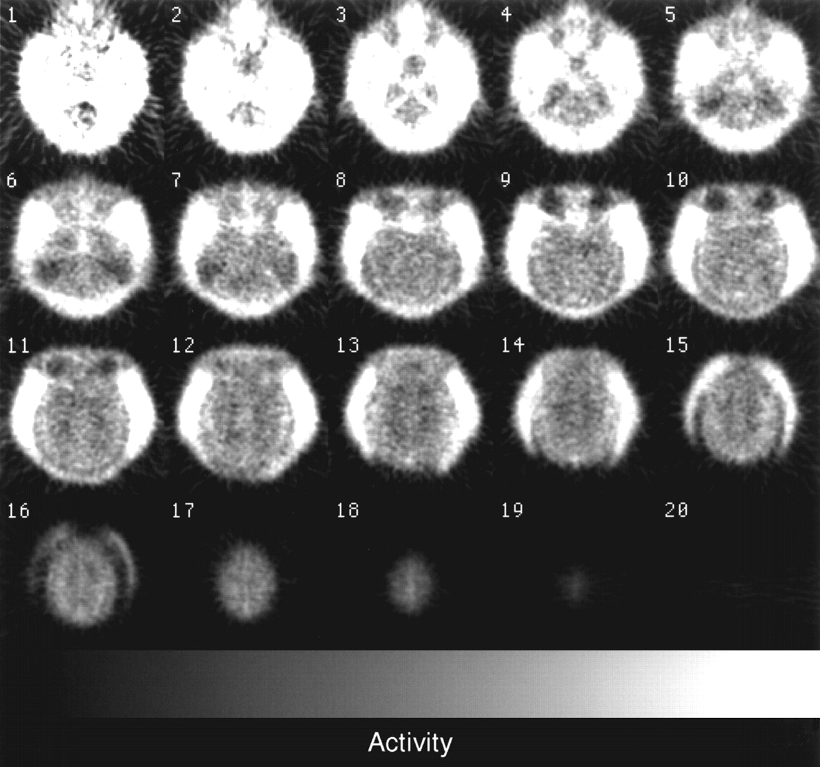
A typical PET image obtained from 30 to 90 min after administration of an 18F-FK960 preparation is shown in Figure 2. No marked focused accumulation was seen in the brain; the distribution was likely according to blood flow but was not distinctly so. The time–concentration curves for the plasma and brain are shown in Figure 3 as mean + SE, averaged over all subjects (n = 3), at each point. No metabolite was found in blood samples at any point. Maximal concentrations in the brain and plasma were 1.11 ± 0.30 × 10⁻⁷ mol/L (at 3.0 ± 0.6 h after administration) and 4.04 ± 1.29 × 10⁻⁷ mol/L (at 2.0 ± 1.1 h after administration), respectively. These mean maximal concentrations were calculated from maximal concentrations determined in each animal.

Typical PET images obtained from 30 to 90 min after administration of 18F-FK960 preparation. Presented images are 3.6-mm transverse slices, and slice 6 corresponds to orbitomeatal plane.
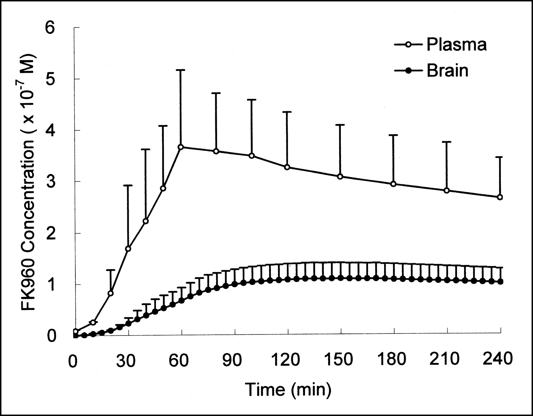
Time–concentration curves for plasma and brain after peroral administration of FK960 (0.1 mg/kg) containing 18F-FK960 (366 ± 14 MBq). Data are presented as mean + SE, averaged over all subjects (n = 3), at each point.
DISCUSSION
In this study, we used PET with 18F-FK960 to measure the FK960 concentration after oral administration to conscious monkeys. PET can measure the radioactivity concentration dynamically in a target organ in a living subject with minimal invasion, and we expected that bridging data between animals and humans could be obtained using PET. Therefore, we used nonhuman primates and studied them while they were conscious and with the drug orally administered. After administration of FK960 at a dose of 0.1 mg/kg, the brain concentration became about 10−7 mol/L 3 h after oral administration. Ours may be the first report of PET measurement of the brain concentration of a drug in conscious monkeys.
Two prerequisites must be observed in applying PET for pharmacokinetic studies of a new drug. First, one must be able to produce the exact molecular structure of the drug in a positron-emitting form. It may be also desirable that the positron-emitting nuclide not be easily eliminated in early metabolism. Second, one must be able to correct for radiometabolites, which are detected quickly by imaging equipment. FK960 has fluorine in the original structure, and the metabolites could be determined by thin-layer chromatography. FK960 is also metabolically stable enough within the scanning period.
FK960 is an antidementia drug, and its mechanism of action might be somatostatin activation (3). FK960 was reported to enhance K+-evoked release of somatostatin from rat hippocampal slices (9). Somatostatin enhanced LTP in the mossy fiber-CA3 pathway in intact guinea pig hippocampal slices (4), and FK960 also enhanced LTP through somatostatin activation (3). Somatostatin also enhanced the release of acetylcholine (10). Therefore, FK960 indirectly activates the release of acetylcholine and has improving effects in animal models of scopolamine-induced amnesia (1,2) and neuronal dysfunction (11).
FK960 has a bell-shaped dose–response relationship, and the dose setting is still more a critical issue. However, determining a clinical dose is difficult because species differences usually exist between experimental animals and humans. Species differences would be expected to be partly due to differences in brain concentration as the result of differences in absorption, distribution, metabolism, and permeability through the blood–brain barrier. The brain concentration could be measured even in humans with a method similar to that of the present study. If any difference in brain concentration between animals and humans were found, a brain concentration would be expected to provide information beneficial to the critical issue; that is, a large discrepancy in brain concentration between animals and humans might suggest a discrepancy in effective dose. In the case of FK960, 0.1 mg/kg was the dose at which FK960 could reverse the cerebral blood flow response to somatosensory stimulation abolished by scopolamine in conscious rhesus monkeys (11). Furthermore, previous in vitro studies revealed that FK960 enhanced K+-evoked release of somatostatin from rat hippocampal slices at more than 10−7 mol/L (9) and enhanced LTP in hippocampal slices with a maximum augmentation at 10−7 mol/L (3). This concentration, 10−7 mol/L, agrees with the brain concentration at a dose of 0.1 mg/kg in the present study. A further human PET study is needed to confirm the brain concentration.
In the present study, no marked focused accumulation of FK960 occurred in the brain. This result was not due to the addition of unlabeled FK960 as a carrier. In our preliminary study (data not shown), we examined whether intravenous administration of a sufficient amount of unlabeled FK960 at 30 min after 18F-FK960 intravenous administration affected the time course of 18F-FK960 itself, and we confirmed that displacement did not occur in any brain regions. The mechanisms by which FK960 enhances memory have been clarified; however, its target tissue in the brain still remains unclear.
CONCLUSION
We succeeded in using PET to measure the brain concentration of FK960 after oral administration to conscious nonhuman primates. The measured value showed an enhancement of LTP in hippocampal slices that is expected to have clinical efficacy. The method of this study is applicable to human PET studies and would also facilitate applications of PET to new drug development.
Acknowledgments
The authors thank Drs. Satoshi Koyama, Nobuya Matsuoka, and Keizo Yoshida, Fujisawa Pharmaceutical Co., Ltd., for their scientific support. The authors also thank Shigeo Hayashi, The Medical and Pharmacological Research Center Foundation, for technical assistance in radiopharmaceutical preparation.
Footnotes
Received Apr. 4, 2002; revision accepted Jul. 26, 2002.
For correspondence or reprints contact: Akihiro Noda, MS, The Medical and Pharmacological Research Center Foundation, Wo-32, Inoyama-machi, Hakui, Ishikawa 925-0613, Japan.
E-mail: anoda{at}mprcf.or.jp


