


Abstract
The purpose of this study was to establish whether 99mTc-mebrofenin could noninvasively assess liver function in Wilson’s disease. Methods: Long–Evans Cinnamon (LEC) rats, which reproduce Wilson’s disease with copper toxicosis, and their normal counterparts, Long–Evans Agouti (LEA) rats, were studied. Scintigraphic findings were correlated with biliary mebrofenin excretion and residual organ counts and with hepatic copper content, histology, copper excretion capacity, and liver test results. Results: Serum alanine aminotransferase (ALT) levels were elevated in some LEC rats, whereas serum bilirubin levels were normal. Liver histology was normal in LEA rats, whereas LEC rats showed multiple abnormalities. Mebrofenin was incorporated rapidly in LEA rats, with a mean time to peak liver activity of 80 ± 30 s, followed by prompt biliary excretion of the tracer. In LEC rats, the mean time to peak activity, 283 ± 190 s, was significantly longer (P = 0.001). The time to half of peak activity, indicating tracer clearance, was significantly greater in LEC rats than in LEA rats (1,825 ± 1,642 s vs. 524 ± 82 s, P = 0.002). Hepatic mebrofenin handling correlated with hepatic copper content, histologic grade, copper excretion capacity, and serum ALT. Conclusion: Correlation of 99mTc-mebrofenin handling with liver morphology, function, and copper accumulation in LEC rats suggests that mebrofenin scintigraphy can be useful for noninvasively monitoring disease progression and therapeutic response in Wilson’s disease. Although the data were obtained in an animal model of Wilson’ disease, these biochemical parameters likely reflect liver damage in general, suggesting that there may be a role for mebrofenin scintigraphy in other chronic liver diseases as well.
Although liver tests such as serum bilirubin, aminotransferase levels, and prothrombin activity estimate global liver function, structure–function correlation in disease states often requires liver biopsy. Noninvasive evaluation of hepatocellular function in chronic conditions such as Wilson’s disease, in which extensive fibrosis develops, has generally been difficult. Sonography, CT, and MRI provide useful morphologic information but are of limited value for assessing hepatocellular function.
Because Wilson’s disease, and chronic liver disease in general, are associated with progressive parenchymal injury, our objective was to identify a noninvasive test that would correlate with hepatic structure and function. We selected hepatobiliary scintigraphy with 99mTc-N-(3-bromo-2,4,6-trimethyacetanilide) iminodiacetic acid (mebrofenin) because this agent is incorporated efficiently by hepatocytes and promptly excreted into bile and because studies indicate that liver disease impairs hepatic mebrofenin handling (1–6). In animals with acute liver injury, correlations have been identified between the hepatic extraction efficiency of 99mTc-mebrofenin, used as a measure of hepatocellular function, and disease severity (5,6). Similarly, abnormal mebrofenin handling was useful in assessing liver disease in rabbits and cats (3,4). This agent was also useful for assessing the function of auxiliary liver transplants in humans (7).
Among the new ways of treating liver disorders, cell-based therapies have attracted significant interest (8). Recent studies have established that transplanted hepatocytes engraft in the liver and that, under suitable circumstances, the diseased liver can be repopulated, with significant proliferation of transplanted cells (9–14). Treatment of chronic liver disease with cell transplantation, however, requires repeated assessment of the survival and function of transplanted cells. Wilson’s disease, which is caused by a mutation in the ATP7B gene necessary for biliary excretion of copper and is characterized by copper toxicosis, is potentially amenable to cell therapy (8,15). Evaluation of liver injury in Wilson’s disease currently requires analysis of hepatic histology by liver biopsy. Radiolabeled copper excretion is helpful in assessing biliary copper excretion (16); however, radiolabeled copper for clinical studies is difficult to obtain. A readily available, easily performed noninvasive test offering structure–function correlation would be especially valuable for assessing disease progression and therapeutic response in this entity. To evaluate the potential of 99mTc-mebrofenin imaging in Wilson’s disease, we studied this agent in an animal model of the disease.
MATERIALS AND METHODS
Animals
The type of animal used was the Long–Evans Cinnamon (LEC) rat, which reproduces Wilson’s disease (17). LEC rats have a naturally mutated ATP7B gene, hypoceruloplasminemia, and progressive copper toxicosis with extensive liver disease (18). The LEC rat is a syngeneic substrain of the Long–Evans Agouti (LEA) rat, which is entirely normal. LEA rats served as controls. Seven LEA rats and 9 LEC rats, ranging in age from 3 to 6 mo, were studied. The animals were housed under a cycle of 14 h of light and 10 h of darkness and were provided with a standard rodent diet containing 11.8 mg of copper per kilogram (Ralston Purina, St. Louis, MO) and with tap water ad libitum. Both LEA and LEC rats received this diet throughout their lives. The institutional Animal Care and Use Committee approved the studies.
Surgical Procedure
The rats were anesthetized with ketamine and xylazine. A midline laparotomy incision was made, and the bile duct was cannulated with PE-50 tubing (Becton Dickinson Labware, Bedford, MA). Bile was collected for 10 min before intrasplenic administration of 12 μmol/L copper-histidine, used to determine copper excretion capacity, and 7.4 MBq 99mTc-mebrofenin. The lower pole of the spleen was mobilized and encircled with a silk ligature followed by insertion of a 23-gauge butterfly needle into the splenic pulp. The ligature was tightened to secure hemostasis. Two milliliters of normal saline were injected at the end of the copper and mebrofenin injections. Aliquots of bile were collected over a 60-min period: every 2 min for the first 10 min and every 10 min for the remaining 50 min. Before administration of copper and mebrofenin, a liver biopsy sample was obtained from the right posterior lobe according to previously described methods (19). At the end of imaging, liver samples were obtained to measure residual 99mTc activity.
Nuclear Medicine Procedure
99mTc-sodium pertechnetate (Choletec; Bracco Diagnostics, Princeton, NJ), 185–222 MBq in 3 mL normal saline, was mixed with mebrofenin according to the manufacturer’s instructions. A gamma camera (370 Digitrac; Siemens, Hoffman Estates, IL) interfaced with a computer and equipped with a low-energy, high-resolution parallel-hole collimator was used for image acquisition. Energy discrimination was accomplished with a 20% window centered on 140 keV. Dorsal images were acquired for 60 min using a 64 × 64 × 16 matrix at a zoom factor of 2. Images were acquired for 5 s per frame for 20 min, followed by 2 min per frame for 40 min. An automated γ-counter (Cobra II; Packard Instrument Co., Meriden, CT) was used for measuring 99mTc activity, which was normalized to liver weight and bile volume as appropriate.
Image analysis used a conventional nuclear medicine workstation (Pegasys; ADAC Laboratories, Milpitas, CA). Separate regions of interest were drawn over the entire heart and the liver. Dynamic hepatic and cardiac time–activity curves were then generated. The time at which maximal hepatic activity occurred, (Tpeak), as well as the time required for peak activity to decrease by 50% (T1/2 peak), was determined. The percentage of hepatic retention of the peak activity at 20 and 60 min after mebrofenin administration was also determined. Residual activity in liver samples was measured at 60 min. Biliary mebrofenin excretion at various intervals was expressed as a percentage of total 99mTc activity excreted during the 60-min collection period (100%).
Because the entire dose of mebrofenin was administered as an intrasplenic bolus and thus directly to the liver, Tpeak of 99mTc activity would reflect hepatic extraction if blood pools, which could affect mebrofenin delivery, remained unchanged. We therefore analyzed the cardiac and hepatic time–activity curves to determine whether blood-pool retention of mebrofenin was different in LEA and LEC rats.
Histologic Analysis
Liver samples were fixed in 10% buffered formalin. Paraffin-embedded sections were prepared and stained with hematoxylin–eosin using standard procedures. Hepatocellular alterations associated with Wilson’s disease include macro- and microvesicular steatosis, polyploidy (the accumulation of megalocytes containing enlarged and abnormal nuclei with greater than diploid DNA content), apoptosis (as the final consequence of cellular injury), and mitosis (indicating regenerative activity to replace lost cells). These alterations were graded by an experienced histopathologist who was unaware of the type of rat from which the samples came. The grades were then summed, with a maximal possible score of 13 (Table 1).
Histologic Grading of Hepatic Lesions
Serum Ceruloplasmin Assay
This assay measures oxidase activity. Dimethoxybenzidine dihydrochloride (o-dianisidine; Sigma Chemical Co., St. Louis, MO) was dissolved in 0.1 mol/L sodium acetate buffer, pH 5.6, at 1 mg/mL. Ten microliters of serum were incubated at 37°C with 20 μg o-dianisidine and the addition of sodium acetate buffer to a 100-μL total volume. Sera containing 0.1 mg sodium azide, which inhibits oxidase reaction, served as negative controls. One hundred microliters of 9 mol/L sulfuric acid were added to reaction tubes after 90 min of incubation. Absorbance was measured at 540 nm, using respective serum blanks as references. Normal rat serum containing a known ceruloplasmin concentration was used as a standard.
Liver Tests
Sera were stored at −20°C, and bilirubin, albumin, alanine aminotransferase (ALT), and alkaline phosphatase were measured with an automated clinical microsystem (Bayer-Chem-1; Bayer Corp., Tarrytown, NY).
Copper Measurement
The livers were desiccated at 65°C under a vacuum for 12 h. Dried liver and bile samples were stored at −20°C. Before analysis, tissue samples were solubilized in nitric acid. Copper was measured by graphite furnace atomic absorption spectroscopy.
Hepatic ATP7B RNA Analysis
Samples were frozen in liquid nitrogen, and RNA was extracted with Trizol reagent (Life Technologies, Grand Island, NY). A commercial kit was used for semiquantitative reverse transcription-polymerase chain reaction (RT-PCR) to simultaneously amplify ATP7B and β-actin mRNAs (Access RT-PCR; Promega Corp., Madison, WI). The ATP7B primers spanning a region absent in LEC rats were 5′CCATCTCCAGTGACATCAG (forward) and 5′AGTCCCAATAGCAATGCC (reverse). Rat β-actin primers were intron spanning: 5′AGGCATACAGGGACAACAC (forward) and 5′GGAGAAGATTTGGCACCAC (reverse). Single-step reverse transcription was for 45 min at 37°C, followed by heat denaturation of reverse transcriptase. cDNA amplification used 40 PCR cycles under denaturation at 94°C for 1 min, annealing at 60°C for 1 min, and extension at 72°C for 2 min. The PCR products were resolved in 1.8% agarose gels.
Statistical Analysis
Data are presented as mean ± SD. Differences were analyzed for significance with, as appropriate, the Student t test, Mann–Whitney test, or Spearman rank correlation after ranking of the data in ascending order. For the Spearman rank correlation, normal parameters in LEA rats were assigned the first rank. To show whether our analysis of mebrofenin-handling parameters was internally consistent and whether these parameters correlated with others indicating structure and function in LEC rats, we examined relationships between mebrofenin-handling parameters; between parameters of hepatic copper, morphology, and functional impairment; and between mebrofenin handling and hepatic structure and function. P < 0.05 was considered significant.
RESULTS
Histopathology
In LEA rats, the mean score of histologic changes was 2 ± 0, which stemmed from the presence of occasional polyploid hepatocytes and cells with mild steatosis. In contrast, LEC rats showed extensive hepatic abnormalities (Fig. 1), with a significantly higher mean score, 11 ± 1 (P < 0.0001, Mann–Whitney test) (Table 2).
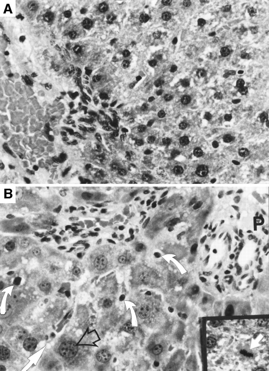
(A) Histologic analysis of tissues in LEA rat liver showed completely normal histology. (B) Liver in LEC rats showed multiple abnormalities, including bile duct proliferation and fibrosis as seen here in portal area (P), as well as apoptosis (curved arrows), fatty change (solid straight arrow), and polyploidy (open arrow), which refers to abnormally enlarged nuclei containing multiple DNA copies. Inset shows metaphase (arrow) indicating increased mitotic activity.
Histologic Analysis of Hepatic Lesions
Laboratory Data
Serum albumin and bilirubin levels were normal in all animals studied. Serum ALT levels were several times higher in LEC rats than in LEA rats. Serum ceruloplasmin was normal in LEA rats but was undetectable in LEC rats. The hepatic copper content was normal in LEA rats but was markedly increased (mean, 68-fold greater) in LEC rats. Similarly, the biliary copper excretion capacity, normal in the LEA group, was markedly impaired in LEC rats. Hepatic ATP7B mRNA, present in all LEA rats, was undetectable in LEC rats (Table 3) .
Differences Between LEA and LEC Rats
Mebrofenin Handling
The cardiac and hepatic blood-pool activities were similar in LEA and LEC rats during the initial 3 min (P = 0.2, not statistically significant, Student t test). In the LEA rat group, prompt uptake of mebrofenin by the liver occurred (Fig. 2A). Activity was distributed uniformly throughout the liver, with prompt clearance. Although activity appeared promptly in the liver of LEC rats, in contrast to LEA rats, peak activity occurred later and clearance was slower (Fig. 2B).
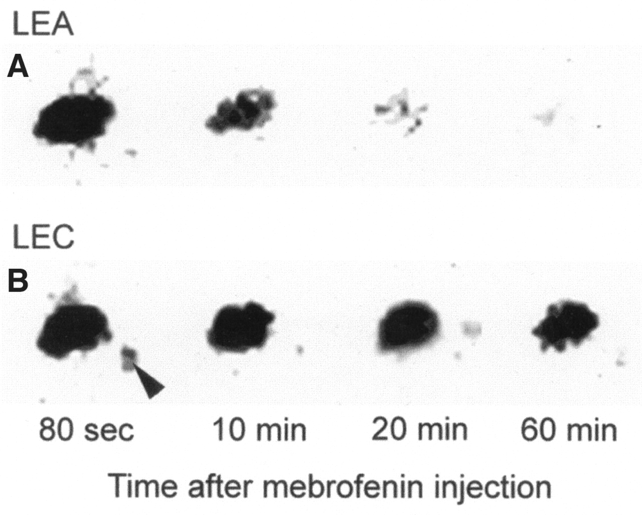
Mebrofenin imaging shows kinetics of hepatic mebrofenin uptake and excretion in representative rats. Sequential images from same animal are shown. (A) LEA rat shows prompt mebrofenin clearance from liver, such that at 20 min after mebrofenin injection, significant amount of activity had cleared. (B) LEC rat shows considerably longer retention of mebrofenin activity in liver. In this animal, exceptionally large amount of activity was present in liver even at 60 min after mebrofenin injection. Arrowhead indicates splenic site of mebrofenin injection.
Mebrofenin was rapidly incorporated by LEA rats, with maximal 99mTc activity persisting briefly before declining subsequent to the onset of biliary excretion. In LEC rats, hepatic tracer incorporation was far less efficient and required much longer times (Fig. 3). Peak hepatic mebrofenin accumulation required a 3.5-fold greater time in LEC rats than in LEA rats. Similarly, the time required for the removal of half-maximal 99mTc counts (T1/2 peak) was 3.5-fold greater in LEC rats than in LEA rats (Table 4).

Representative hepatic time–activity curves derived from mebrofenin imaging. Data are from 1 LEA rat (A) and 1 LEC rat (B). For superior graphic representation, data are restricted to initial 20 min after mebrofenin administration. Mebrofenin activity declined earlier in LEA rat than in LEC rat.
Hepatic Mebrofenin Handling
In LEA rats, 90% ± 8% of total activity excreted during the 60-min study was excreted during the first 20 min after injection, and 98% ± 1% was excreted by 40 min. In LEC rats, 64% ± 24% of the activity was excreted during the first 20 min, and 84% ± 14% was excreted during the first 40 min. These differences were significant (P = 0.005 and 0.002, respectively). Measurement of retained hepatic activity, after the animals were killed at 1 h, revealed that the liver of LEC rats contained 21-fold greater activity than the liver of LEA rats.
The Tpeak and T1/2 peak of hepatic 99mTc-mebrofenin activity correlated positively with each other (r = 0.83; P < 0.001) (Fig. 4), and both correlated with hepatic mebrofenin retention. Tpeak showed a correlation of 0.93 with hepatic mebrofenin retention at 20 min (P < 0.001) and of 0.83 at 60 min (P < 0.001). Similarly, T1/2 peak showed a correlation of 0.97 with hepatic mebrofenin retention at 20 min (P < 0.001) and of 0.93 at 60 min (P < 0.001).
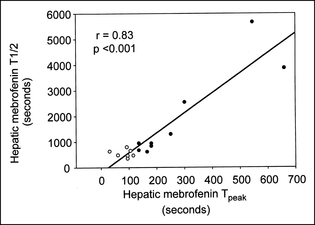
Mebrofenin-handling parameters were internally consistent, showing excellent positive correlation between Tpeak of mebrofenin uptake and T1/2 peak (T1/2) of mebrofenin retention in liver. ○ = LEA rats; • = LEC rats.
Hepatic Copper Handling, Liver Structure–Function Relationships, and Mebrofenin Handling
We first determined whether hepatic copper content correlated with our histologic grading of liver injury and with impairment of biliary copper excretion capacity. The analysis included all LEA and LEC rats studied, such that the parameters analyzed represented a range that would not have been possible if the animals had been analyzed as separate groups. The histologic grade correlated significantly with hepatic copper content (r = 0.83; P < 0.001) (Fig. 5). The correlation between hepatic copper content and biliary copper excretion capacity was also significant, albeit relatively less strong (r = 0.55; P < 0.02).

Correlation between hepatic copper content and graded evaluation of liver histology. Data are from all LEC and LEA rats studied. Highly significant association existed between these parameters. Additional relationships were observed between hepatic copper and other parameters. These findings indicate that comparison of mebrofenin-handling parameters with selected parameters of liver disease in LEC rats would be useful. ○ = LEA rats; • = LEC rats.
Elevations in serum ALT levels, the only abnormal liver finding in LEC rats, correlated well with hepatic copper content (r = 0.76; P = 0.02) and with copper excretion capacity (r = −0.79; P = 0.003). Serum ALT levels did not correlate well with histologic findings (r = 0.51; P = 0.10).
Table 5 shows correlations between mebrofenin handling and hepatic structure–function analysis. Hepatic copper content correlated positively with multiple parameters dealing with hepatic mebrofenin handling, including Tpeak. Hepatic copper content correlated negatively with biliary mebrofenin excretion. These correlation coefficients ranged from 0.8 to 0.9 and indicated that low hepatic copper content was associated with efficient mebrofenin handling in LEA rats, whereas LEC rats with marked copper accumulation handled mebrofenin less well.
Structure–Function Correlations
The histologic grade showed positive correlations, of an approximate range of 0.7–0.9, with mebrofenin incorporation (Tpeak) and mebrofenin excretion. Similarly, serum ALT levels showed significant correlations between mebrofenin accumulation and excretion parameters, ranging from 0.6 to 0.7. Stimulated bile copper excretion also showed a limited negative correlation with mebrofenin retention and a positive correlation with biliary mebrofenin excretion.
DISCUSSION
This investigation offers insights into the hepatic handling of mebrofenin in Wilson’s disease. Multiple parameters concerning hepatic mebrofenin uptake and excretion, including Tpeak, T1/2 peak, hepatic retention, and fractional biliary excretion, were abnormal in LEC rats. The greater mebrofenin activity retained in the liver of LEC rats was related to copper accumulation in the liver, to morphologically apparent abnormalities in hepatocytes, and to hepatic injury, as reflected by an increased serum ALT level in LEC rats. A lack of correlation between serum ALT and histologic findings was not surprising, because the magnitude of ALT elevation may not correlate directly with the magnitude of histologically apparent hepatitis.
Multiple mebrofenin imaging parameters were identified that correlated with various aspects of Wilson’s disease. The Tpeak (mebrofenin incorporation), T1/2 peak (mebrofenin excretion), and mebrofenin retention at 20 and 60 min provided useful information and should be helpful in translating the findings into clinical applications. In several ways, these parameters are dependent on one another. Tpeak represents the efficacy of the initial mebrofenin incorporation, which has been validated by 2-compartment modeling, taking into account fluxes between systemic and hepatic blood pools and hepatocyte uptake (1,2). Hepatic extraction fraction, measured by deconvolutional analysis, is a true measure of first-pass extraction, because the blood pool is considered. Although deconvolutional analysis was not performed as part of this investigation, hepatic and cardiac blood pools of both LEA and LEA rats were analyzed, and a reasonable approximation of hepatic extraction could thus be made. Such analysis showed that hepatic extraction efficiency for mebrofenin follows first-order kinetics and is extraordinarily high. The similar depletion of the reference systemic (cardiac) blood pool in both groups, at early times before the commencement of biliary excretion, indicates that hepatic extraction of the agent was efficient in both LEA and LEC rats. Our findings of efficient mebrofenin extraction are similar to those of previous studies in which mebrofenin was administered either into the systemic circulation through the inferior vena cava or into the portal circulation through the superior mesenteric vein (1,5,6).
Mebrofenin excretion into bile was clearly impaired in LEC rats. T1/2 peak of mebrofenin excretion, as well as mebrofenin accumulation at late periods, represented this process. Abnormalities in Tpeak of mebrofenin incorporation correlated strongly with parameters of mebrofenin excretion, suggesting that somehow these 2 processes are linked. If so, T1/2 peak reflects the balance between hepatic extraction, retention, and biliary mebrofenin excretion. Although organic anions related to mebrofenin (20), have been found to follow paradigms for carrier-dependent transport in hepatocytes, the identity of specific transporters involved in both hepatic uptake and excretion of mebrofenin is unknown. Therefore, it is not currently possible to further define the basis of our observation concerning abnormality in Tpeak and bile excretion parameters of mebrofenin. However, the fact that T1/2 peak of mebrofenin excretion correlated extremely strongly with hepatic mebrofenin retention and impairment of biliary mebrofenin excretion offers several convenient parameters for clinical image analysis. The stimulated copper excretion test showed some correlation between mebrofenin excretion into bile, further emphasizing the potential of mebrofenin imaging in evaluating Wilson’s disease.
Of course, no single available test provides information about global liver function. To assess the severity of liver disease in LEC rats, we attempted to study multiple parameters, including serum bilirubin, which was normal in all animals, and serum ALT, which was abnormal in most LEC rats. We devised an arbitrary grading system to rank the severity of the specific histologic manifestations observed in Wilson’s disease (Table 1). This grading, in agreement with its efficacy, correlated highly with hepatic copper and with mebrofenin excretion and copper excretion capacity. A previous study established that hepatocellular abnormalities precede the onset of other hepatic lesions, such as cholangiofibrosis and oval cell activation in LEC rats (18). We believe that our histologic criteria reflect cellular injury arising from copper-induced free radical injury and lipid peroxidation (21–23). Studies by Chavez-Cartaya et al. (5), Daniel et al. (6), and Matwichuk et al. (24) showed correlations between mebrofenin handling and measures of hepatic dysfunction in response to ischemia-reperfusion injury or drug toxicity. These findings and our results suggest that analysis of hepatic mebrofenin handling should be useful in chronic liver disease in general.
CONCLUSION
Currently, assessment of Wilson’s disease requires an initial liver biopsy to determine hepatic copper content and histologic changes. Noninvasive tests providing reliable estimates of functional liver capacity are not available. Correlation of 99mTc-mebrofenin handling with liver morphology, function, and copper accumulation in LEC rats suggests that mebrofenin scintigraphy can be useful for noninvasively monitoring disease progression and therapeutic response in Wilson’s disease. Although the data were obtained in an animal model of Wilson’ disease, these biochemical parameters likely reflect liver damage in general, suggesting that there may be role for mebrofenin scintigraphy in other chronic liver diseases as well.
Acknowledgments
This study was supported in part by grants RO1 DK46952, P30 DK41296, P30 CA13330, and MO1 RR12248 from the National Institutes of Health; by grant 3894 from the Long Island Jewish Medical Center; by the Wilson’s Disease Association; and by The National Center for the Study of Wilson’s Disease, Inc.
Footnotes
Received Jul. 16, 2001; revision accepted Oct. 22, 2001.
For correspondence or reprints contact: Kuldeep K. Bhargava, PhD, Long Island Jewish Medical Center, 270-05 76th Ave., New Hyde Park, NY 11040.
E-mail: bhargava{at}lij.edu
REFERENCES
In this issue


Jump to section
Related Articles
Cited By...
- Quantitative Assessment of Hepatic Function During Liver Regeneration in a Standardized Rat Model
- Nuclear Imaging Techniques for the Assessment of Hepatic Function in Liver Surgery and Transplantation
- Comparison Between the Values of the Hepatic Uptake Rate Obtained by 2 Methods, Using Hepatobiliary Scintigraphy in Patients with Nonalcoholic Steatohepatitis
- 99mTc-GSA Scintigraphy with SPECT for Assessment of Hepatic Function and Functional Volume During Liver Regeneration in a Rat Model of Partial Hepatectomy
- Regulation of Hepatobiliary Transport Activity and Noninvasive Identification of Cytokine-Dependent Liver Inflammation