


Abstract
99mTc-d,1-hexamethylpropyleneamine oxime (HMPAO) is widely used as a labeling agent for leukocytes in the diagnosis of inflammatory or infectious foci. Cytotoxicity studies have indicated that intracellular labeling of leukocytes with 111In compounds may have severe detrimental effects on the cells. Methods: In this study, the radiotoxic effects on mixed lymphocytes after labeling with 99mTc-HMPAO was investigated using the cytokinesis-blocked micronucleus assay and chromosomal aberration assay. Results: Whereas negligible numbers of chromosome abnormalities were noted in unlabeled lymphocytes, the labeled lymphocytes showed multiple aberrations of various types, including dicentric, tricentric, and fivecentric chromosomes; centric rings; chromosome and chromatid type breaks; and acentric fragments. Conclusion: Heavily aberrant lymphocytes are seen in 99mTc-HMPAO–labeled mixed leukocytes after routine clinical procedures. It is unlikely, however, that this would cause detrimental effects, such as lymphoid malignancy, as these cells would normally be eliminated through apoptosis or phagocytosis.
Radiolabeled autologous leukocytes are used to image infection, inflammation, and other immunogenic processes. Various cytotoxicity studies have indicated that intracellular 111In labeling of leukocytes may have detrimental effects on white cells (1). These detrimental effects are mainly caused by Auger electrons. In vitro studies have shown that the dose received by cells is highly dependent on whether an Auger electron emitter is internalized by the cells or not (2). As an alternative to lipophilic chelates of 111In, 99mTc-d,1-hexamethylpropyleneamine oxime (HMPAO) labeling is routinely performed on mixed leukocyte preparations. This agent diffuses through the cell membrane and binds to intracellular bodies because of its lipophilicity. However, the decay (deexcitation) of 99mTc-labeled nuclei also results in the emission of electrons. These electrons may have intermediate energy (internal conversion) or low energy (Auger electrons). The latter electrons have a short range equal to or less than the cell diameter. The intracellular energy deposit of Auger electrons may cause chromosomal abnormalities. The current study reports on chromosomal damage caused by the 99mTc labeling of lymphocytes.
MATERIALS AND METHODS
Cell Separation and Labeling
Blood samples (50 mL) from 22 volunteers with no history of recent illness were collected in syringes with citric acid dextrose A. Cell counting of the samples was performed by CTKS (Cincinnati, OH) Coulter system (mean white blood cell count, 6,200 ± 1,080 cells per milliliter). Cell-rich plasma was obtained after sedimentation for 45–60 min at 37°C in the presence of 2.5 mL hydroxyethyl starch solution (6%) (Plasmasteril; Fresenius Laboratories, Hamburg, Germany). After centrifugation at 200g for 10 min, leukocytes were isolated on Ficoll gradient and divided into 2 equal fractions. For mixed leukocyte labeling, 518–740 MBq (14–20 mCi; mean, 17.6 ± 0.40 SE) of freshly prepared 99mTc-HMPAO in 1 mL 0.9% NaCl, a dose commonly used in clinical practice, were added to the leukocyte suspension. The unlabeled fraction of the cell suspension was used as a control. 99mTc-HMPAO (Ceretec; Amersham, Chalfont, U.K.) was prepared according to manufacturer instructions, after which a standard quality control was performed. This commercially available kit contains 0.5 mg exametazim (HMPAO), 7.6 μg stannous (II) chloride, and 4.5 mg sodium chloride per ampule.
After incubation for 15 min at room temperature, the labeled cells were washed and resuspended in cell-poor plasma. After centrifugation and removal of the unbound activity, the labeling efficiency was determined and expressed as cell-bound activity/(bound + unbound activity).
The resulting pellet was resuspended in 2 mL Roswell Park Memorial Institute (RPMI) 1640 culture medium (Gibco, Paisley, U.K.). Of this suspension, 0.2 mL was used for cell counting and 1.8 mL was divided into 2 fractions for the micronucleus assay and cytogenetic analysis. The number of lymphocytes in the cell suspensions ranged between 2.1 × 10⁶/mL and 3.5 × 10⁶/mL.
Cytogenetic Analysis
After labeling of the mixed leukocytes with 99mTc-HMPAO, cytogenetic damage in lymphocytes was assessed using the cytokinesis-blocked micronucleus and chromosomal aberration assays.
Micronucleus Assay
Lymphocytes were cultured at 37°C for 70 h at an initial density of 2 × 10⁶ cells per milliliter. Culture medium consisted of RPMI 1640 with 2 mmol/L l-glutamine and N-(2-hydroxyethyl)piperazine-N′-(2-ethanesulfonic acid) buffer (Gibco) supplemented with 15% fetal calf serum (Gibco) and antibiotics (10,000 IU/mL penicillin and 10 mg/mL streptomycin in 0.9% NaCl). Phytohemagglutinin (M form; Sigma Chemical Co., St. Louis, MO) at a final concentration of 5 μg/mL was used to stimulate cell division. Cytochalasin B (Sigma) in a final concentration of 3 μg/mL was added to culture medium 42 h after initiation of the cultures to block cytokinesis. At 72 h after culture initiation, cells were harvested, treated with a hypotonic solution containing 0.075 mol/L KCl, and fixed with a mixture of methanol:glacial acetic acid (6:1). Slides were stained with Giemsa (Merck; Darmstadt, Germany). Micronuclei were assessed in binucleated cytokinesis-blocked cells by 2 experienced cytogeneticists. One thousand binucleated cells from various slides were scored per subject.
Chromosomal Aberration Assay
Lymphocytes from labeled and unlabeled preparations were cultured at 37°C for 48 h in McCoy’s medium (Gibco) supplemented with phytohemagglutinin (5 μg/mL, M form; Sigma), and streptomycin (10 mg/mL; Gibco). Colchicine (10 μg/mL; Irvine Scientific, Santa Ana, CA) at a concentration of 0.25 mmol/L was added at 2 and 5 h before the end of the culture time. The cells were given a hypotonic shock with KCl and then fixed in a 1:3 acetic acid:ethanol solution. Thirty well-spread metaphase plates for each subject–treatment combination were evaluated for the presence of chromosomal aberrations after Giemsa staining.
Statistical Analysis
The paired t test was used for statistical analysis of MN scoring. A probability value of 0.05 was considered significant. Statistical analysis for the evaluation of chromosomal aberrations was not performed because of the very large number of aberrations in the labeled cells. Values after frequencies, efficiencies, and other results are SEM.
RESULTS
For the 22 blood samples, the labeling efficiency, defined as the 99mTc activity in the final cellular suspension in relation to the initial 99mTc activity, expressed as a percentage, amounted to 49.5% ± 2.2%. The mean cell-bound activity in the cell pellet was 325.6 ± 18.5 MBq (8.8 ± 0.5 mCi). The radiochemical purity was 85.2% ± 1.7%, and the cell viability assessed by Trypan blue exclusion was 95.5% ± 1.8%.
The mean number of micronuclei observed in lymphocytes from unlabeled preparations was 5.5 ± 1.0/1,000 binucleated cells. The number of micronuclei observed in labeled lymphocytes was 272.2 ± 2.5/1,000 binucleated cells, which was significantly higher than that of unlabeled preparations (P < 0.001). All investigated lymphocytes in the labeled preparations carried multiple aberrations: dicentrics, tricentrics, fivecentrics, centric rings, and chromosome and chromatid type breaks, as well as acentric fragments. None of the metaphase plates was considered normal. The number and types of aberrations per cell were not quantified because of the presence of multiple breaks. In some metaphases, triradial configurations (as observed in chromosomal break syndromes) and complex chromosomal rearrangements were observed. Figures 1 and 2 show examples of lymphocytes from labeled preparations in which the chromosomes show multiple asymmetric aberrations. The chromosomes in unlabeled lymphocytes showed virtually no aberrations.

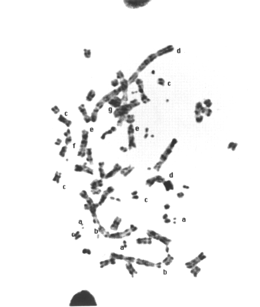
Chromosomes of labeled lymphocyte with multiple aberrations. a = chromosome fragments; b = fivecentric chromosome; c = tricentric chromosome; d = chromatid break; e = acentric fragment; f = dicentric chromosome.
Metaphase with multiple aberrations. a = chromosome fragments; b = chromatid break; c = acentric fragment; d = fourcentric chromosome; e = dicentric chromosome; f = chromosome break.
DISCUSSION
The results of this study show that the chromosome-breaking effect of 99mTc-HMPAO labeling on lymphocytes (mixed leukocytes) is considerable and leads to various types of chromosome and chromatic aberrations. Judging from the presence of multiple aberrations in all metaphase plates studied and the tremendous increase in frequency of micronucleated and binucleated cells, it is estimated that the radiation dose received by the labeled cells was at least 5 Gy. The aberrations observed in this study are typically radiation induced and, in our opinion, most likely not caused by constituents of the labeling kit or the labeling procedure itself. This is confirmed by experiments by Van de Wiele et al. (3), which indicated that unlabeled cells and cells labeled with cold HMPAO showed normal chemotactic and apoptotic behavior, whereas 99mTc-HMPAO–labeled cells showed considerable abnormalities. Moreover, experiments by Schmidt et al. (4) and de Labriolle-Vaylet et al. (5) have shown that lymphocyte viability and the frequency of chromosomal aberrations in unlabeled cells and cold-HMPAO–labeled cells were similar. In contrast, lymphocytes labeled with 99mTc-HMPAO showed numerous chromosomal abnormalities. In addition, studies on [3H]-thymidine incorporation to assess DNA synthesis showed the impaired function of 99mTc-HMPAO lymphocytes, whereas no abnormalities were noticed in lymphocytes incubated in saline or labeled with cold HMPAO (5). It should be noted that all the above-mentioned authors used the same labeling kit and labeling procedure as were used in the current study. We believe, therefore, that neither the kit constituents nor the labeling techniques, such as cell separation and centrifugation, could have caused the chromosomal aberrations observed in our study, and that these aberrations were almost certainly caused by radiation from 99mTc.
It has been pointed out that, with stable cellular uptake over time, the radiation dose at the cellular level may be underestimated considerably compared with conventional dosimetry if the energy of the emitted electrons is <10 keV (2). Dose-dependent depression of the proliferative capability of lymphocytes, pronounced aberrations in lymphocyte migration patterns, and serious chromosomal aberrations have been observed using 111In labeling of mixed white cells (1,6). Data on the effects of 99mTc-HMPAO labeling are scarce. In a microautoradiographic study, it was shown that labeling was heterogeneous, which prevented calculation of the mean absorbed dose. In that study, mainly dicentric and ring chromosomal abnormalities were observed (5).
In the current study, multiple aberrations were observed in labeled cell preparations, whereas virtually no abnormalities were seen in unlabeled cell preparations. The aberrations were observable because the lymphocytes were stimulated to divide in culture (in vitro), with the consequence that radiation-induced DNA single- or double-strand breaks could be converted into chromosomal aberrations. As a result of this induced DNA damage, the circulating lymphocytes of a patient would likely be eliminated from the bloodstream through apoptosis or phagocytosis. This is in agreement with recent experiments by Thierens et al. (7), who reported on the longer-term fate of 111In- and 99mTc-labeled lymphocytes. Their experiments indicated that within a few days after injection, most of the radioactivity was transferred to noncirculating cells, presumably macrophages present in the spleen and lymph nodes. The observation of unstable chromosomal aberrations in circulating lymphocytes is a sign of damage caused by ionizing radiation. The scoring of these unstable aberrations is used as a biologic dosimeter (8).
Apart from the induction of unstable chromosome aberrations, there was undoubtedly also a simultaneous induction of stable chromosome aberrations such as translocations. Such stable aberrations could theoretically represent a cancer risk through activation of oncogenes or deactivation of cancer suppression genes. Because this study clearly showed the presence of multiple unstable aberrations in all tested labeled lymphocytes, there is, in our opinion, no possibility that stable aberrations in the same cells would be able to manifest themselves. Thus, the type of diagnostic procedures used in this study would not represent a risk for patients to develop long-term adverse consequences, such as cancer.
CONCLUSION
Heavily aberrant lymphocytes were seen in 99mTc-HMPAO–labeled mixed leukocytes after routine clinical procedures. It is considered unlikely, however, that these aberrations would cause detrimental effects such as lymphoid malignancy, as the aberrant cells would likely be eliminated through apoptosis or phagocytosis.
Acknowledgments
The authors acknowledge the scientific support of Dr. Ad D. Tates, Department of Radiation Genetics and Chemical Mutagenesis, Leiden University Medical Center, The Netherlands. The authors are indebted to Tilly P.M. Hagendoorn for her excellent secretarial assistance. This study was financially supported by the Research Foundation of Osmangazi University, Eskişehir, Turkey.
Footnotes
Received Apr. 9, 2001; revision accepted Oct. 15, 2001.
For correspondence or reprints contact: Ilknur Ak, MD, Department of Nuclear Medicine, Medical Faculty, Osmangazi University, Eskisehir, Turkey.
E-mail: t.hagendorn{at}lumc.nl


