
Main menu

  • Home
  • Content
    • Current
    • Ahead of print
    • Past Issues
    • JNM Supplement
    • SNMMI Annual Meeting Abstracts
    • Continuing Education
    • JNM Podcasts
  • Subscriptions
    • Subscribers
    • Institutional and Non-member
    • Rates
    • Journal Claims
    • Corporate & Special Sales
  • Authors
    • Submit to JNM
    • Information for Authors
    • Assignment of Copyright
    • AQARA requirements
  • Info
    • Reviewers
    • Permissions
    • Advertisers
  • About
    • About Us
    • Editorial Board
    • Contact Information
  • More
    • Alerts
    • Feedback
    • Help
    • SNMMI Journals
  • SNMMI
    • JNM
    • JNMT
    • SNMMI Journals
    • SNMMI

User menu

  • Subscribe
  • My alerts
  • Log in
  • My Cart

Search

  • Advanced search
Journal of Nuclear Medicine
  • SNMMI
    • JNM
    • JNMT
    • SNMMI Journals
    • SNMMI
  • Subscribe
  • My alerts
  • Log in
  • My Cart
Journal of Nuclear Medicine

Advanced Search

  • Home
  • Content
    • Current
    • Ahead of print
    • Past Issues
    • JNM Supplement
    • SNMMI Annual Meeting Abstracts
    • Continuing Education
    • JNM Podcasts
  • Subscriptions
    • Subscribers
    • Institutional and Non-member
    • Rates
    • Journal Claims
    • Corporate & Special Sales
  • Authors
    • Submit to JNM
    • Information for Authors
    • Assignment of Copyright
    • AQARA requirements
  • Info
    • Reviewers
    • Permissions
    • Advertisers
  • About
    • About Us
    • Editorial Board
    • Contact Information
  • More
    • Alerts
    • Feedback
    • Help
    • SNMMI Journals
  • View or Listen to JNM Podcast
  • Visit JNM on Facebook
  • Join JNM on LinkedIn
  • Follow JNM on Twitter
  • Subscribe to our RSS feeds
Meeting ReportPhysics, Instrumentation & Data Sciences - Data Sciences

Automated segmentation of lesions in [18F]FDG PET/CT images of lung cancer patients: external evaluation of an AI-driven lesion segmentation tool (LION)

Mathilde Droguet, Lalith Kumar Shiyam Sundar, Manuel Pires, Narinée Hovhannisyan Baghdasarian, Nicolas Captier, Marie Luporsi, Erwin Woff, Christophe Nioche, Frédérique Frouin, Thomas Beyer, Nicolas Girard, Irene Buvat and Fanny Orlhac
Journal of Nuclear Medicine June 2024, 65 (supplement 2) 241927;
Mathilde Droguet
1Institut Curie, Inserm, Université PSL, U1288 LITO
  • Find this author on Google Scholar
  • Find this author on PubMed
  • Search for this author on this site
Lalith Kumar Shiyam Sundar
2Medical University of Vienna
  • Find this author on Google Scholar
  • Find this author on PubMed
  • Search for this author on this site
Manuel Pires
3Medical University of Vienna, Austria
  • Find this author on Google Scholar
  • Find this author on PubMed
  • Search for this author on this site
Narinée Hovhannisyan Baghdasarian
4LITO, Institut Curie
  • Find this author on Google Scholar
  • Find this author on PubMed
  • Search for this author on this site
Nicolas Captier
5Institut Curie
  • Find this author on Google Scholar
  • Find this author on PubMed
  • Search for this author on this site
Marie Luporsi
6Curie Institute
  • Find this author on Google Scholar
  • Find this author on PubMed
  • Search for this author on this site
Erwin Woff
7Institut Jules Bordet, Anderlecht, Belgium
  • Find this author on Google Scholar
  • Find this author on PubMed
  • Search for this author on this site
Christophe Nioche
5Institut Curie
  • Find this author on Google Scholar
  • Find this author on PubMed
  • Search for this author on this site
Frédérique Frouin
1Institut Curie, Inserm, Université PSL, U1288 LITO
  • Find this author on Google Scholar
  • Find this author on PubMed
  • Search for this author on this site
Thomas Beyer
2Medical University of Vienna
  • Find this author on Google Scholar
  • Find this author on PubMed
  • Search for this author on this site
Nicolas Girard
  • Find this author on Google Scholar
  • Find this author on PubMed
  • Search for this author on this site
Irene Buvat
4LITO, Institut Curie
  • Find this author on Google Scholar
  • Find this author on PubMed
  • Search for this author on this site
Fanny Orlhac
1Institut Curie, Inserm, Université PSL, U1288 LITO
  • Find this author on Google Scholar
  • Find this author on PubMed
  • Search for this author on this site
  • Article
  • Figures & Data
  • Info & Metrics
Loading

Abstract

241927

Introduction: Reliable and automatic lesion segmentation adds quality to the exploration of prognostic image-based biomarkers by reducing delineation time and inter/intra-observer variability. We investigated the performance of an automatic lesion segmentation tool based on deep learning (LION, Medical University of Vienna, Austria) to segment lesions on whole-body [18F]FDG-PET/CT images of lung cancer patients.

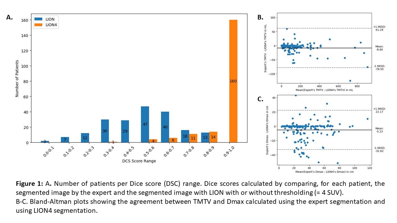
Methods: LION has been trained using an open-source dataset (autoPET challenge) comprising more than 1,000 [18F]FDG-PET studies of patients with malignant melanoma, lymphoma, or lung cancer, and of control subjects without any cancer (). We tested LION on an independent database of baseline [18F]FDG-PET/CT images from patients with advanced Non-Small Cell Lung Cancer (NSCLC) subsequently treated with immunotherapy in our institution. The reference segmentation was performed manually by an experienced nuclear medicine physician (expert). To refine lesion delineation and ensure comparability between segmentations, we applied a threshold of 4 SUV to the segmentations produced both by LION (called LION4) and by the expert. The Dice score (DSC) was calculated for each patient. Two PET biomarkers were derived from the segmentations using LIFEx v.7.4.2 [Nioche et al. Cancer Res. 2018]: Total Metabolic Tumour Volume (TMTV) and Maximum Distance between two lesions (Dmax). Bland-Altman analyses were performed to characterize the differences in TMTV and Dmax between the two segmentations. The ability of features to stratify patients according to Overall Survival (OS) was compared using a Kaplan-Meier analysis. Patients were stratified in 2 groups using a cut-off value that maximized the log-rank test statistic for OS.

Results: A total of 196 patients were included in this study. LION segmentation yielded a mean (±1 sd) DSC of 0.5±0.2 (range: [0.0-0.9], Figure 1) across all patients before applying a threshold of 4 SUV. The mean DSC increased to 0.9±0.1 ([0.3-1.0]) after thresholding, with 82% of patients with DSC higher than 0.9. The Bland-Altman analysis showed that TMTV and Dmax measured from LION4 tended to be larger than those from the expert with a mean difference equal to 9±36 mL (Figure 1) and 5±14 cm respectively. Visual inspection showed that in cases of disagreement, LION4 had either incorrectly included regions with atypical physiological uptake, or omitted tumor regions with an uptake higher than 4 SUV. TMTV and Dmax extracted from the LION4 segmentations could both distinguish two survival profiles in patients (log-rank test: p<0.001, Figure 2). Results were similar to those obtained with the expert segmentations. Median survivals were the same between the expert and LION4 for TMTV in low- and high-risk groups (low-risk: 1082 days (d), high-risk: 489 d) and were close for Dmax (low-risk: 1286 d for the expert vs not reached for LION4; high-risk: 535 d vs 650 d respectively).

Conclusions: Automated lesion segmentation such as that provided by LION has the potential to improve inter and intra-reader reproducibility as evidenced by equivalent TMTV and Dmax values compared to the expert segmentation for lung cancer patients. Nonetheless, further training of the algorithm is ongoing to help reduce the differences still observed when comparing with expert segmentation.

Figure
  • Download figure
  • Open in new tab
  • Download powerpoint
Figure
  • Download figure
  • Open in new tab
  • Download powerpoint
Previous
Back to top

In this issue

Journal of Nuclear Medicine
Vol. 65, Issue supplement 2
June 1, 2024
  • Table of Contents
  • Index by author
Article Alerts
Sign In to Email Alerts with your Email Address
Email Article

Thank you for your interest in spreading the word on Journal of Nuclear Medicine.

NOTE: We only request your email address so that the person you are recommending the page to knows that you wanted them to see it, and that it is not junk mail. We do not capture any email address.

Enter multiple addresses on separate lines or separate them with commas.
Automated segmentation of lesions in [18F]FDG PET/CT images of lung cancer patients: external evaluation of an AI-driven lesion segmentation tool (LION)
(Your Name) has sent you a message from Journal of Nuclear Medicine
(Your Name) thought you would like to see the Journal of Nuclear Medicine web site.
Citation Tools
Automated segmentation of lesions in [18F]FDG PET/CT images of lung cancer patients: external evaluation of an AI-driven lesion segmentation tool (LION)
Mathilde Droguet, Lalith Kumar Shiyam Sundar, Manuel Pires, Narinée Hovhannisyan Baghdasarian, Nicolas Captier, Marie Luporsi, Erwin Woff, Christophe Nioche, Frédérique Frouin, Thomas Beyer, Nicolas Girard, Irene Buvat, Fanny Orlhac
Journal of Nuclear Medicine Jun 2024, 65 (supplement 2) 241927;

Citation Manager Formats

  • BibTeX
  • Bookends
  • EasyBib
  • EndNote (tagged)
  • EndNote 8 (xml)
  • Medlars
  • Mendeley
  • Papers
  • RefWorks Tagged
  • Ref Manager
  • RIS
  • Zotero
Share
Automated segmentation of lesions in [18F]FDG PET/CT images of lung cancer patients: external evaluation of an AI-driven lesion segmentation tool (LION)
Mathilde Droguet, Lalith Kumar Shiyam Sundar, Manuel Pires, Narinée Hovhannisyan Baghdasarian, Nicolas Captier, Marie Luporsi, Erwin Woff, Christophe Nioche, Frédérique Frouin, Thomas Beyer, Nicolas Girard, Irene Buvat, Fanny Orlhac
Journal of Nuclear Medicine Jun 2024, 65 (supplement 2) 241927;
Twitter logo Facebook logo LinkedIn logo Mendeley logo
  • Tweet Widget
  • Facebook Like
  • Google Plus One
Bookmark this article

Jump to section

  • Article
  • Figures & Data
  • Info & Metrics

Related Articles

  • No related articles found.
  • Google Scholar

Cited By...

  • No citing articles found.
  • Google Scholar

More in this TOC Section

  • Artificial Intelligence-Accelerated Parametric Imaging of Myocardial Blood Flow with Rubidium-82 PET
  • PET-Disentangler: PET lesion segmentation via disentangled healthy and disease feature representations
  • Comparative analysis of large language models: ChatGPT and Google Bard answer non-expert questions related to the diagnostic and therapeutic applications of prostate-specific membrane antigen (PSMA) in patients with prostate cancer
Show more Physics, Instrumentation & Data Sciences - Data Sciences

Similar Articles

SNMMI

© 2025 SNMMI

Powered by HighWire